Radiographic Contrast Agents And Contrast-induced Nephropathy
All contrast agents have a basic structure of a benzene ring, which is composed of 6 joined carbon atoms, each of which has an attached hydrogen atom.
Contrast media consist of triiodinated benzene rings, whereby 3 hydrogen atoms are replaced with attached iodine atoms.
Monomers contain 1 triiodinated benzene ring, and dimers contain 2 triiodinated benzene rings
Contrast induced nephropathy (CIN) is agenerally reversible form of acute kidney injury (AKI) that occurs soon after the administration of radiocontrast media.
1. Radiocontrast agents, also known as contrast media, are substances used to improve the visibility of internal organs and structures during medical imaging. The most common types are iodine-based agents used for computed tomography and angiography, and gadolinium-based agents used for magnetic resonance imaging.
2. Contrast-induced nephropathy (CIN) refers to acute kidney injury caused by radiocontrast agents in patients with underlying renal impairment or risk factors. Preventing CIN involves identifying at-risk patients, minimizing contrast volume, using iso-osmolar or low-osmolar agents, intravenous hydration before and after exposure, and holding nephrotoxic drugs like metformin.
3
This document summarizes the Simplicity HTN-2 and Simplicity HTN-3 clinical trials that evaluated the effectiveness of renal denervation for treating resistant hypertension. The Simplicity HTN-2 trial found that renal denervation significantly reduced blood pressure in patients with resistant hypertension compared to a control group. However, the larger Simplicity HTN-3 trial, which was blinded and sham-controlled, did not find a significant reduction in blood pressure between the renal denervation and sham groups at 6 months.
CT calcium scoring can detect asymptomatic coronary artery disease by identifying coronary artery calcification. Calcium starts accumulating early in atherosclerosis and increases as the disease progresses. While a calcium score of zero does not rule out disease, it is associated with a very low risk of cardiac events. Higher calcium scores correlate with increased risk. CT calcium scoring provides individualized risk assessment and can guide aggressive risk factor modification in high-risk patients.
Contrast echocardiography uses microbubble ultrasound contrast agents to improve image quality. These microbubbles remain in the intravascular space and allow for assessment of cardiac structure, function, and perfusion. Second generation contrast agents use an inert gas encapsulated by albumin or phospholipid shells. They interact with ultrasound by reflecting at fundamental frequencies and resonating to produce harmonic frequencies. Continuous infusion provides steady contrast levels needed for perfusion assessment. Contrast echocardiography is a non-invasive technique that improves evaluation of the heart.
This document discusses fractional flow reserve (FFR), which is a technique used to functionally assess the significance of coronary artery stenosis. FFR is defined as the ratio of maximum blood flow in a stenotic artery to maximum blood flow if there was no stenosis. It is calculated as the ratio of mean distal coronary pressure (Pd) to mean aortic pressure (Pa) during maximal hyperemia induced by pharmacological agents. An FFR value below 0.75 is associated with inducible ischemia, while a value above 0.80 indicates an insignificant stenosis in most cases. FFR has advantages over angiography alone in evaluating stenosis as it accounts for vessel characteristics like length and takes collateral flow into consideration.
Intraluminal coronary thrombus aspiration in patients with STEMI. Prof. Andre...Chaichuk Sergiy
Intraluminal coronary thrombus aspiration in patients with STEMI was studied in randomized trials. Results showed thrombus aspiration before stenting improved myocardial perfusion scores and ST-segment resolution compared to conventional PCI alone. Meta-analyses found manual thrombus aspiration reduced distal embolization and improved angiographic and electrocardiographic outcomes, while its effect on mortality is unclear. Larger randomized trials are still needed to definitively establish the benefits of routine thrombus aspiration in STEMI.
How to deal with CALCIFIED CORONARY ARTERY LESIONS .Coronary artery calcification (CAC) is highly prevalent in patients with coronary heart disease (CHD) and is associated with major adverse cardiovascular events. There are two recognized type of CAC—intimal and medial calcification, and each of them have specific risk factors. Several theories about the mechanism of vascular calcification have been put forward, and we currently believe that vascular calcification is an active, regulated process. CAC can usually be found in patients with severe CHD, and this asymptomatic phenomenon make early diagnosis of CAC important. Coronary computed tomographic angiography is the main noninvasive tool to detect calcified lesions. Measurement of coronary artery calcification by scoring is a reasonable metric for cardiovascular risk assessment in asymptomatic adults at intermediate risk. To date, effective medical treatment of CAC has not been identified. Several strategies of percutaneous coronary intervention have been applied to CHD patients with CAC, but with unsatisfactory results. Prognosis of CAC is still a major problem of CHD patients. Thus, more details about the mechanisms of CAC need to be elucidated in order to improve the understanding and treatment of CAC.
The document discusses various coronary artery anomalies including anomalies of origination, course, and intrinsic anatomy. Some key points include:
- Coronary artery anomalies have a global incidence of 5.64% and incidence of sudden death is 0.6%
- Anomalous origination of the left main coronary artery from the pulmonary artery (ALCAPA) is a rare but serious anomaly if left untreated
- Certain anomalous coronary artery courses, such as between the aorta and pulmonary artery, are associated with higher risks of sudden cardiac death
- Other anomalies discussed include single coronary arteries, coronary hypoplasia, ectasia/aneurysms, and intramural coronary arteries
1) Natriuretic peptides like BNP and NT-proBNP are the most extensively studied and used biomarkers in heart failure. They are useful for diagnosis, assessing prognosis, and monitoring response to treatment.
2) Other biomarkers like troponins, ST2, galectin-3, and inflammatory markers can provide additional prognostic information beyond natriuretic peptides.
3) Biomarkers reflect different pathophysiological processes in heart failure like myocyte injury, stress, remodeling, neurohormonal activation, and inflammation. Used together, they can improve risk stratification and guidance of therapy.
Cardiac catheteriztion, Oximetery study in a patient with VSDPRAVEEN GUPTA
In this ppt i am going to discuss how to do cardiac catheterisation study, oximetry study and how to analyse its data in a patient with VSD who came to our hospital
Raja Lahiri provides an overview of coronary angiography. Key points include:
- Coronary angiography is the current gold standard for visualizing the coronary arteries through X-ray imaging with contrast injection.
- The history of coronary angiography began in the 1920s-1940s with early experiments in cerebral and cardiac catheterization.
- Modern techniques involve accessing arteries typically through the femoral or radial arteries to insert a catheter for contrast injection into the coronary arteries under X-ray imaging.
- Multiple angiographic views are needed to visualize different segments of the left and right coronary arteries. Coronary angiography is used to evaluate coronary artery disease, graft patency, and left ventricular function.
This document discusses contrast-induced nephropathy (CIN). It describes the types of contrast agents used, risk factors for CIN, methods for prevention including volume expansion with intravenous or oral hydration, and diagnostic criteria for CIN. The nephrotoxic effects of contrast agents increase with higher osmolality, larger volume, repeated or intra-arterial administration, and can be prevented through adequate hydration before and after exposure.
Intravascular ultrasound (IVUS) uses sound waves to visualize the inside of arteries. There are two types of IVUS systems - mechanical systems using a rotating internal cable and solid-state systems using externally mounted transducers. Both produce 360-degree images with a resolution of 100-150 μm. IVUS is used to assess plaque, vessel dimensions, stent deployment, and more. It produces cross-sectional images showing the lumen, layers of the artery wall, and plaque composition and size. Measurements include diameters, areas, plaque burden, and indices of eccentricity. IVUS helps identify vulnerable plaque and has diagnostic and interventional applications.
This document discusses contrast agents used in angiography and factors related to contrast-induced nephropathy (CIN). It notes that the route of contrast administration, volume administered, and properties of the specific contrast agent can influence CIN risk. Low-osmolar contrast agents are associated with less adverse effects than ionic agents. Hydration before and after the procedure can help reduce CIN incidence. Gadolinium may be considered as an alternative to iodinated contrast for patients at high risk of CIN.
This document discusses fractional flow reserve (FFR), a technique used during coronary catheterization to measure pressure differences across a coronary stenosis and determine if it is causing myocardial ischemia. An FFR value below 0.75 is considered functionally significant while a value above 0.80 rules out ischemia. FFR is useful for evaluating single-vessel disease, left main stenosis, tandem lesions, diffuse disease, grafts, and ostial lesions. Limitations include inability to assess plaque morphology.
Fractional flow reserve (FFR) is a technique that evaluates the hemodynamic significance of coronary artery stenoses. It is defined as the ratio of maximal flow achievable in the stenotic coronary artery to the maximal flow achievable if the artery was normal. An FFR value ≤ 0.80 is considered hemodynamically significant. Several clinical trials including DEFER and FAME have found that FFR-guided revascularization reduces major adverse cardiac events compared to angiography-guided procedures alone by helping to identify which intermediate lesions are functionally significant. Guidelines recommend using FFR to guide revascularization decisions, especially for intermediate lesions, multivessel disease, and acute coronary syndromes.
The document discusses newer advancements in heart failure device therapy. It summarizes that device therapies have greatly improved outcomes for heart failure patients. Some key devices discussed include implantable cardioverter defibrillators (ICDs) which reduce sudden cardiac death, cardiac resynchronization therapy which improves heart function, and left ventricular assist devices (LVADs) which are increasingly being used as long term support devices or as a destination therapy for end stage heart failure patients. The document provides details on the development, indications, benefits and risks of these various heart failure devices.
This document discusses Eisenmenger syndrome, a condition where pulmonary hypertension develops due to increased blood flow through defects between the systemic and pulmonary circulations. It provides details on causes, clinical features, pathology findings, and treatments. Key points include:
- Eisenmenger syndrome is caused by defects like VSDs, ASDs, and PDA that allow high blood flow to the lungs and cause pulmonary hypertension over time.
- Common causes of death include hemoptysis from pulmonary artery ruptures, heart failure, and complications from attempted defect repair surgery.
- Pathological findings show thickened pulmonary arteries that resemble the fetal pattern and contribute to high pulmonary vascular resistance.
- Medical treatments are generally ineffective once int
Management of AF patients with ACS undergoing PCI.pptxPraveen Nagula
- The AUGUSTUS trial compared apixaban to vitamin K antagonist (VKA) for prevention of thromboembolic events in patients with atrial fibrillation undergoing percutaneous coronary intervention or experiencing acute coronary syndrome.
- Over 4,600 patients were randomized to apixaban 5mg twice daily or adjusted-dose VKA plus a P2Y12 inhibitor with or without aspirin for 6 months.
- The primary outcome was major or clinically relevant non-major bleeding. Apixaban resulted in a 31% relative risk reduction in the primary outcome compared to VKA.
Optical coherence tomography (OCT) provides high-resolution cross-sectional images of tissue structures on the micron scale in situ and in real time. It uses near-infrared light instead of sound like IVUS. OCT images are generated by measuring the echo time delay and intensity of light reflected or backscattered from internal structures using interferometry techniques. OCT can characterize atherosclerotic plaque composition and identify thin fibrous caps. Studies have shown OCT can detect plaque rupture and intracoronary thrombus with higher accuracy than IVUS or angiography.
Contrast-induced nephropathy (CIN) is a type of acute kidney injury caused by iodinated contrast media used in medical imaging procedures. The document defines CIN and discusses its risk factors, pathophysiology, prevention, and management. It summarizes that CIN risk increases with reduced kidney function, diabetes, and other comorbidities. Prevention focuses on identifying at-risk patients, using lower contrast volumes and iso-osmolar agents when possible, and intravenous fluid administration before and after the procedure. Sodium bicarbonate and N-acetylcysteine may provide additional protective effects. For higher risk patients, alternative imaging should be considered to avoid CIN.
This document discusses carotid artery stenting (CAS) as an alternative to carotid endarterectomy (CEA) for treatment of carotid artery stenosis. It provides details on patient selection criteria and describes the CAS procedure, including diagnostic arteriography, embolic protection device placement, stent placement, and post-procedure care. Several major clinical trials are summarized that demonstrated CAS to be non-inferior to CEA for reducing risk of stroke in both symptomatic and asymptomatic patients.
Assessment of prosthetic valve functionSwapnil Garde
This document discusses the assessment of prosthetic valve function through various imaging modalities. It begins with an introduction to prosthetic valves and outlines topics to be covered, including classification of valve types. Evaluation methods like chest x-ray, fluoroscopy, echocardiography, and CT are described. Parameters assessed on each modality and guidelines for evaluation are provided. Complications of prosthetic valves and 3D imaging advances are also mentioned.
The document provides an overview of right ventricular assessment using echocardiography. It discusses normal RV anatomy, segmental nomenclature, and coronary supply. Key metrics for evaluating RV size, wall thickness, function, and pressures are outlined. Normal values and technical aspects of measuring RV dimensions, area/fractional area change, tricuspid annular plane systolic excursion, myocardial velocity, and diastolic function are summarized. Hemodynamic assessment of pulmonary pressures is also reviewed.
Stress echocardiography uses ultrasound imaging during exercise or pharmacologic stress testing to detect ischemia-induced changes in heart wall motion. It has several advantages over nuclear stress testing including better visualization of cardiac structures and no radiation exposure. Various stress agents can be used including exercise, dobutamine, dipyridamole, and adenosine. Detection of new or worsening wall motion abnormalities or improvement after revascularization indicates viable myocardium. Factors like image quality, timing of acquisition, and operator experience can impact test sensitivity. Stress echo is an established technique for diagnosing coronary artery disease and assessing myocardial viability.
Contrast media are agents used to improve visualization of internal structures that otherwise cannot be seen clearly. There are two main types - positive contrast agents that are radiopaque, and negative contrast agents that are not. Iodinated contrast media are most commonly used today. They can be ionic monomers, ionic dimers, non-ionic monomers or non-ionic dimers. The ideal contrast medium has high solubility, stability, low toxicity and viscosity, and is excreted selectively like by the kidneys. Ultrasound contrast agents contain microbubbles that enhance echogenicity and improve tissue contrast. They act by resonating within the ultrasound beam. Newer agents use more stable low solubility gases and encapsulation to increase
1) The document discusses the historical development of radiographic contrast media from its earliest uses in the late 19th century to the development of tri-iodinated benzene derivatives and modern low-osmolar non-ionic contrast agents.
2) It covers the ideal characteristics, classification, and basic chemistry of iodinated contrast media including differences between ionic and non-ionic monomers and dimers.
3) The risks of contrast media administration are discussed including predictable chemo-toxic reactions affecting tissues and organ systems as well as unpredictable anaphylactoid reactions.
Contrast media are agents used to improve visualization of internal structures in medical imaging. They work by increasing the contrast between tissues. Contrast media can be classified as positive or negative based on whether they appear bright or dark on images. The main types of contrast media used are barium sulfate for x-rays and iodinated compounds for CT and angiography. Ultrasound contrast agents contain microbubbles that enhance backscatter and improve tissue contrast on ultrasound images. They allow better evaluation of organ perfusion and detection of abnormalities. Contrast media are important tools that improve diagnostic accuracy in medical imaging.
Contrast agents are substances used in radiography to improve visualization of internal structures. They can be radiopaque (positive contrast) or non-radiopaque (negative contrast). Iodinated contrast media are commonly classified based on their ionicity, osmolality, and viscosity. Low osmolar contrast media including non-ionic dimers and monomers are preferred due to their favorable safety profile. Ultrasound contrast agents contain microscopic gas-filled bubbles that strongly reflect ultrasound waves, increasing echogenicity and tissue contrast.
This document discusses the history and use of contrast media and emergency drugs for radiology procedures. It begins by outlining the early discoveries of contrast agents from the 1890s to the 1920s. It then describes how contrast agents are administered and their properties like osmolality and viscosity. The document discusses common contrast agents like barium, iodine-based agents, and categorizes them as high vs low osmolar. It outlines risks of contrast media and expected adverse reaction rates. The document concludes by listing emergency equipment needed to treat potential severe reactions.
1. Contrast agents are substances that have different atomic numbers or electron densities than surrounding tissues, allowing visualization of internal organs on imaging.
2. Early contrast agents included barium sulfate, iodinated compounds, and air or CO2. Modern agents are classified as ionic or non-ionic monomers and dimers with varying osmolalities and viscosities.
3. Contrast agents are used with multiple imaging modalities like CT, MRI, ultrasound, and fluoroscopy. They are administered orally, intravenously, or directly into structures. The choice depends on the physical properties and safety of the specific agent and the anatomy being imaged.
The document discusses the history and types of contrast media and emergency drugs used in radiology. It provides details on:
- The development of contrast media from early toxic substances like bismuth and sodium iodide to safer iodine-based agents.
- How contrast media are classified based on osmolality as high osmolar, low osmolar, and iso-osmolar, and whether they are ionic or non-ionic.
- Common reactions to contrast media ranging from mild nausea to life-threatening emergencies, and the emergency equipment and drugs needed to treat reactions.
Iodinated contrast media are widely used in radiology to enhance images. They ideally have properties like water solubility, stability, and safety. Contrast agents have evolved from early sodium iodide to include low and iso-osmolar agents. They are excreted renally and can interact with other drugs. Guidelines recommend using the lowest necessary dose and selecting low-osmolar media for high-risk patients. While generally safe, adverse reactions still occur occasionally and require prompt treatment.
Unit 9: Critical care Analytes and Electrolytes & water balanceDrElhamSharif
1) The document discusses electrolyte and water balance, providing definitions of key terms like osmolality, anion gap, and cation.
2) It describes the regulation of osmolality and blood volume through hormones like vasopressin, angiotensin II, and aldosterone which act on the kidneys to influence water and sodium reabsorption.
3) Imbalances in electrolytes, water, and these regulatory systems can lead to conditions like hypo-osmolality and hyponatremia if renal excretion of water is impaired.
This document provides an overview of radiographic contrast media. It discusses how contrast media enhance images by increasing the absorption of x-rays in certain tissues. It describes the ideal properties of contrast media and classifications such as iodinated versus non-iodinated, ionic versus non-ionic, monomer versus dimer. Examples are given for different types of contrast media including barium sulfate, iodinated monomers and dimers, oil-soluble agents, and MRI contrast agents containing gadolinium. The document covers the history, properties, advantages, disadvantages and examples of various contrast media used in radiology.
1. The document discusses balanced fluid therapy and compares various intravenous fluid solutions. It outlines the evolution of infusion solutions from saline to more balanced solutions like Ringer's lactate, Plasmalyte, and Sterofundin.
2. Unbalanced solutions like saline have limitations as they do not contain all essential electrolytes, have electrolyte concentrations different than plasma, are not always isotonic, and lack buffered base. This can lead to issues like hyperchloremic acidosis, dilutional acidosis and disturbances in acid-base balance.
3. More balanced solutions mimic the electrolyte composition of plasma, are isotonic, and contain metabolizable buffers. Studies have found that restrictive use of chloride-lib
This document discusses the history of anticoagulants used for blood transfusion and storage. It describes some of the key developments including:
- Early attempts in the 1800s using defibrinated blood or direct transfusion before anticoagulants were discovered.
- The first chemical anticoagulant experimented with was sodium phosphate by John Braxton Hicks in 1868.
- Important early anticoagulants developed were sodium citrate in 1914 and acid-citrate-dextrose solution in 1943 which allowed blood to be stored for longer periods.
- Common anticoagulants now used include sodium citrate, heparin, EDTA, and oxalates
This document discusses various contrast agents used in medical imaging. It begins by defining contrast agents and describing their classification. It then focuses on water soluble iodinated contrast agents, describing their physiology and classifications including conventional high osmolar agents, low osmolar agents, and iso-osmolar agents. The document also discusses ultrasound contrast agents, their generations and mechanisms of action. It concludes by covering MR contrast agents such as gadolinium chelates and their uses and properties.
This document discusses contrast agents used in medical imaging. It begins by outlining the aims of discussing contrast agents, including why they are used and desirable features. The main types of contrast agents are then described - positive contrast agents like iodine and barium sulfate which increase attenuation, and negative contrast agents like air which decrease attenuation. Methods of administration and examples of examinations using contrast are provided. Risks associated with contrast agents like reactions and nephrotoxicity are also summarized.
The document discusses electrolyte balance and acid-base balance in urology. It begins by defining electrolytes as ions that are able to carry electrical currents when dissolved in solution. It then discusses the major electrolytes in the body - sodium, potassium, chloride, bicarbonate, calcium, and phosphate. It explains their roles in fluid balance, acid-base balance, distribution between intracellular and extracellular compartments, and neuromuscular function. The document focuses on sodium balance, describing the renal and extra-renal mechanisms that regulate sodium excretion and concentration. It discusses causes and management of hyponatremia and hypernatremia.
The document defines key terms related to contrast agents used in radiology, including their chemical properties and classifications. It discusses:
- Osmosis, dialysis, and tonicity classifications like isotonic, hypotonic, and hypertonic solutions and their effects on red blood cells.
- Properties of contrast agents like osmolality, viscosity, and ionicity. Early agents were ionic monomers and dimers while later non-ionic agents had fewer particles per iodine atom.
- Classifications of iodine-based contrast media by contrast agent ratio and comparisons of osmolality and viscosity. The safest have lower osmolality and viscosity.
- Descriptions of barium sulfate
This document discusses different types of glucose biosensors. The first generation uses glucose oxidase enzyme and oxygen as an oxidizing agent. It has drawbacks like oxygen fluctuations and enzyme deactivation. The second generation uses redox mediators instead of oxygen to overcome oxygen limitations, but mediators can leach out. The third generation allows direct electron transfer between the enzyme's active site and electrode using molecules like TCNQ and TTF. Non-enzymatic sensors use metal nanoparticles for glucose oxidation. Another method uses carbon nanotube electrode ensembles with immobilized glucose oxidase for selective glucose detection.
This document discusses various anticoagulants used in hematology. It describes the characteristics anticoagulants should have and provides details on commonly used anticoagulants including EDTA, oxalates, heparin, sodium citrate, and sodium fluoride/potassium oxalate mixtures. The anticoagulants are classified as calcium chelators or non-calcium chelators and the mechanisms of action, concentrations, advantages, and disadvantages of each type are outlined.
This document discusses different types of contrast agents used in medical imaging. It describes:
1. Positive contrast agents like iodine and barium that appear white on images as they attenuate x-rays more than soft tissue. Negative contrast agents like air and CO2 appear dark as they attenuate x-rays less.
2. Iodinated contrast agents are most commonly used. They are classified as high-osmolar ionic monomers, low-osmolar ionic dimers and non-ionic monomers/dimers based on their molecular structure and osmolarity.
3. Important factors for contrast agents include water solubility, stability, biocompatibility, and renal excretion.
Optical coherence tomography (OCT) uses near-infrared light to generate high-resolution images of coronary arteries in vivo.
The near-infrared light with a wavelength of about 1.3 μm is invisible to the human eye.
To generate cross-sectional images, OCT uses low-coherence interferometry by measuring the echo time delay and intensity of the light reflected from internal structures in tissue.
Tetralogy of Fallot (TOF) is a congenital heart defect characterized by four anatomical features - ventricular septal defect, right ventricular outflow tract obstruction, overriding aorta, and right ventricular hypertrophy. It is the most common cyanotic heart defect in children beyond infancy. Patients typically present with cyanosis in infancy which worsens with activity due to right-to-left shunting through the ventricular septal defect. On examination, a systolic murmur may be heard due to the outflow tract obstruction. Without surgical repair, long-term outcomes are poor.
Brugada Syndrome is a genetic cardiac condition characterized by an abnormal ECG pattern and increased risk of sudden cardiac death. It is caused by mutations that result in loss of function of cardiac ion channels, most commonly sodium channels encoded by SCN5A. The ECG typically shows ST segment elevation in leads V1-V3. Brugada Syndrome presents variably from asymptomatic to sudden cardiac death, usually during sleep. Drug challenges may help diagnose when the ECG is unclear. An ICD is recommended for those with symptoms or inducible arrhythmias on electrophysiological study to prevent sudden death.
The funny current, also known as the If current or pacemaker current, underlies the generation of the diastolic depolarization phase of the cardiac action potential and is responsible for repetitive cardiac activity. It is a mixed sodium/potassium current that is activated during membrane hyperpolarization in the diastolic range. The If current is modulated by intracellular cyclic AMP levels and its main channel subunits are HCN channels. Ivabradine is a specific inhibitor of the If current that reduces heart rate by decreasing the slope of diastolic depolarization without substantially changing action potential duration.
- Koch's triangle delineates the location of the atrioventricular node. It is bounded posteriorly by the tendon of Todaro, anteriorly by the tricuspid valve septal leaflet, and inferiorly by the coronary sinus ostium.
- The atrioventricular node and His bundle are located near the apex of the triangle where the His bundle penetrates the central fibrous body. Catheter ablation for atrioventrial nodal reentrant tachycardia often targets the slow pathway region within the triangle.
- The dimensions and structures within Koch's triangle can vary between individuals, which is clinically relevant for catheter ablation procedures guided by anatomic landmarks in this region.
This document discusses bifurcation lesions and interventions. It begins by defining bifurcation lesions as coronary artery narrowings adjacent to or involving the origin of a significant side branch. True bifurcations involve narrowings in both the main branch and side branch. Classification systems like the Medina classification are used to describe bifurcation lesions. The preferred approach for most lesions is provisional stenting of the main branch first, followed by stenting the side branch only if needed. Techniques like wiring both branches, main branch stenting, proximal optimization, and kissing balloon inflations are described. Clinical trials have shown provisional stenting to have similar outcomes to two-stent approaches.
- T-wave alternans (TWA) refers to beat-to-beat variability in the T-wave morphology and amplitude on ECGs and reflects temporal heterogeneity in ventricular repolarization, an important mechanism for arrhythmias.
- TWA can be measured using frequency or time domain analyses and has been associated with increased risk of sudden cardiac death and mortality in various populations, though limitations exist.
- TWA testing can yield positive, negative, or indeterminate results but its use in guiding therapy such as ICD implantation requires further evidence.
Coronary intravascular lithotripsy and lasers/ IVLYogesh Shilimkar
1. The document discusses coronary intravascular lithotripsy (IVL), a new technique for treating severely calcified coronary lesions by using low-pressure shockwaves to fracture calcium deposits.
2. Early studies found IVL to be safe and effective for facilitating stent delivery and expansion in calcified lesions, with low rates of complications.
3. Larger trials confirmed IVL's safety and ability to modify calcium, with most lesions experiencing calcium fracture and acute gains in lumen size post-IVL.
The left atrial appendage (LAA) is a remnant of the left atrium that can be a source of thrombus and stroke in patients with atrial fibrillation. Several percutaneous devices have been developed to occlude the LAA to prevent thrombus formation and reduce the risk of stroke, including the Watchman device. The Watchman is a nitinol frame covered with PET fabric that is implanted via transseptal puncture and deployed in the LAA orifice. Correct placement is confirmed using TEE and fluoroscopy to ensure the device is properly positioned, anchored, sized, and sealing the LAA opening.
This document discusses percutaneous mitral valve interventions for mitral regurgitation. It begins by describing the anatomy of the mitral valve and causes of mitral regurgitation. It then discusses the natural history of mitral regurgitation and indications for surgery. Current percutaneous options are described including the MitraClip device, which is the only FDA approved one. The MitraClip procedure involves grasping the leaflets edges to reduce regurgitation. Early results show high rates of procedural success for MitraClip in patients at high risk for surgery. Complications are usually low at 15-19% and include bleeding, partial clip detachment, and stroke.
This document summarizes the history and mechanisms of action of SGLT2 inhibitors, a class of diabetes medications. Key points include:
- SGLT2 inhibitors work by blocking glucose reabsorption in the kidney, inducing glucosuria and lowering blood glucose levels.
- Several large clinical trials demonstrated SGLT2 inhibitors effectively lower HbA1c and blood glucose with a low risk of hypoglycemia compared to other diabetes medications.
- Beyond glycemic control, SGLT2 inhibitors provide additional benefits like weight loss, blood pressure reduction, and improved cardiac outcomes.
Rasamanikya is a excellent preparation in the field of Rasashastra, it is used in various Kushtha Roga, Shwasa, Vicharchika, Bhagandara, Vatarakta, and Phiranga Roga. In this article Preparation& Comparative analytical profile for both Formulationon i.e Rasamanikya prepared by Kushmanda swarasa & Churnodhaka Shodita Haratala. The study aims to provide insights into the comparative efficacy and analytical aspects of these formulations for enhanced therapeutic outcomes.
Adhd Medication Shortage Uk - trinexpharmacy.comreignlana06
The UK is currently facing a Adhd Medication Shortage Uk, which has left many patients and their families grappling with uncertainty and frustration. ADHD, or Attention Deficit Hyperactivity Disorder, is a chronic condition that requires consistent medication to manage effectively. This shortage has highlighted the critical role these medications play in the daily lives of those affected by ADHD. Contact : +1 (747) 209 – 3649 E-mail : sales@trinexpharmacy.com
Does Over-Masturbation Contribute to Chronic Prostatitis.pptxwalterHu5
In some case, your chronic prostatitis may be related to over-masturbation. Generally, natural medicine Diuretic and Anti-inflammatory Pill can help mee get a cure.
Travel vaccination in Manchester offers comprehensive immunization services for individuals planning international trips. Expert healthcare providers administer vaccines tailored to your destination, ensuring you stay protected against various diseases. Conveniently located clinics and flexible appointment options make it easy to get the necessary shots before your journey. Stay healthy and travel with confidence by getting vaccinated in Manchester. Visit us: www.nxhealthcare.co.uk
These lecture slides, by Dr Sidra Arshad, offer a simplified look into the mechanisms involved in the regulation of respiration:
Learning objectives:
1. Describe the organisation of respiratory center
2. Describe the nervous control of inspiration and respiratory rhythm
3. Describe the functions of the dorsal and respiratory groups of neurons
4. Describe the influences of the Pneumotaxic and Apneustic centers
5. Explain the role of Hering-Breur inflation reflex in regulation of inspiration
6. Explain the role of central chemoreceptors in regulation of respiration
7. Explain the role of peripheral chemoreceptors in regulation of respiration
8. Explain the regulation of respiration during exercise
9. Integrate the respiratory regulatory mechanisms
10. Describe the Cheyne-Stokes breathing
Study Resources:
1. Chapter 42, Guyton and Hall Textbook of Medical Physiology, 14th edition
2. Chapter 36, Ganong’s Review of Medical Physiology, 26th edition
3. Chapter 13, Human Physiology by Lauralee Sherwood, 9th edition
Integrating Ayurveda into Parkinson’s Management: A Holistic ApproachAyurveda ForAll
Explore the benefits of combining Ayurveda with conventional Parkinson's treatments. Learn how a holistic approach can manage symptoms, enhance well-being, and balance body energies. Discover the steps to safely integrate Ayurvedic practices into your Parkinson’s care plan, including expert guidance on diet, herbal remedies, and lifestyle modifications.
Histololgy of Female Reproductive System.pptxAyeshaZaid1
Dive into an in-depth exploration of the histological structure of female reproductive system with this comprehensive lecture. Presented by Dr. Ayesha Irfan, Assistant Professor of Anatomy, this presentation covers the Gross anatomy and functional histology of the female reproductive organs. Ideal for students, educators, and anyone interested in medical science, this lecture provides clear explanations, detailed diagrams, and valuable insights into female reproductive system. Enhance your knowledge and understanding of this essential aspect of human biology.
share - Lions, tigers, AI and health misinformation, oh my!.pptxTina Purnat
• Pitfalls and pivots needed to use AI effectively in public health
• Evidence-based strategies to address health misinformation effectively
• Building trust with communities online and offline
• Equipping health professionals to address questions, concerns and health misinformation
• Assessing risk and mitigating harm from adverse health narratives in communities, health workforce and health system
2. HISTORY AND CHEMICAL PROPERTIES OF
CONTRAST MEDIA
• In 1896, Wilhelm Roentgen invented radiographs.
• Shortly thereafter, Hashek and Lindenthal used calcium and mercury
compounds to perform the first angiography in an amputated hand.
• Initial investigators realized that elements with high atomic numbers would
enhance tissue on x-ray images.
• Bismuth, lead, barium salts, and other elements initially were evaluated as
CM but all were abandoned due to toxicity.
2
3. • A solution of sodium iodide was first used by Cameron in 1918.
• In the early 1920s, Osborne and colleagues noted that urine of
syphilis patients treated with iodine compounds was radiopaque.
• Uroselectan and diodrast, iodinated derivatives of iodopyridine (a 5-
carbon ring molecule), were the earliest contrast media used
worldwide.
• In 1951, Wallingord and Swick introduced a 6-carbon benzene ring as
the iodine carrier leading to the development of acetrizoate (Urokon),
the first true iodinated benzoic acid derivative.
3
5. • All contrast agents have a basic structure of a benzene ring, which is
composed of 6 joined carbon atoms, each of which has an attached
hydrogen atom.
• Contrast media consist of triiodinated benzene rings, whereby 3
hydrogen atoms are replaced with attached iodine atoms.
• Monomers contain 1 triiodinated benzene ring, and dimers contain 2
triiodinated benzene rings.
5
6. • Attachment at the first carbon atom differentiates ionic from
nonionic contrast agents, with sodium or meglumine,
attached in ionic agents and an amide group attached in
nonionic agents.
• The iodine molecule is attached at carbon atoms 2, 4, and 6.
• Iodine has a tight bond to the carbon atoms, which augments
attenuation by increasing the linear coefficient of radiation.
• Side chains containing OH groups are attached at carbon
atoms 1, 3, and 5 and functions to raise solubility and
decrease protein binding.
6
7. • Iodinated contrast agents are hydrophilic and quickly distribute
throughout the extravascular space, do not cross lipid membranes
and, therefore, remain extracellular.
• The circulatory half-life - 1 to 2 hours, with primarily renal excretion
via glomerular filtration.
• Ionic, high-osmolar contrast agents have calcium-chelating properties
as they are preserved in EDTA, which contributes to their
hemodynamic and electrophysiologic side effects such as
bradyarrhythmias and ventricular fibrillation.
• Therefore, calcium was added to contrast formulations to reduce these
adverse effects.
7
8. Iodine
• Atomic number - 53 , Atomic weight - 127
• Radioopacity depends on:
• iodine concentration of the solution, so dependent on number of
iodine atoms in each molecule of the contrast medium.
• Iodine particle ratio:
• the ratio of number of iodine atoms per molecule to the number
of osmotically active particles per molecule of solute in solution
8
9. Why Iodine?
• The iodine atom, with its relatively high atomic weight, attenuates x-rays.
• Iodine’s K shell binding energy of 33.2 keV is ideal for x-ray photon
absorption.
• Covalent bonding of 3 iodine atoms to a benzene ring at the 2, 4, and 6
position allows iodine to be delivered intravascularly, free of the side
effects of free iodine, and also increases the effective molecular
concentration of iodine, improving the ability to attenuate x-rays.
• An iodine concentration of 320 to 370 mg/mL is optimal for angiographic
studies.
9
10. High Osmolar contrast media
• Also known as Ratio- 1.5 ionic compounds/Conventional
contrast media
• These early CM are ionic, containing a sodium or meglumine
atom that dissociate in aqueous solution from a benzene ring
molecule carrying 3 iodine atoms.
• Because these ionic CM require 2 osmotically active particles
to deliver 3 iodine atoms, this resulted in extremely high
osmolality of approximately 2000 mOsm/L; thus, these CM
have been termed, HOCM.
• Their utility is limited because of cardiotoxicity related to
calcium binding and repolarization charges.
10
11. • These agents have
• sodium concentration roughly equal to blood,
• pH titrated between 6.0 and 7.0, and
• a low concentration (0.1 to 0.2 mg/mL) of calcium
disodium EDTA.
• Higher or lower sodium concentrations may
contribute to ventricular arrhythmias during coronary
injection, and calcium binding by sodium citrate may
cause greater myocardial depression.
11
12. • Examples of high osmolar contrast media:
• Renografin (Bracco),
• Hypaque (Nycomed), and
• Angiovist (Berlex) ,
• These are mixtures of the meglumine and sodium salts of
diatrizoic acid.
• Functionally similar agents are based on
• iothalamic acid (Conray) or
• metrizoic acid (lsopaque).
12
13. Low Osmolar contrast media
• High osmolality of high osmolar CM was reduced by substituting the
ionized carboxyl radical in the benzene ring with a nonionized amide
(CONH2) radical, reducing the osmolality of the CM molecule by 50%.
• Subsequently the ratio-3 lower-osmolality contrast materials (LOCM)
were introduced.
• These include:
• Ionic dimers
• Nonionic monomers
13
14. Ionic dimers
• Ioxaglate (Hexabrix)
• Mixture of sodium and meglumine salts
• Two benzene rings (each with 3 iodine atoms) are linked by a bridge to
form a large compound, carries only one carboxyl group, so known as
monoacid dimers
• Iodine particle ratio is= 6:2 or 3:1
• Molecular weight is= 1269
• Osmolality ~ 580 mOsmol/kg H2O
14
15. Nonionic monomers
• Carboxyl group (-COOH) at C-1 is replaced by non ionising radical &
CONH2
• Iodine particle ratio= 3:1
• Molecular weight= 600-800
• Osmolality ̴ 600 mOsmol/kg H2O
15
16. • These low-osmolality contrast agents are water-soluble in a
noncharged form, without an associated cation.
• Examples Nonionic monomers:
• iopamidol (Isovue),
• iohexol (Omnipaque),
• metrizamide (Amipaque),
• Ioversol (Optiray), and
• ioxilan ( Oxilan).
16
17. • LOCM resulted in significantly less pain and less
hypotension than high osmolar contrast media, with
no difference in radiographic properties.
• The LOCM agents resulted in
• less cardiac depression during angiography,
• Less blood volume increase,
• Less renal toxicity, and
• Less anaphylactic reactions.
17
18. Iso-osmolar contrast media
• In order to reduce osmolality further, a new contrast was developed by
attaching 2 nonionic benzene rings to produce a dimer, each containing 3
atoms of iodine, resulting in each osmotically active molecule having 6
iodine atoms.
• Due to the reduction in molecules needed to deliver the optimal iodine
concentration, the osmolality of this CM was reduced and is iso-osmolar
(290 mOsm/kg H2O) to plasma, termed, iso-osmolar CM (IOCM).
• High viscosity.
18
19. • The only IOCM approved by the FDA (approval 1996)
is iodixanol (Visipaque).
• Iodine particle ratio= 6:1
• Molecular weight= 1550-1626
• Osmolality = 290 mOsmol/kg H2O
19
20. High
Osmolar
Ionic
monomers
3 iodinated molecules with 2 osmotically active particles or ratio 1.5 (3:2)
agents
Low viscosity
Diatrizoate (Hypaque and Renografin); osmolality: 1500–1800 mosmol/kg
water
Low
Osmolar
Ionic
dimers
6 iodine molecules for 2 osmotically active particles; 6:2 agents, or ratio 3.0
Low viscosity
Ioxaglate (Hexabrix); osmolality: 580 mosmol/kg water
Non-ionic
monomers
3 iodine atoms with 1 osmotically active particle; 3:1 agents, or ratio 3.0
Intermediate viscosity
Iohexol (Omnipaque): 322–844 mosmol/kg water
Ioversol (Optiray): 502–792 mosmol/kg water
Iopadimol (Isovue): 524–794 mosmol/kg water
Ioxilan (Oxilan): 585–695 mosmol/kg water
Iso-osmolar Non-ionic
dimer
6 iodine atoms for 1 osmotically active particle; 6:1 agents, or ratio 6.0
High viscosity
Iodixanol (Visipaque); osmolality: 290 mosmol/kg water
20
21. PHYSIOLOGIC EFFECTS
• Hemodynamic effects include
• a mild and transient decrease in ventricular function and increase in ventricular filling
pressures, effects that are greater with high-osmolar than with low- or iso-osmolar
agents.
• Contrast administration also increases intravascular volume, again more
profoundly with high-osmolar than low- or iso-osmolar agents.
• These effects are important in patients with heart failure.
• Another hemodynamic effect of intra-arterial contrast administration is
transient arteriolar vasodilation, resulting in decreased vascular resistance,
increased blood flow, and potentially decreased systemic pressure.
21
22. • Electrophysiologic effects include
• transient changes on the surface electrocardiogram such as QRS
prolongation, axis shift, ST-segment depression, PR prolongation,
and QT prolongation.
• Bradyarrhythmias - often transient, generally respond to coughing
or to IV atropine.
• These arrhythmias are more common following injection of
the right coronary artery and may reflect a vagal response as
well as a direct effect on the sinoatrial node.
22
23. • Contrast also lowers the myocardial ventricular fibrillation
threshold.
• The bradycardia and ventricular fibrillation potential are
exacerbated by concomitant ischemia.
• All of these electrophysiologic effects are more common
with the high-osmolar agents than the low- or iso-osmolar
agents.
23
24. COAGULATION ISSUES
• Both ionic and non-ionic agents have anticoagulant and antiplatelet effects,
these being pronounced with ionic agents.
• With the introduction of non-ionic contrast media, there was a concern for
potential thrombus formation in angiographic catheters.
• In initial EPIC trial, GUSTO IIB trial & RESTORE trial, there was less
refractory ischemia & fewer ischemic events with ionic agents as compared
to nonionic agents.
• However, later COURT trial, VIP trial and VICC trial demonstrated no
significant difference between ionic or nonionic agents regarding
thrombotic complications of either type of agent.
24
28. Gadolinium
• Gadolinium is a rare earth metal that has paramagnetic properties.
• These properties allow movement of the metal ions within a
magnetic field.
• In its salt form, gadolinium is very toxic.
• However, through a process of chelation by which large molecules
create a complex surrounding gadolinium ions, less toxic gadolinium
compounds have been developed and are currently used for imaging.
28
29. • It is used in radiographic imaging in patients with severe iodine
allergy.
• In the recommended concentration of 0.4 mmol/kg body weight, only
small doses can be injected in the coronary circulation, and image
quality is inferior to iodinated contrast, but is still diagnostic.
• The viscosity of the agent can cause cardiac arrhythmias, in particular
ventricular fibrillation.
• This adverse effect is enhanced by catheter dampening of the
coronary artery.
29
30. • Gadolinium does not prevent contrast-induced nephropathy.
• It is well tolerated in patients with serum creatinine levels of <2.0 mg/dL
but can worsen renal function in patients with creatinine levels >2.0 mg/dL.
• In patients with renal insufficiency (estimated glomerular filtration rate <30
mL/min) and end-stage renal disease, the heavy metal accumulates in body
tissues, causing nephrogenic systemic fibrosis.
• Therefore , the current recommendation is to avoid the use of gadolinium
• in patients with advanced renal failure (GFR < 30 ml/min/1.73 m2) ,
• in patients on dialysis , and
• in patients with hepatorenal syndrome.
30
31. Carbon Dioxide
• Carbon dioxide, given via an injector, in the vasculature is absorbed over a
2-minute period by the blood, where it is converted into sodium
bicarbonate and excreted by the lungs as carbon dioxide.
• It produces radiologic contrast and does not cause an allergic reaction &
nephrotoxicity.
• The image quality is less than iodinated contrast and requires
enhancement by digital subtraction angiography.
• Carbon dioxide imaging is used in
• below-the-diaphragm visceral, renal, and lower extremity angiography,
• renal stent placement, and
• endovascular aortic repair for abdominal aortic aneurysms.
31
32. • Delivery of carbon dioxide is through special injectors and connecting
tubes, free of air contamination.
• Generally, 20 to 40 mL of carbon dioxide is injected through 3- to 4-Fr
catheters.
• It is not used in coronary or pulmonary angiography because it
produces a “vapor lock” that resolves over a 2-minute period of
absorption of the gas, and in the coronary circulation, this may cause
myocardial ischemia similar to coronary air embolism.
• In the pulmonary circulation, this vapor lock is similar to pulmonary
air embolism, resulting in hypotension.
32
34. INTRODUCTION
• Contrast-induced nephropathy (CIN):
• responsible for one third of all hospital-acquired acute kidney injury
• affects between 1% and 2% of the general population and
• up to 50% of high-risk subgroups following coronary angiography (CAG)
or percutaneous coronary intervention (PCI).
• Varying terminology to describe CIN has been used, e.g.,
• contrast-induced acute kidney injury [CI-AKI],
• contrast nephropathy,
• contrast-associated AKI.
Mehran R, Nikolsky E. Contrast-induced nephropathy: definition, epidemiology, and patients at risk.
Kidney Int Suppl 2006: S11–15
34
35. DEFINITION AND DIAGNOSTIC CRITERIA OF CIN
• Most commonly, CI-AKI is defined as
• an absolute serum creatinine (sCr) increase of ≥0.5 mg/dL (44
μmol/L) or
• a ≥25% relative increase in sCr from baseline within 48 to 72 hours
of CM exposure.
• Kidney Disease Improving Global outcomes (KDIGO) working
group criteria:
• an increase in sCr ≥0.3 mg/dL (≥26.5 μmol/L) within 48 hours; or
• an increase in sCr ≥1.5 times the baseline value within 7 days; or
• a urine volume less than 0.5 mL/kg/h for 6 hours.
35
36. • Definition proposed by Harjai et al aims to classify CIN according to
three grades corresponding to three relative and absolute creatinine
rise cut-offs, including a group with only minor rise (<25% or 0.5
mg/dL), which also correlates with long-term adverse outcomes
36
37. MECHANISMS UNDERLYING CIN
• CI-AKI has three potential pathophysiologic
mechanisms:
(1) Direct toxicity of nephrons by iodinated contrast
material
(2) Microshowers of atheroemboli to the kidneys, and
(3) Intrarenal vasoconstriction induced by contrast material
or atheroemboli.
37
40. Procedure related risk factors
• The total volume of contrast (>350 mL or >4 mL/kg) and previous
contrast exposure within 72 h are directly related to the development
of CIN.
• Ratio of the volume of contrast media to creatinine clearance (V/CrCl)
greater than 3.7:1 correlates strongly with the risk of developing CIN
in patients with moderate CKD undergoing CA.
• Periprocedural haemodynamic instability requiring the use of
inotropic agents or intra-arterial balloon pump therapy is particularly
high-risk feature.
40
41. • In ACS patients who underwent invasive management, RA was associated
with a reduced risk of AKI compared with FA.
• It is hypothesized that a reduction of atheroemboli to the renal circulation may
have been one of the contributing factors that led to the reduction of AKI
associated with radial compared to femoral access
41
43. • A number of risk factors have been integrated into a well-known post-
procedure risk scoring system and validated in a large cohort study by
Mehran et al
43
45. • A limitation of this scoring systems is that calculation is only
possible after contrast has been administered.
• However, it is clinically desirable to be able to predict the risk
of CIN before the patient is exposed to contrast allowing
appropriate precautionary measures to be taken.
• Such a pre-procedural CIN risk score has been proposed by
Maioli et al, following validation in a prospective cohort
study.
45
47. Biomarkers for Risk Prediction and Early
Detection of Contrast-Induced Acute
Kidney Injury
47
48. Problems with SCr
• Relatively insensitive to the rapid GFR changes seen in AKI,
particularly in patients with normal baseline renal function.
• SCr levels are influenced by age, sex, muscle mass,
nutritional status, and hydration.
• Elevations in SCr typically take 2–3 days to reach the
current diagnostic threshold following an acute renal
insult, thus reducing its usefulness as a marker of AKI.
48
49. • Studies suggest that serum Cystatin C (sCyC) increases earlier (as early
as 8 hours after contrast application) and is less influenced by non-
renal factors compared to sCr.
49
50. • This study identified a
15% increase of sCyC as
the optimal cutoff to
predict CI-AKI.
50
51. • This study reported that
• a CyC increase <10% at 24 hours is a reliable marker for ruling out CI-AKI and
• a CyC increase ≥10% at 24 hours is an independent predictor of 1-year MAE.
• In both studies an increase of sCyC predicted major adverse events;
patients with a rise in both sCyC and sCr had the highest risk.
51
52. Neutrophil Gelatinase–Associated Lipocalin
• NGAL is a reliable marker for
the early detection of CI-AKI
and prediction of 1-year
major adverse events.
• Serum and urine NGAL levels
rise within 2 and 4 hours,
respectively, after contrast
administration.
• Urine NGAL less than 20 ng/mL
and serum NGAL less than 179
ng/mL at 6 hours were
identified as cutoffs to rule out
CI-AKI.
52
53. • Other biomarkers that have been investigated for the
early detection and risk assessment of CI-AKI include:
• serum liver fatty acid-binding protein (L-FABP),
• serum kidney injury marker 1 (KIM-1), and
• urine interleukin 18 (IL-18).
53
54. PROGNOSIS OF CONTRAST-INDUCED ACUTE
KIDNEY INJURY
• CIN is often regarded in clinical practice as a transient event.
• In up to 80% of cases, SCr levels normalise after
approximately 1–3 weeks.
• However, CIN is of clinical importance as a number of clinical
trials have revealed that it portends a multitude of short-
term and long-term adverse events.
54
55. • In the analysis of the NCDR database by Tsai et al.
• the rate of new required dialysis was 0.3%.
• The rate of new required dialysis was 4.3% in patients with CKD and
• 7.2% in patients with ST-segment elevation myocardial infarction (STEMI).
55
56. The incidence of death, bleeding, and MI stratified by the absence and
presence of AKI and the presence of dialysis in NCDR analysis
56
57. • Resolution of an AKI
episode within 48
hours is associated with
better outcomes than
longer durations of AKI.
• Patients with AKI
episodes persisting
beyond 7 days are
more likely to develop
CKD.
57
60. • For all patients referred for procedures using contrast, a CIN
risk assessment should be performed which includes
• baseline measurement of SCr and
• calculation of eGFR using a suitable formula, for example,
Modification of Diet in Renal Disease or Chronic Kidney Disease–
Epidemiology.
• If patients are identified as being at risk of CIN, particularly if
eGFR is <40 mL/min, clinical indications for the contrast
procedure should be reviewed and preventative measures
instigated.
60
61. Minimising contrast volume
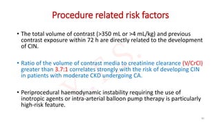
• The maximal acceptable contrast dose (MACD) which is defined as
• “5 mL×body weight [kg])/baseline serum creatinine [mg/dL]”
• This value should not be exceeded.
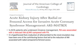
• A single invasive approach should ideally be adopted, with CAG followed
by ad hoc PCI to reduce the risk of atheroembolic complications while
minimizing contrast volumes to <4 mL/kg or V/CrCl <3.7:1.
• A slight reduction in contrast volume was documented with the use of
an automated contrast injector system, although it did not impact the
occurrence of CI-AKI.
61
62. • The lower the eGFR, the less contrast material may cause CI-
AKI.
• In general, it is desirable to limit the contrast medium to less
than 30 mL for a diagnostic procedure and less than 100 mL
for an interventional procedure.
• If staged procedures are planned and CI-AKI has occurred
with the first procedure, it is advantageous to have more
than 10 days between the first and second contrast
exposures.
62
63. “Ultra-low contrast coronary angiography”
• It uses meticulous contrast sparing techniques such as
• Use small diameter catheters (i.e., 5–6 French) without side-holes
• Limit the volume of contrast injected from the catheter to 1–2 cm3 per injection
using a 3-cm3 syringe.
• During PCI, prior to exchange of devices such as balloon catheters, remove contrast
from the guide catheter by back bleeding contrast out of the ‘‘Y’’ connector.
• If available, display previous angiographic images alongside active fluoroscopy screen
as a reference to use as guidance during guide wire, balloon, stent and ultrasound
passage.
• Absolutely no contrast ‘‘puffing’’ during the procedure.
• Use IVUS liberally for pre-PCI assessment of the lesion, selection of therapeutic
modalities, and post-PCI result assessment
• These techniques can reduce contrast load during diagnostic and PCI
procedures to under 10 to 20 cc.
63
64. • Dedicated devices to reduce overall CV have been developed.
• The DyeVert PLUS system is a device that is connected between the
injection syringe and the manifold via a 4-way stopcock, allowing
diversion of excess contrast during manual injection.
• The fraction of CM that would not contribute to coronary
opacification, but rather would reflux into the aortic root, is diverted
into a reservoir chamber.
• In initial trial, there were no differences in the rates of CI-AKI.
64
65. • The next-generation DyeVert system was evaluated in a multicenter
single-arm pilot study, in which up to 40% of CV was saved (p <
0.0001).
• Future research is needed to assess whether this reduction in CV
translates into clinically meaningful benefit.
65
66. • The type of CM plays an important role in the development
of CI-AKI.
• Low-osmolar CM (LOCM) compared to high-osmolar CM
(HOCM) has decreased the risk of CI-AKI regardless of pre
existing CKD; therefore, LOCM is favored in current clinical
practice.
• A benefit of iso-osmolar CM (IOCM) over LOCM with regard
to CI-AKI is uncertain due to mixed results of available
studies on this topic.
66
67. • CONCLUSION:
• The incidence of CIN in patients with diabetes and chronic kidney
disease was not significantly different after CT using iopamidol 370
(796 mOsm/kg)or iodixanol 320- Visipaque (290mOsm/kg).
67
68. • Conclusion:
• The rate of contrast-induced nephropathy is not statistically different after the
intraarterial administration of iopamidol or iodixanol to high-risk patients,
with or without diabetes mellitus.
68
69. • The nephrotoxicity associated with iodixanol was not significantly different
from that observed with ioversol in CKD patients undergoing coronary
angiography, although in diabetic patients, mean peak percentage change
in baseline serum creatinine was significantly lower in the iodixanol group.
69
70. • Nephropathy induced by contrast medium may be less likely to
develop in high-risk patients when iodixanol (visipaque) is used rather
than a low-osmolar, nonionic contrast medium.
70
71. • The IOCM
iodixanol was
significantly
less
nephrotoxic
than
ioxaglate, an
ionic, dimeric
LOCM.
71
72. • Compilation of pooled odds ratios from head-to-head trials for IA, IV,
and mixed IA and IV metaanalyses of the incidence of CI-AKI
72
73. • Patients should be advised to stop all non-essential nephrotoxic
medications for 24h prior to and for 48 h following the contrast
procedure pending SCr measurement.
• It is also recommended that patients receiving intra-arterial contrast
with an eGFR of <60 mL/min/1.73 m2, or those receiving intravenous
contrast with an eGFR of <45 mL/min/1.73 m2, discontinue
metformin for 48 h prior to contrast exposure and restart once a 48 h
SCr measurement excludes CIN.
• This is to mitigate the risk of lactic acidosis due to reduced renal
clearance of metformin that may occur following a potential CIN
episode, rather than metformin nephrotoxicity per se.
73
74. Periprocedural Hydration
• The most effective prophylactic intervention prior to contrast exposure.
• Supplementing intravascular volume ensures renal blood flow is
maintained and acts to dilute contrast in both blood plasma and tubular
filtrate.
• In lower risk ambulant patients, the oral route may be appropriate if
adequate fluid intake is assured.
• However, in moderate/higher risk or in hospitalised patients intravenous
hydration with a crystalloid fluid is preferred over oral hydration as it
guarantees delivery of appropriate fluid volumes and has been
demonstrated as superior in clinical trials.
74
75. • The choice of which crystalloid to use is, however, less clear.
• When compared with isotonic (normal) saline (0.9%), intravenous sodium
bicarbonate (1.26%) may have additional ROS scavenging properties
mediated through urine alkalinisation and lacks chloride ions that are
thought to exacerbate renal vasoconstriction.
• In view of the current lack of evidence supporting the use of sodium
bicarbonate 1.26% and with some studies offering conflicting evidence, the
current ESC guidelines recommend pre-hydration with sodium chloride
0.9%.
• For elective day case patients and for those with CCF in whom large
volumes of intravenous fluid may provoke pulmonary oedema, it is also
reasonable to use an alternative protocol delivering a shorter duration and
volume of sodium chloride 0.9%.
75
76. • In AMACING (A Maastricht Contrast-Induced Nephropathy Guideline) randomized
trial, intravenous 0.9% sodium chloride was directly compared with no
prophylaxis in 660 patients with an estimated GFR of 30–59mL/min/1.73m2
undergoing an elective procedure requiring iodinated contrast material.
• Within 2 - 6 days after contrast exposure, no prophylaxis was non-inferior to
intravenous hydration for the prevention of CIN and cost-saving.
• However, the validity of this finding is diminished by substantial underenrollment,
low rates of intraarterial procedures (48%) and interventional procedures (16%),
and moderate chronic kidney disease in a majority of patients.
• Given the known impact of low effective circulating volumes on the risk of CIN,
there is still consensus that adequate hydration is needed to prevent CIN.
76
77. IVF Infusion Rate to Prevent CIN
• IVF = 1 ml/kg/hr (max 100 ml/hr), 12 hours pre and upto 24 hours
post procedure
• CHF (NYHA > 2) or LVEF ≤ 35% = 0.5 ml/kg/hr (Max 50 ml/hr)
• Emergent procedure? (Suggested regimen)
Fluid bolus of 3ml/kg prior to procedure. Hydration during
procedure and 12 hrs after if possible
77
79. • The optimum rate and volume of intravenous fluid delivery a
significant challenge:
• underhydration increases CIN risk, whereas
• overhydration may precipitate acute pulmonary oedema in
vulnerable patients with severe CKD and CCF.
• Two hydration optimisation strategies have been trialled in
CIN:
• LV end diastolic pressure (LVEDP) guided volume expansion and
• high urine output matched fluid replacement (RENALGUARD).
79
80. Prevention of Contrast Renal Injury with Different
Hydration Strategies (POSEIDON) study
• In this study, fluid administration to prevent CI-AKI was guided by the LVEDP.
• Total of 396 patients with an eGFR of 60 mL/min/1.73 m2 and one additional
risk factor.
• Randomized to
• either a standard hydration protocol (1.5 mL/kg/h) or
• the LVEDP-guided strategy with isotonic saline:
• 5 mL/kg/h for left ventricular end-diastolic pressure lower than 13 mm Hg,
• 3 mL/kg/h for pressure of 13–18 mm Hg, and
• 1·5 mL/kg/h for pressure higher than 18 mm Hg.
• The overall rate of CI-AKI was significantly lower in the LVEDP-guided
hydration group compared to the control (6.7% vs. 16.3%; relative risk 0.41;
95% CI 0.22 to 0.79; P = .005).
80
81. RenalGuard system
• The concept of the RenalGuard system is
based on measuring urine output by a
Foley catheter and simultaneously
infusing an equivalent volume of isotonic
saline.
• After a 250 mL bolus of saline at 90
minutes prior to contrast application, a
continuous infusion of IV furosemide 0.25
to 0.5 mg/kg is started.
• The system automatically adjusts the rate
of the continuous IV saline infusion to
maintain a urine output rate greater than
300 mL/h throughout the procedure and
for 4 hours thereafter.
81
82. • In REMEDIAL II trial, a strategy of controlled forced diuresis using the
RenalGuard system was investigated.
• The study demonstrated superiority of the RENALGUARD system plus oral
NAC in preventing CIN (11%, 16/146) against a control group receiving
sodium bicarbonate plus oral NAC (20.5%, 30/146) (p value – 0.025, OR
0.47; 95% CI 0.24 to 0.92).
82
84. N-Acetyl Cysteine and Sodium Bicarbonate
• N-acetyl cysteine (NAC) is assumed to limit CI-AKI by acting as a
scavenger of ROS and promoting vasodilatory effects in the renal
medulla.
• It has been widely investigated for the prevention of CI-AKI with
mixed results.
• Earlier trials with small sample sizes suggested a potential benefit of
NAC, whereas larger studies failed to show a significant reduction in
CI-AKI.
84
85. • Acetylcysteine does not reduce the risk of contrast-induced acute
kidney injury or other clinically relevant outcomes in at-risk patients
undergoing coronary and peripheral vascular angiography.
85
86. • Similarly, isotonic sodium bicarbonate (NaHCO3) was
hypothesized to prevent CI-AKI due to inhibition of
free-radical formation by alkalinizing the renal tubular
fluid.
• Although smaller studies suggested a benefit of
NaHCO3 administration for CI-AKI prevention, these
findings were not confirmed in larger studies.
86
87. • Among patients at high risk for renal complications who were
undergoing angiography, there was no benefit of intravenous sodium
bicarbonate over intravenous sodium chloride or of oral
acetylcysteine over placebo for the prevention of death, need for
dialysis, or persistent decline in kidney function at 90 days or for the
prevention of contrast-associated acute kidney injury.
87
88. • As such the ESC guidelines recommend that NAC is not to be
used alone, although it may be used in addition to standard
intravenous hydration regimes.
• KDIGO guidelines recommend using oral NAC together with
IV isotonic crystalloids, in patients with increased risk of CI-
AKI.
• Routine use of NAC for CI-AKI prevention is class III in the
ACC/AHA guidelines.
88
89. Statins
• More recently high-dose statin therapy (eg, rosuvastatin 40/20 mg,
atorvastatin 80 mg or simvastatin 80 mg) has shown efficacy in
preventing CIN in statin-naïve patients in several clinical studies and
as such is regarded as reasonable preventative therapy in the current
ESC guidelines.
• Initial PROMISS trial investigated short-term pretreatment with high-
dose simvastatin in patients with baseline renal insufficiency
undergoing coronary angiography and did not show a benefit of this
strategy with regard to deterioration of renal function.
89
90. • PRATO-ACS trial showed that high-dose rosuvastatin (40 mg
loading and 20 mg/day maintenance) compared to no statin
significantly reduced CI-AKI and 30-day rates of
cardiovascular and renal events (death, dialysis, MI, stroke,
or persistent renal damage) in statin-naïve patients
undergoing PCI.
• TRACK-D study was the largest trial investigating the role of
statins in CI-AKI prevention.
• A significant reduction of CI-AKI was reported for the statin group
compared to the control (2.3% vs. 3.9%; P = .01).
90
92. • Conclusion:
• Oxygenation, a simple, nonpharmacological strategy, may be beneficial when
using contrast media in patients with impaired renal function from
noninvasive angiography to emergency catheterization.
92
96. • Hemofiltration: The benefit of hemofiltration in the high risk groups, needs
to be studied further.
• Hemodialysis: Patients on hemodialysis need not be volume loaded before
contrast administration and dialysis after the procedure is useful only if
patient has evidence of volume overload.
• Repeat contrast exposure: No studies have been conducted for the ideal
timing for a repeat contrast exposure, but since in majority of CIN patients
renal function is restored in 3 weeks, this time period is usually advised.
• Other pharmacological agents such as dopamine, fenoldopam have shown
no benefit in the prevention of CIN, forced diuresis with mannitol or
furesomide may even be harmful.
96
99. Management
• Decline in renal function:
• The most common manifestation of CIN is asymptomatic transient decline in renal
function, which usually normalizes within 10-14 days.
• Serial creatinine:
• Patients at high risk should especially be followed up with serial creatinine
measurements daily for 5 days.
• Electrolyte and fluid balance:
• If oliguric renal failure occurs, the management should be similar to as that for acute
renal failure due to other causes, including judicious acid-base, electrolyte and fluid
balance.
• Dialysis:
• Temporary dialysis may be required in severe cases, with a minority of patients
requiring permanent dialysis
99
100. European Society of Cardiology CIN prevention guidelines, 2018
Recommendation Detail Class Level
Patients undergoing coronary angiography or MSCT
It is recommended that all patients
are assessed for the risk of contrast
induced
nephropathy.
I C
Adequate hydration is recommended. I C
100
101. Recommendation Detail Class Level
Patients with moderate or severe CKD (National Kidney Foundation stages 3b and 4)
Use of low-osmolar or iso-osmolar
contrast media is recommended.
I A
It is recommended that the volume of
contrast media be minimized.
Total contrast volume/GFR <3.7 I B
In statin-naive patients, pre-treatment
with high dose statins should be
considered.
Rosuvastatin 40/20 mg or atorvastatin
80 mg.
IIa A
Pre- and post-hydration with isotonic
saline should be considered if the
expected contrast volume is >100 mL
1 mL/kg/h 12 h before and continued
for 24 h after the procedure (0.5
mL/kg/h if LVEF ≤ 35% or NYHA > 2).
IIa C
As an alternative to the pre- and post-
hydration regimen, tailored hydration
regimens may be considered.
IIb B
101
102. Recommendation Detail Class Level
Patients with severe CKD (National Kidney Foundation stage 4)
Prophylactic haemofiltration
6 h before complex PCI may
be Considered
Fluid replacement rate
1000 mL/h without
negative loss and saline
hydration continued for
24 h after the procedure.
IIb B
Haemodialysis is not
recommended as a
preventive measure.
III B
102
Anemia: baseline hematocrit value 39% for men and 36% for women;
CHF congestive heart failure class III/IV by NYHA classification and/or history of pulmonary edema;
Hypotension: systolic BP 80 mm Hg for at least 1 h requiring inotropic support with medications.
CyC is a 122–amino acid, nonglycosylated protein that is a member of the family of cysteine proteinase inhibitors.
CyC concentration is independent of age, sex, changes of muscle mass, and nutrition.
Its serum concentration is determined by glomerular filtration and it does not undergo tubular secretion and appears in the urine solely through filtration.
For these reasons, CyC has the potential to be a useful marker in detecting both chronic and acute changes in GFR.
The shorter (1.5-hour) half-life of CyC compared with creatinine accounts for the more rapid rise and the earlier attainment of a new steady state,
The ADQI proposed the term acute kidney disease for AKI stage 1 or greater that persists beyond 7 days after the exposure.
It may eventually result in CKD, which is defined as abnormalities in kidney function that last greater than 90 days
All contrast injections require simultaneous cine angiogram, i.e., ‘‘no dye without the cine’s eye.’’
Two meta-analyses have demonstrated a modest reduction in CIN when using intravenous sodium bicarbonate 1.26% as compared with isotonic saline, although no significant mortality benefit has been demonstrated.