
Recommended
Recommended
More Related Content
What's hot
What's hot (20)
Similar to EQUIPMENT FOR CRANIAL AND DENTAL RADIOGRAPHY.pptx
Similar to EQUIPMENT FOR CRANIAL AND DENTAL RADIOGRAPHY.pptx (20)
More from RukamaneeYadav
More from RukamaneeYadav (13)
Recently uploaded
Recently uploaded (20)
EQUIPMENT FOR CRANIAL AND DENTAL RADIOGRAPHY.pptx
- 2. Record of an image produced by transmission of x-rays through an cranium and dental that is called cranial and dental radiography . First dental radiography discovered in 1896 by O walk Hoff . Cranial and dental radiography are easier, faster and generally more accurate procedure . These examination are use high technical standard .
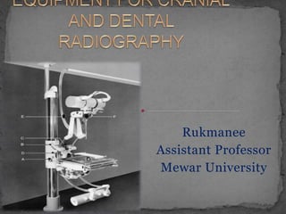
- 3. Cranial table is much more readily appreciated from practical observation than from written description. Fixed column provided support to object table and x- ray tube. Object table which essential is a bucky tray. Anode to film distance 90 cm.
- 4. Base design- T or round -shaped Floor – ceiling mounting Floor – wall mounting Double semi circular arch – x-ray tube counter weight
- 5. Upper and lower sections are made up of transparent “Plexiglas ”. Upper surface- large circular protractor scaled in four quadrants of 90⁰ angle. Anatomy of interest for positioning. Under the table are a pair of lighted mirrors to look for accurate body. Arches and object table are mounted together on the column by a single carriage.
- 6. The couch is neither an integral part of the equipment nor as a rule in a fixed relationship to it . It can be raised or lowered hydraulically and its usually built in three hinged sections. a) Tilting 1/3rd reclining angle- for back rest nearest to skull table. b) Middle 1/3rd is horizontal surface- forms a seat. c) Remaining 1/3rd dropped vertically downwards- supports the knees and calve.
- 7. The couch can be converted into a chair and can be used for the examination of the patients in sitting position. For patient immobilization- arm rest , head rest, shoulder rest and sling for the chin.
- 8. Whole device of object table & x- ray tube can be moved up and down supporting the column . Whole device can be rotated 360 degrees. The x-ray tube can be tilted independently on its own axis + - 30 degree. a) Angulation of x-ray tube b) Tilting the object table c) A combination of putting some of the tilt on the table and some on the x-ray tube.
- 9. The upper and lower sections of the table are made of transparent Plexiglas. Upper surface are etched a pair of cross lines intersecting at the centre of the field. Under the table are a pair of lighted mirrors. It permits the x- ray beam to be centred accurately on surface anatomical landmarks which are near to the film and ordinarily lost to sight because of this .
- 10. Immobilizing devices - band , head clamps may be used which fix to the side of table. Beam limiting & centring devices – usually limitation of x-ray beam is obtain by removable diaphragms which are slotted into the tubehead . Visual indication of the direction of the primary beam is provided either by a centre finding pointer or by varay lamps. Spring loaded cassette boxes – spring loaded cassette boxes for cerebral angiography. the Bucky mechanism is removed & replaced with the box which will be employed for the anteroposterior projection, the box for the lateral projection is fitted to the edge of the table in the same position as the cassette holder described earlier .
- 11. There are three significant parts of the dental equipment – 1.Tubehead 2.Tubestand 3.Timer
- 12. Tube head is Oil filled & vacuum sealed. Tube head are present high tension transformer, filament transformer, x-ray tube, oil -expansion diaphragm. Tube head mounted in a contrivance known as gimbals in which it is free to rotate in two planes. Localising cone is provided for the tubehead giving an anode - skin distance of 18- 23 cm (7 – 9 inch).
- 13. Tube head is mounted on a tube stand. Tube stand is mounted in one of the following ways: 1.On the wall by means of a bracket. 2.On a mobile pedestal which moves on four large castors. 3.On the dentist's pedestal control.
- 14. The timer is of the electronic type. Provides maximum exposure interval of 5 sacs. A red pilot lamp indicates when x-rays are 'on' and the exposure push- button is designed to prevent any operation as the result of accidental pressure.
- 15. The cylinder (or cone) is a fixed to the tube head and is used to align the tube head with the patient and the X-ray film. It is open ended and composed of lead laminated material that establish the minimum distance from the x-ray source to patients skin.
- 16. Cephalostats Cephalostat is an apparatus which permits a precise correlation position to be established between an x-ray film, head of patient and the anode of x-ray tube . Cephalostat is a wall mounted & of a strong rigid structure. Patient is laterally pinned by the ears by means of ear plugs in a fixed relationship to a cassette-holder X-ray tube which is shown in neither illustration is at another fixed point.
- 17. The Orbital indicator, nasal positioner & scales showing certain distances all play a necessary part in the indication recording and subsequent reproduction of a particular radiographic situation. Filter prevent over penetration on radiograph of the nasal bone and soft tissue. Generators can provide max current 7 – 12 mA and voltage 50 – 55 kvp .
- 21. Pantomography equipment provide panoramic radiographs of jaw and face that is all teeth together with mandibles and maxillae are seen on single film 15cm. X 30cm. These are use smaller radiation dose. These are use demonstration of temporomadibular joint, fracture of facial bone, developed abnormalities, dental cyst. Duration of exposure varies for different equipments normally it takes 15 seconds for whole examination.
- 22. Effective tube focal area 0.6mm x 0.6mm. Total filtration is equivalent to 3mm Al. Cassette is curved and these convex surface is facing the tube. Beam is collimated by : 1) primary diaphragm . 2) secondary slit panel .
- 23. Patient supporting/immobilizing device – 1) bite block 2) chin support 3) forehead support 4) RT and LT temporal support 5) RT and LT Perspex face plates