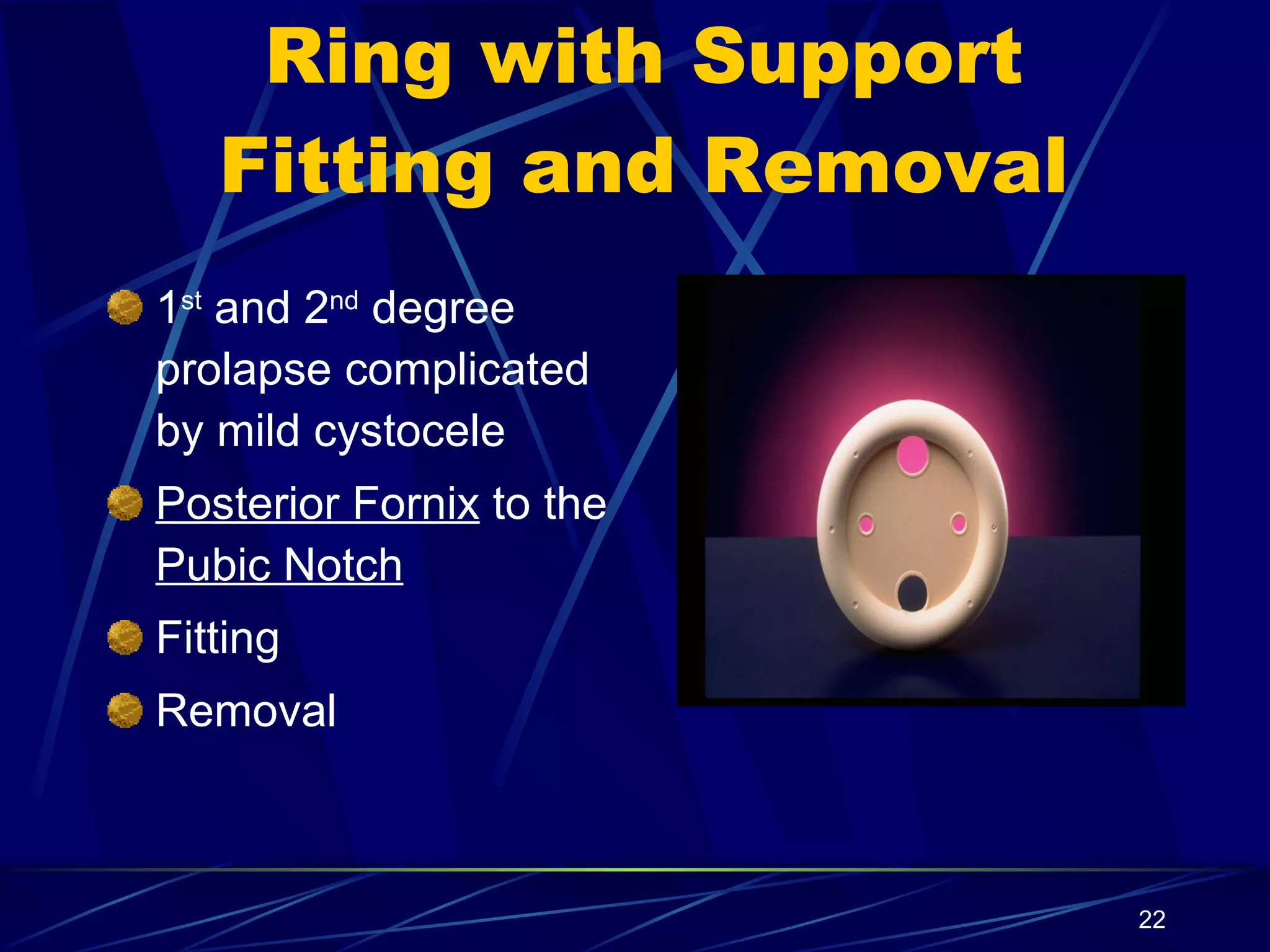
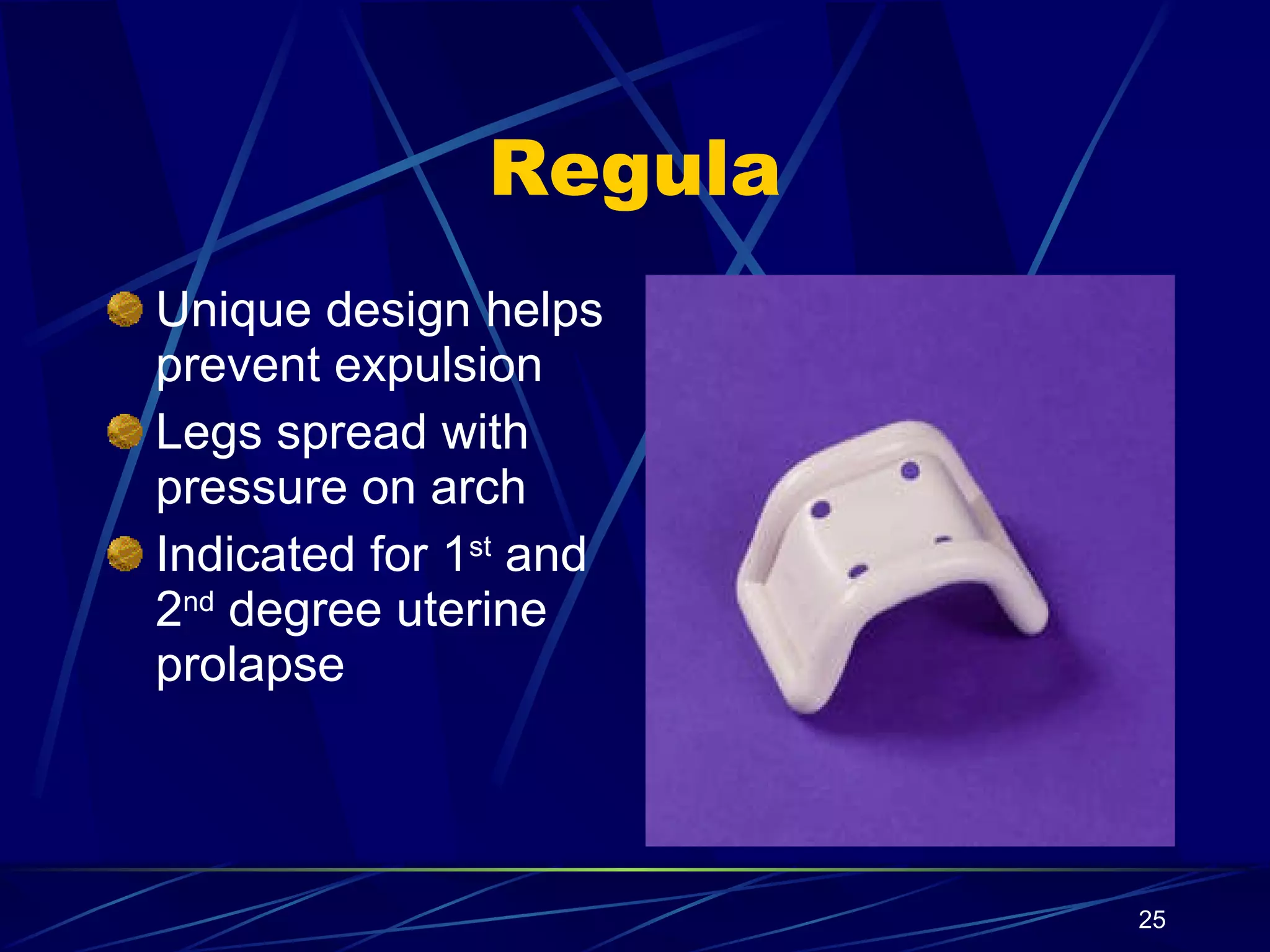
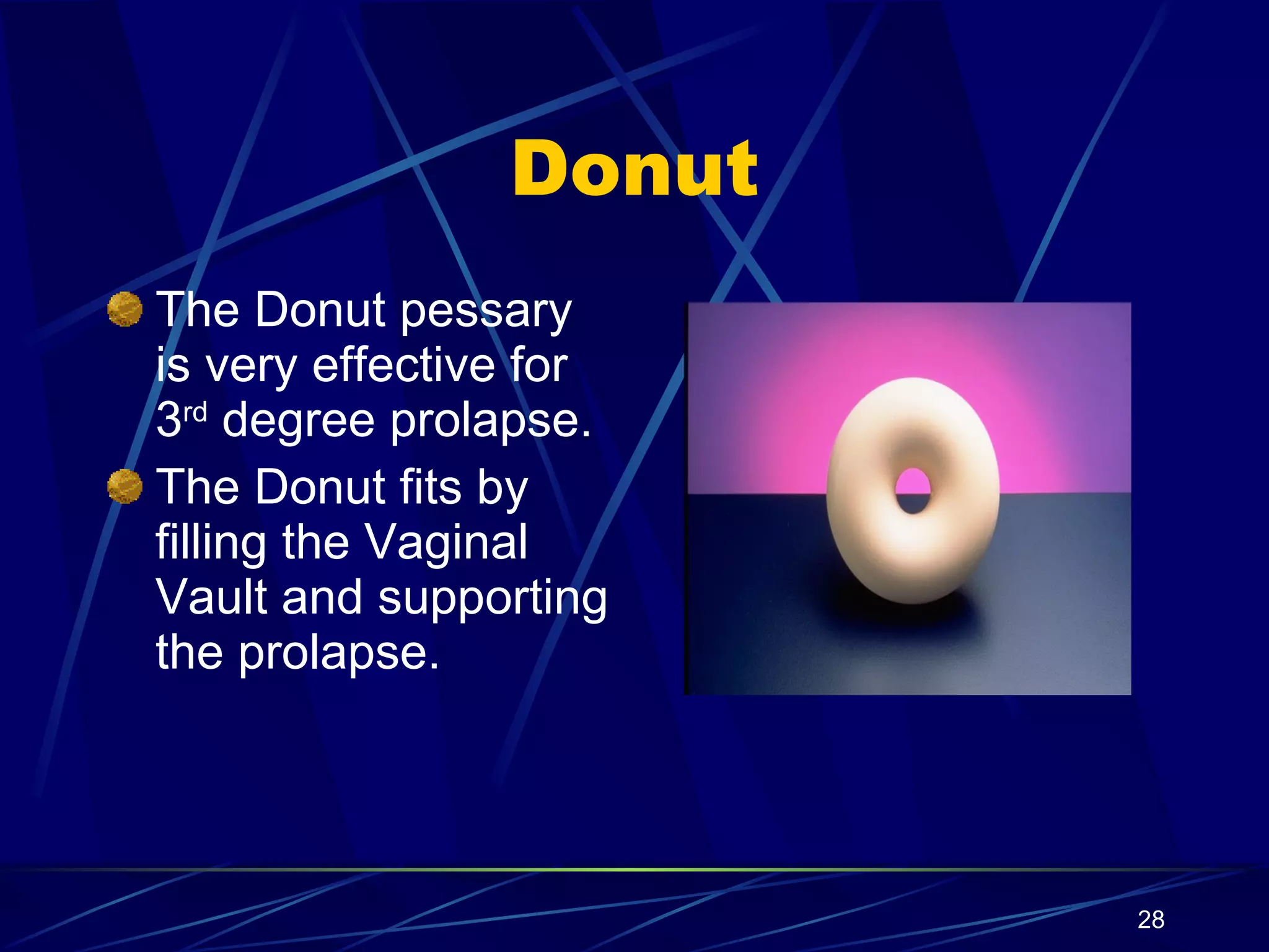
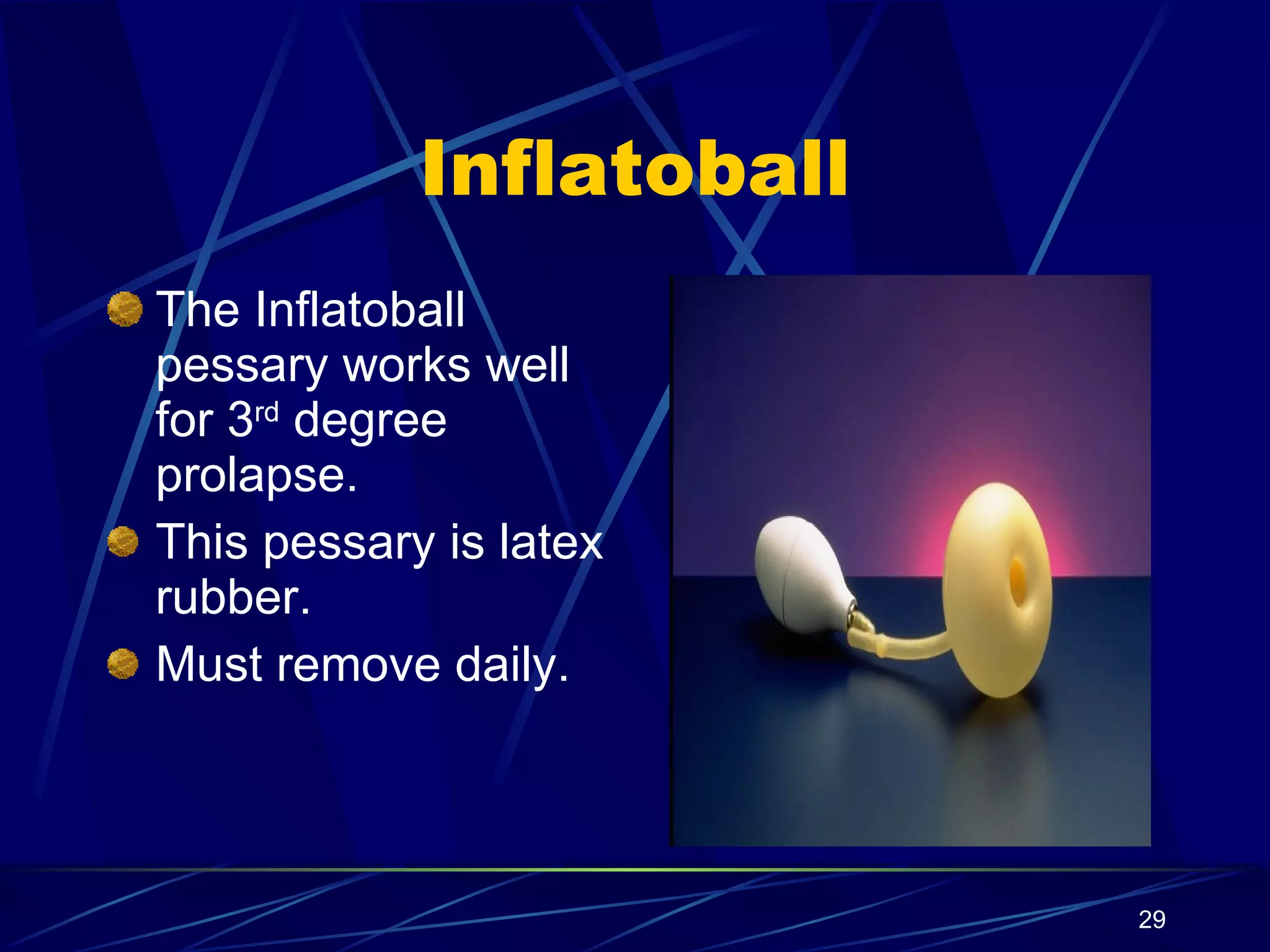
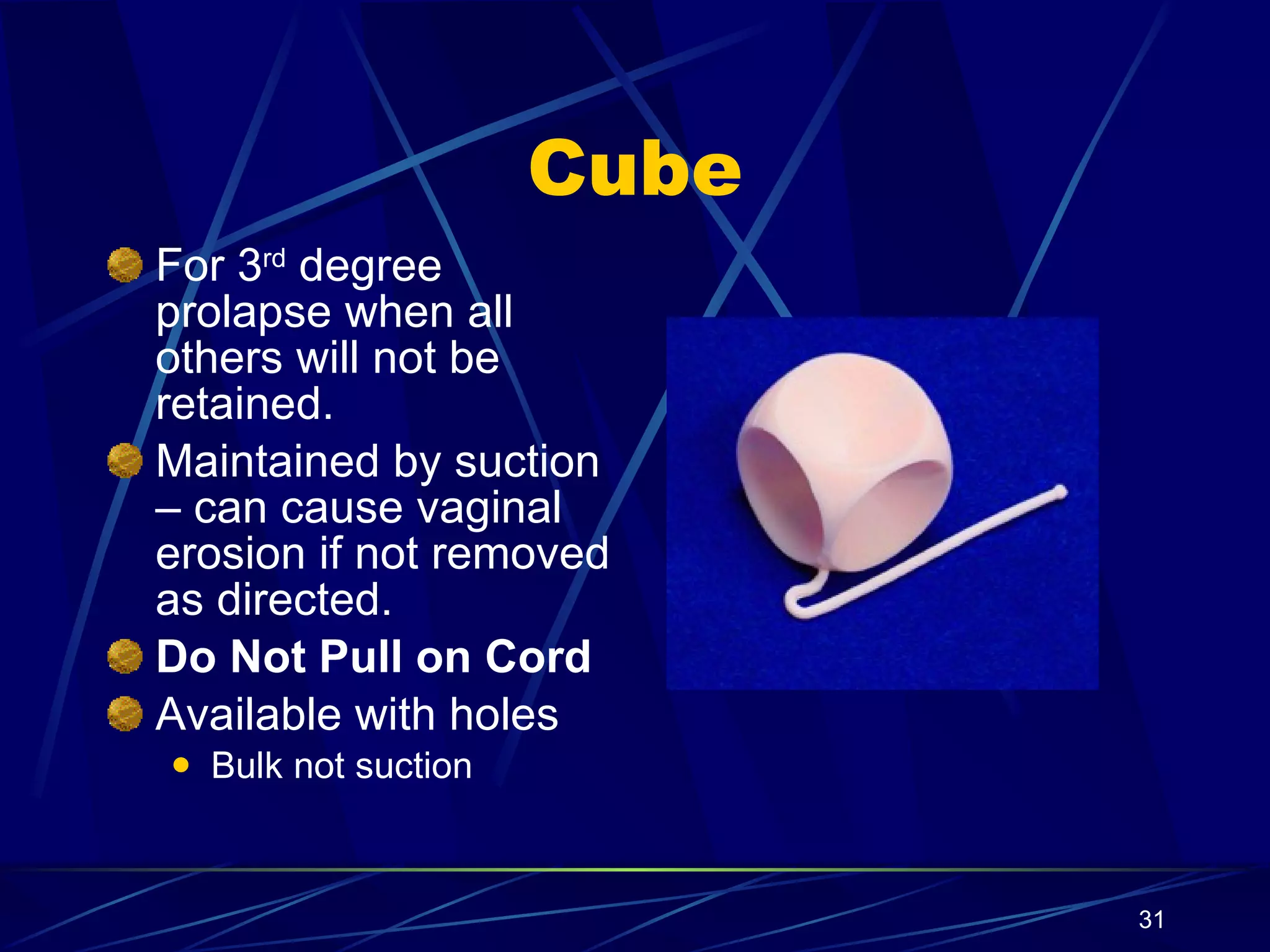
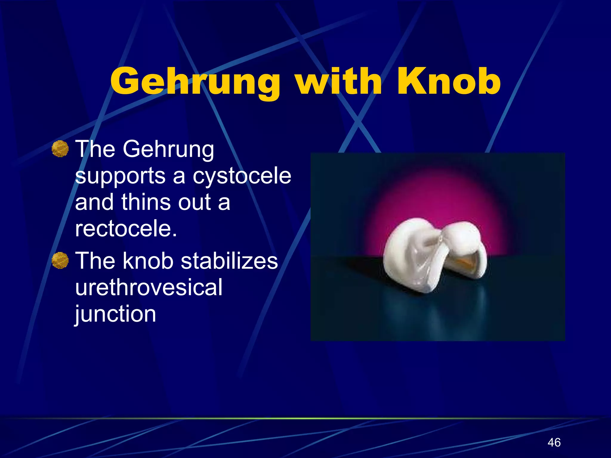
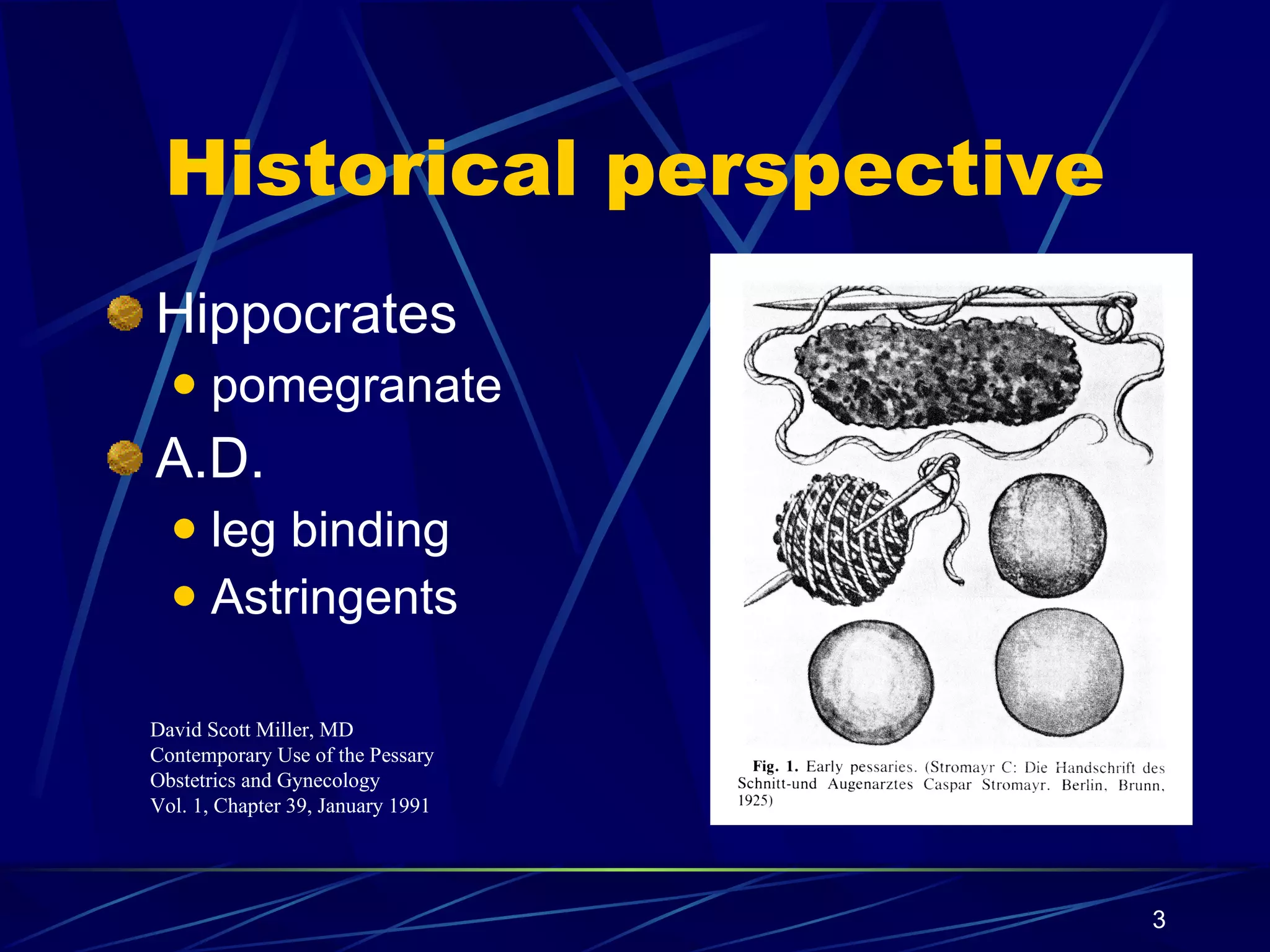
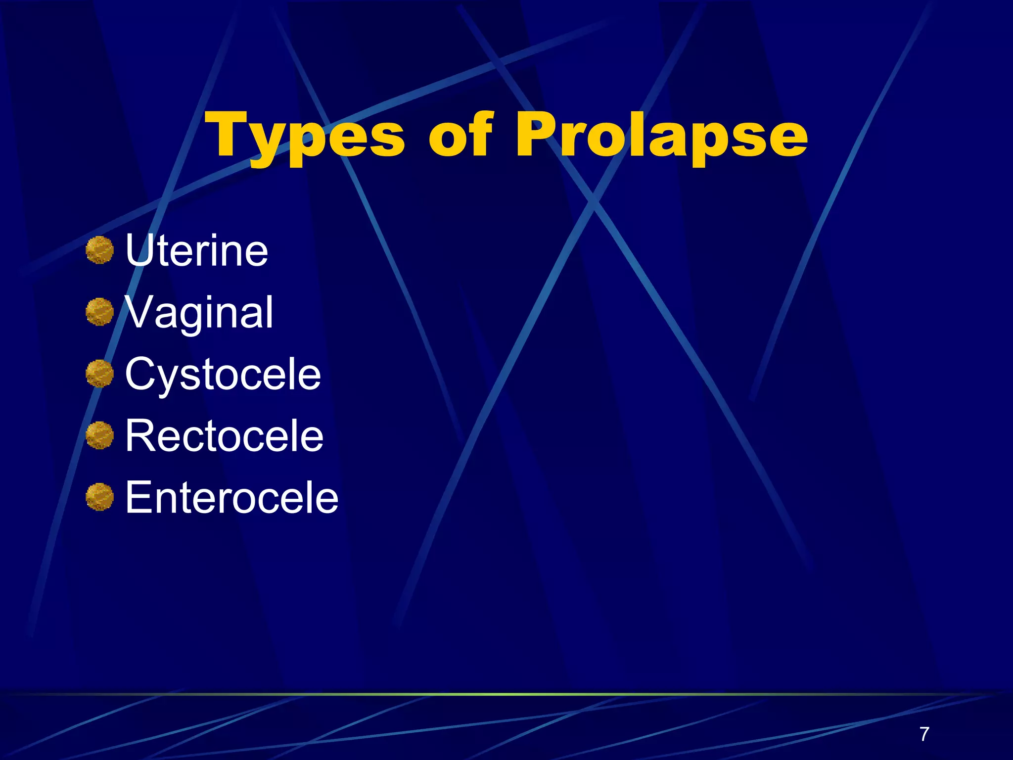
The document provides an overview of pessary use, including their historical origins, types, fitting procedures, and care instructions. It discusses various pessary designs and their uses for conditions like uterine prolapse, cystocele, rectocele, urinary incontinence, and cervical incompetence. Guidelines are provided on patient education, fitting procedures, follow-up care, and reimbursement codes.
Dilatation and curettage (D & C) is a procedure to remove tissue from inside the uterus. Doctors perform D & C to diagnose and treat certain uterine conditions — such as a heavy bleeding — or to clear the uterine lining after an abortion or miscarriage.
Bivalve speculum (Cusco's speculum) The two-bladed, or bivalve, speculum is the most common type of instrument gynecologists use to examine the vagina and cervix. ...
Pediatric speculum. ...
Huffman speculum. ...
Pederson speculum. ...
Graves speculum.
nurses/doctor will insert two lubricated, gloved fingers into your vagina with one hand, while the other hand presses gently on the outside of your lower abdomen. During this part of the exam, your doctor will check the size and shape of your uterus and ovaries, noting any tender areas or unusual growths.
BURN REHABILITATION
It include definition, purposes, stages, skin graft, rehabilitation, role and responsibility
for more details follow
www.vipinpatidar.wordpress.com
It is a composite graphical recording of cervical dilatation and descent of head against duration of labour in hours.
It also gives information about fetal and maternal condition that are all recorded on single sheet of paper.
Please find the power point on Utero-Vaginal Prolapse. I tried to present it on understandable way and all the contents are reviewed by experts and from very reliable references. Thank you
Uterine prolapse occurs when weakened or damaged muscles and connective tissues such as ligaments allow the uterus to drop into the vagina. Common causes include pregnancy, childbirth, hormonal changes after menopause, obesity, severe coughing and straining on the toilet.
Pelvic organ prolapse
Etiology of pelvic organ prolapse
Vaginal vault prolapse
Etiological factors of vault prolapse
Signs and symptoms of vaginal vault prolapse
Diagnosis of vaginal vault prolapse
Treatment measures
1. Contemporary Use of the Pessary Indications - Fitting Instructions Milex Products, Inc. Chicago, Illinois 60634-1403 Copyright August 2002 All Rights Reserved
2.
3.
4.
5.
6.
7.
8.
9. Ordinal Staging of Pelvic Organ Prolapse tvl=total vaginal length Maximal prolapse point protrudes the length of the vagina (2 cm) beyond the hymen. Complete eversion of the vagina +/- cervix ([value >/= + [tvl-2]) IV Maximal prolapse point protrudes beyond 1 cm above hymen but less than 2 cm less than the total vaginal length. (value >+1 but <+(tvl-2)) III Maximal prolapse point protrudes to or beyond 1 cm above hymen but not more than 1 cm below hymen (value >-1 to <+1) II All points are more than 1 cm above hymen (value<-1) I No prolapse: Apex of cervix is at a position above the hymen that equals to or is within +/- 2 cm of vaginal length (value </= (tvl-2)) No prolapse: All points are 3 cm above the hymen (value=-3) 0 Leading Edge of Prolapse: Location of Apex of Vagina or Cervix (Value of Point C or D) Leading edge of Prolapse: Location of the Most Distal Point of the Anterior or Posterior Vaginal Wall (any points Aa, Ap, Ba, Bp) Stage