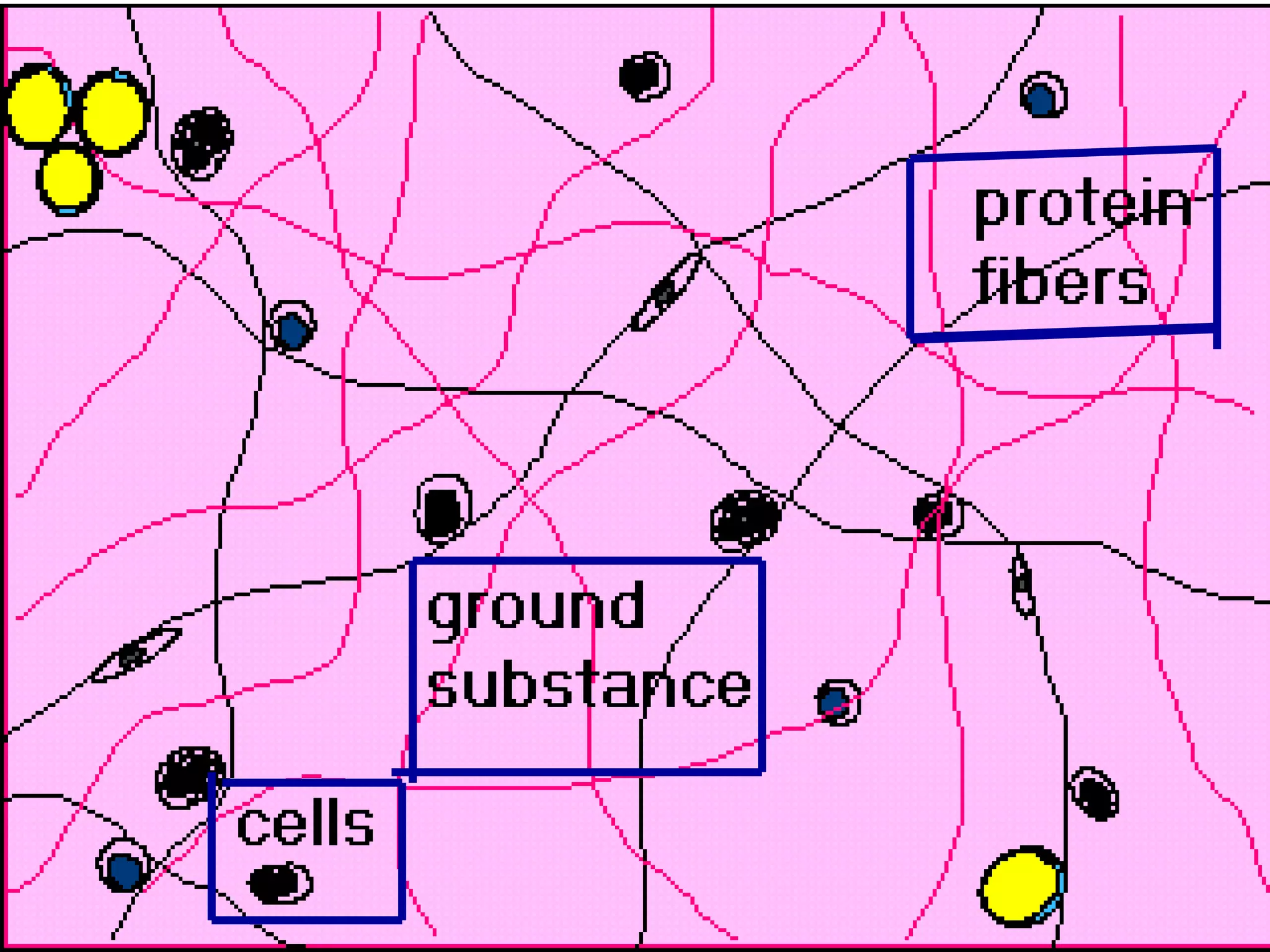
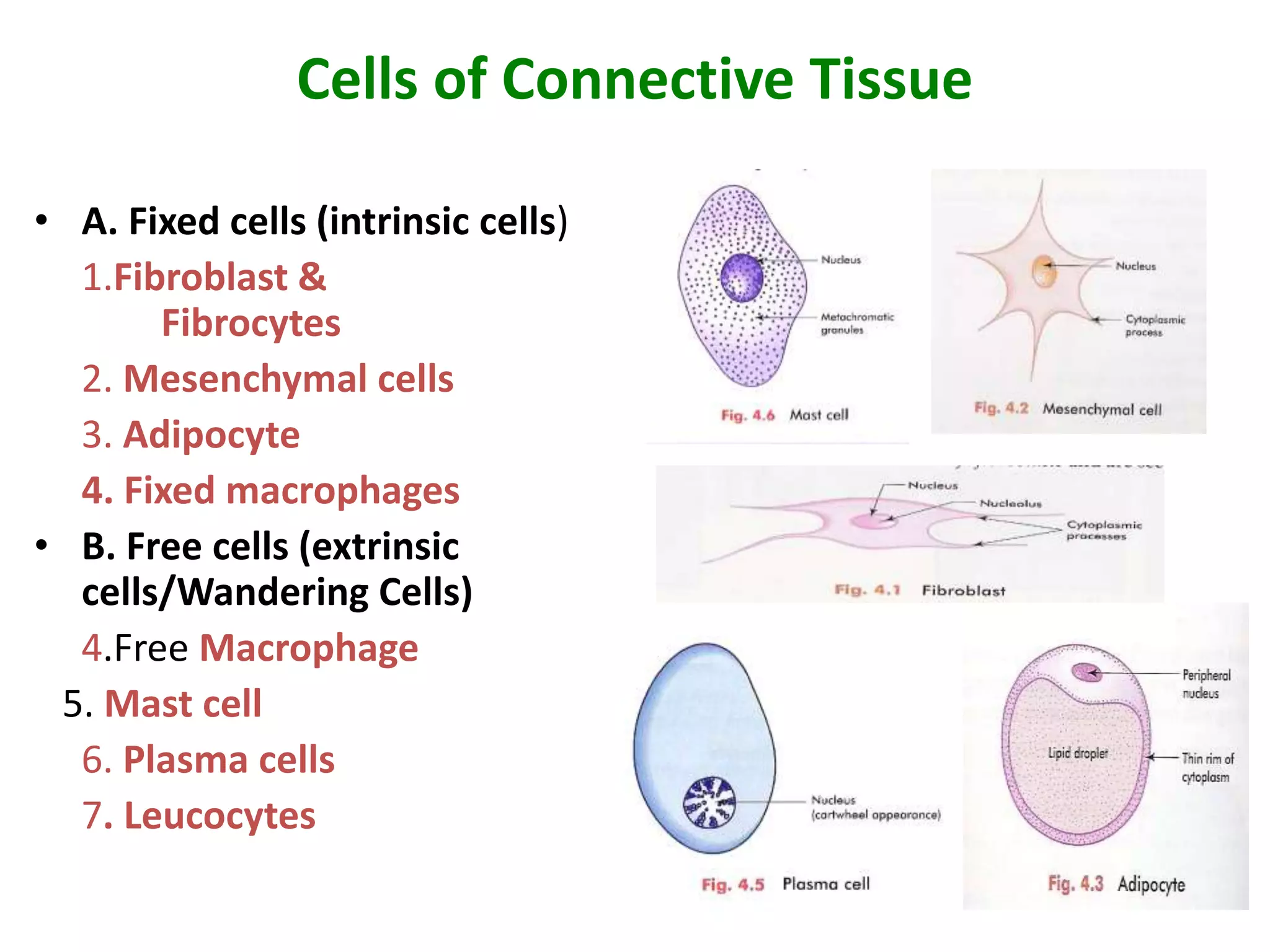
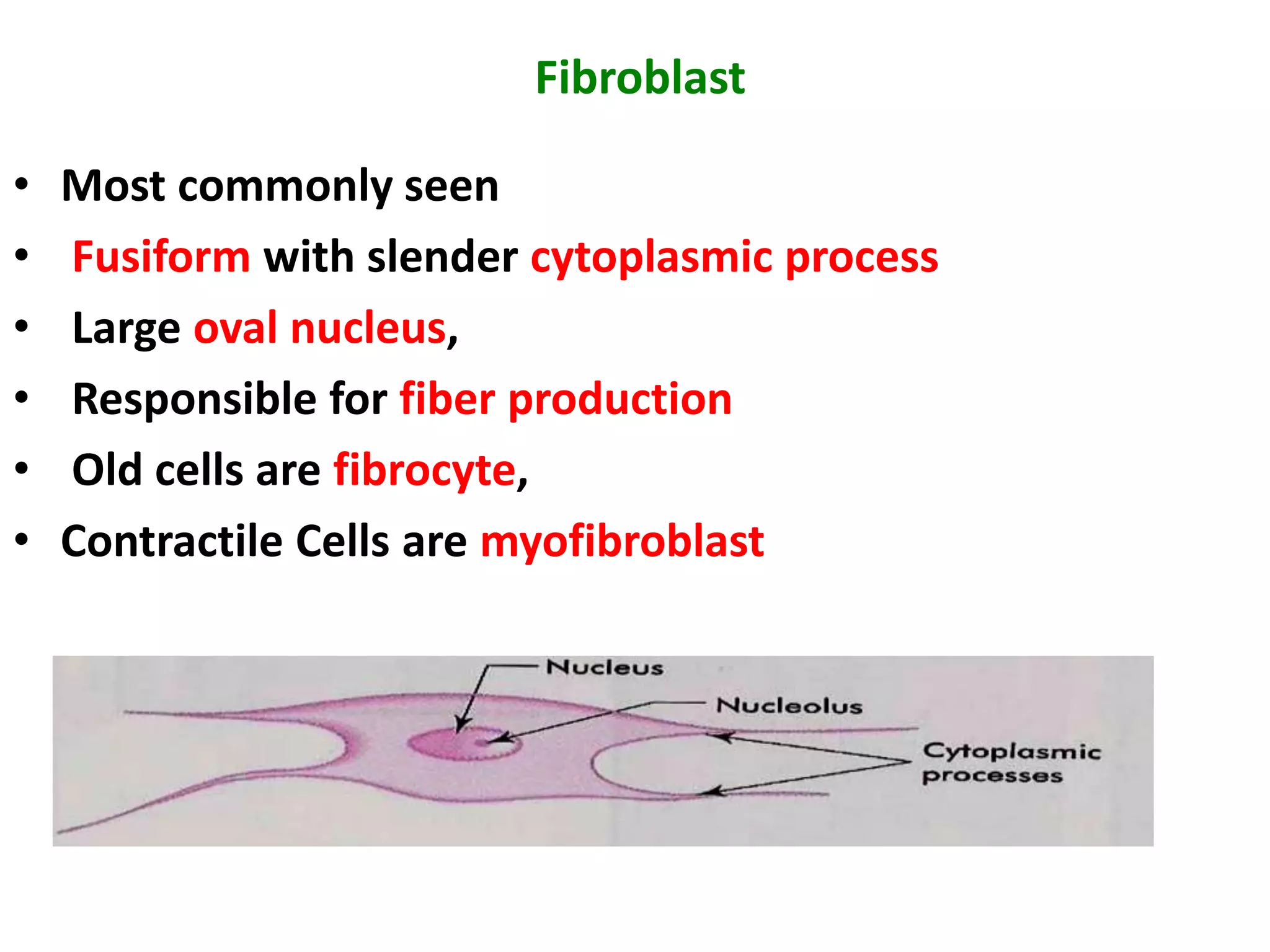
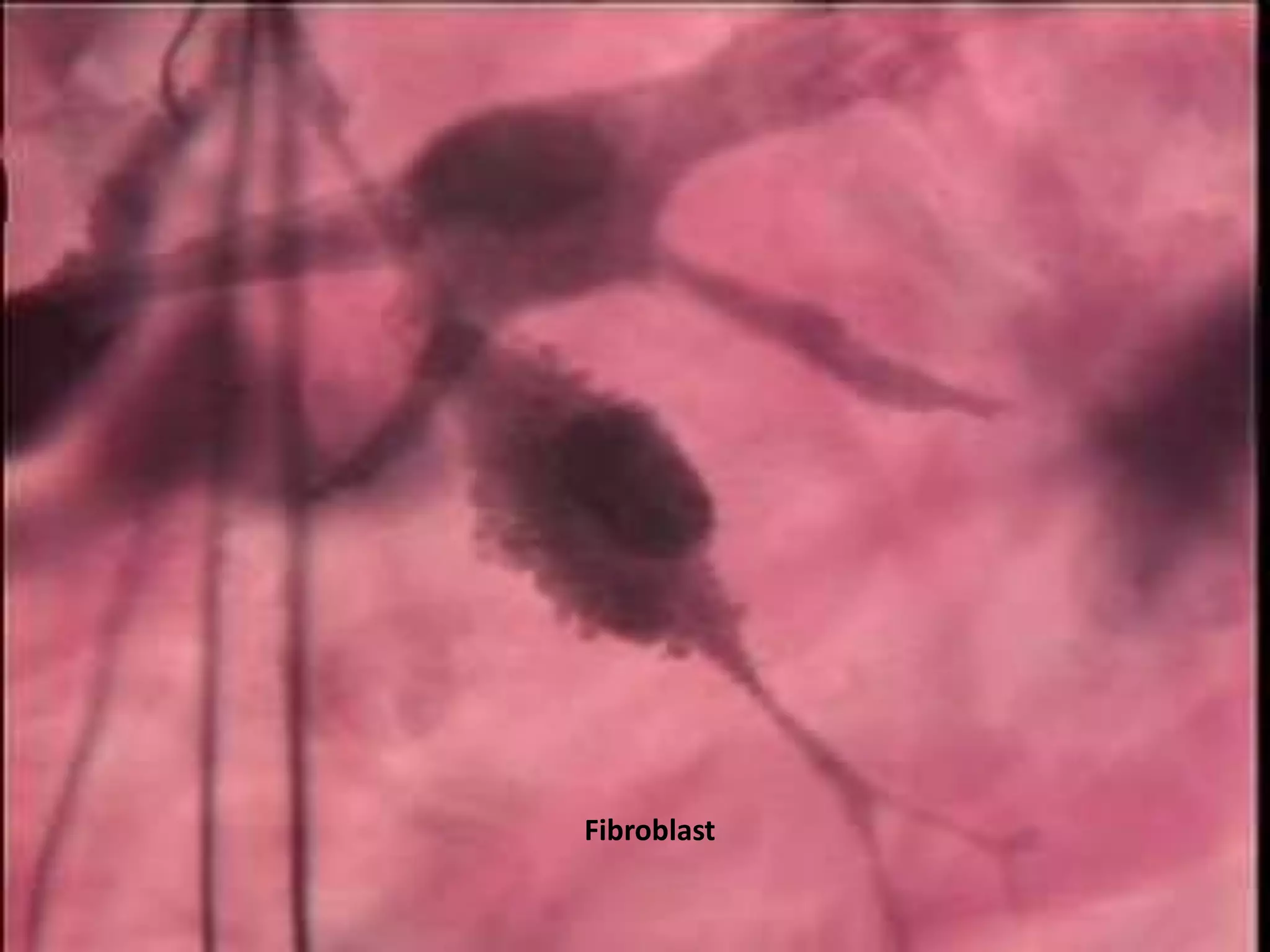
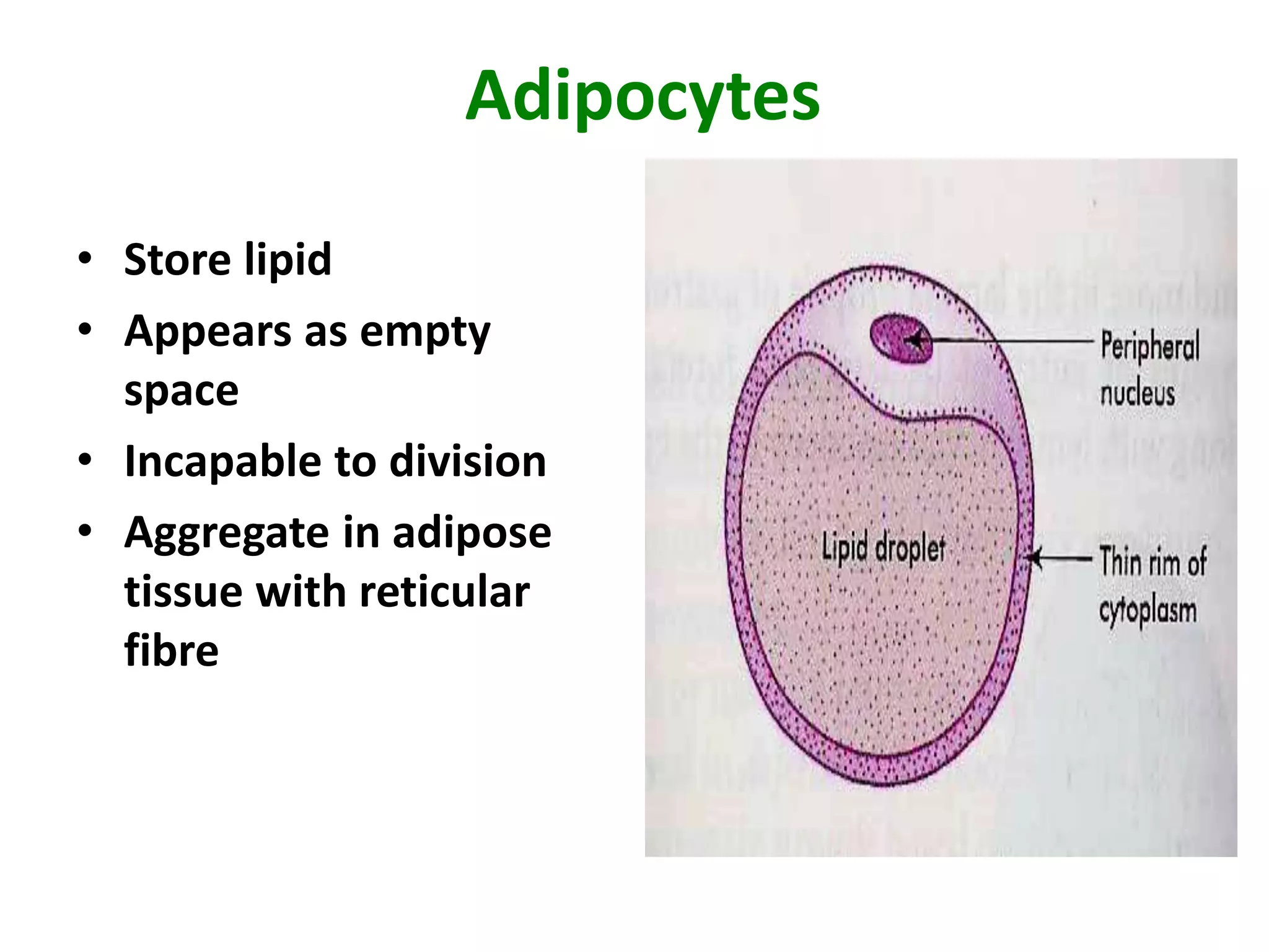
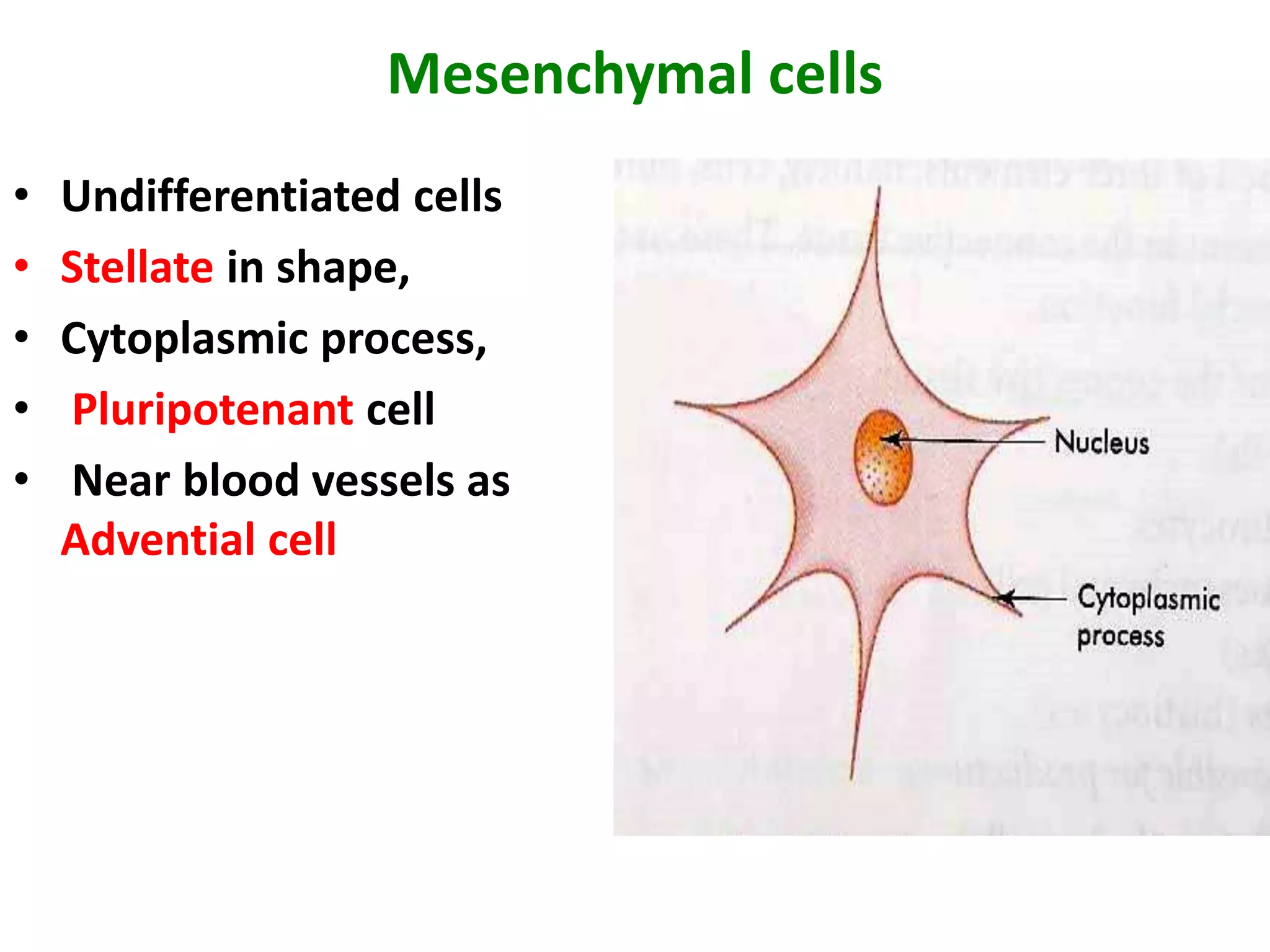
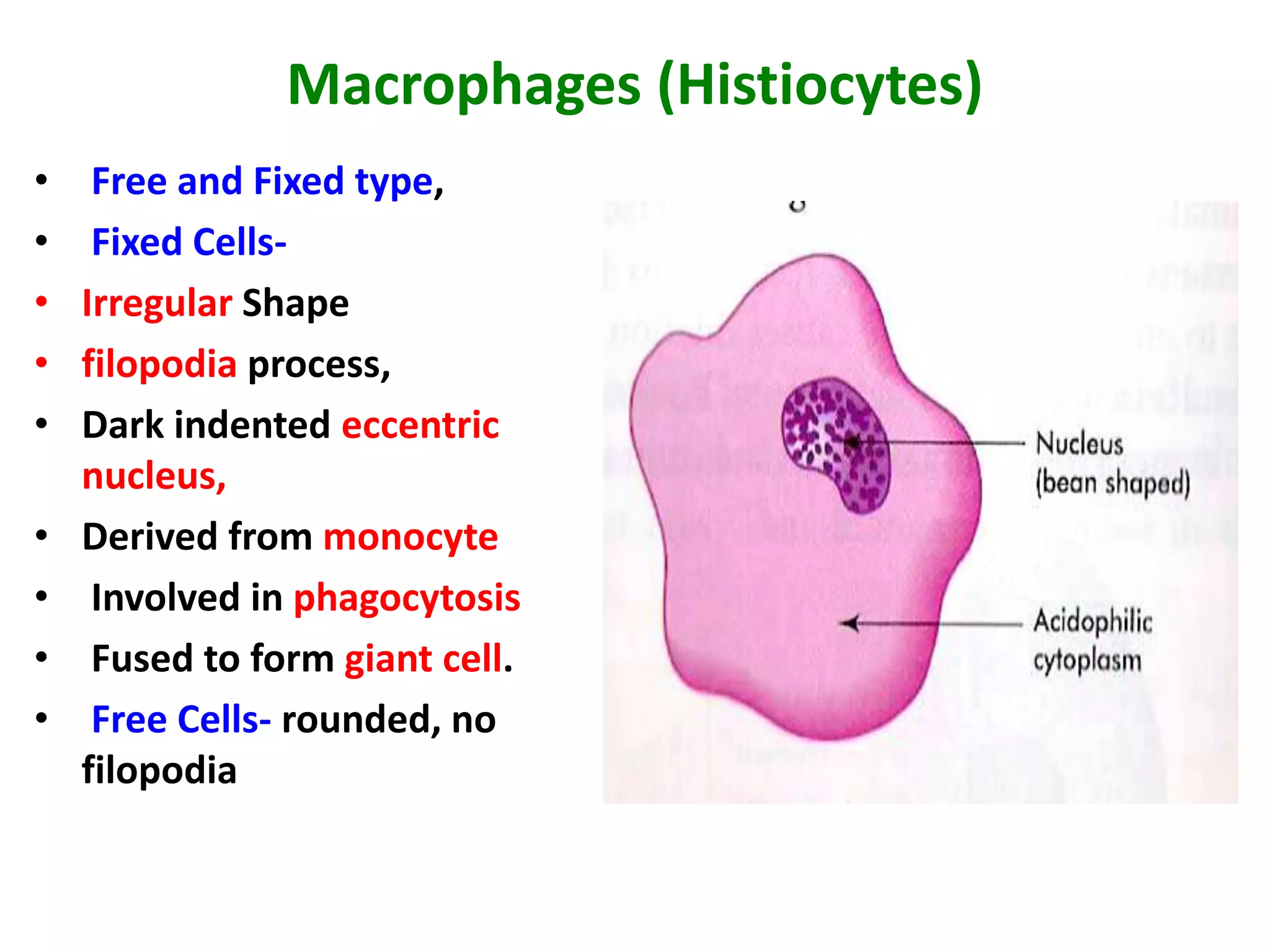
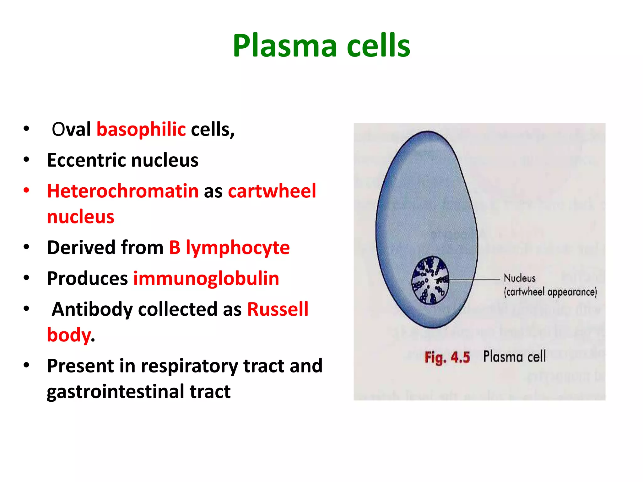
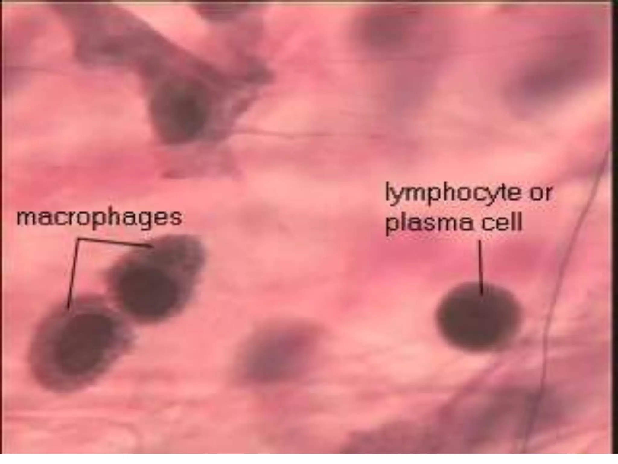
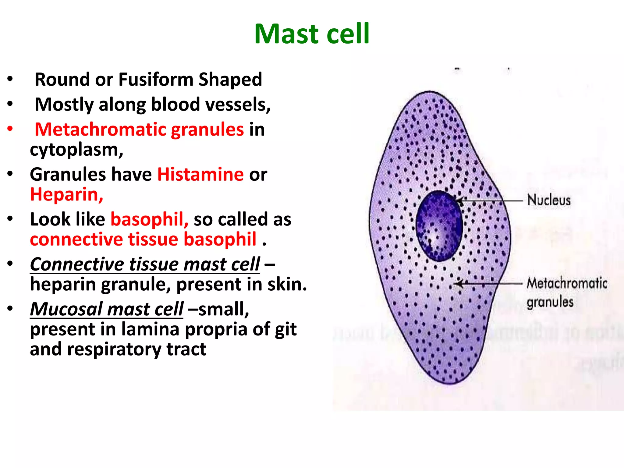
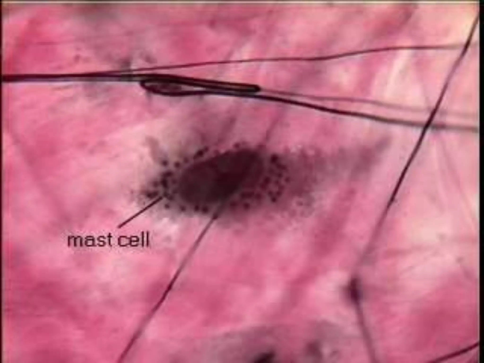
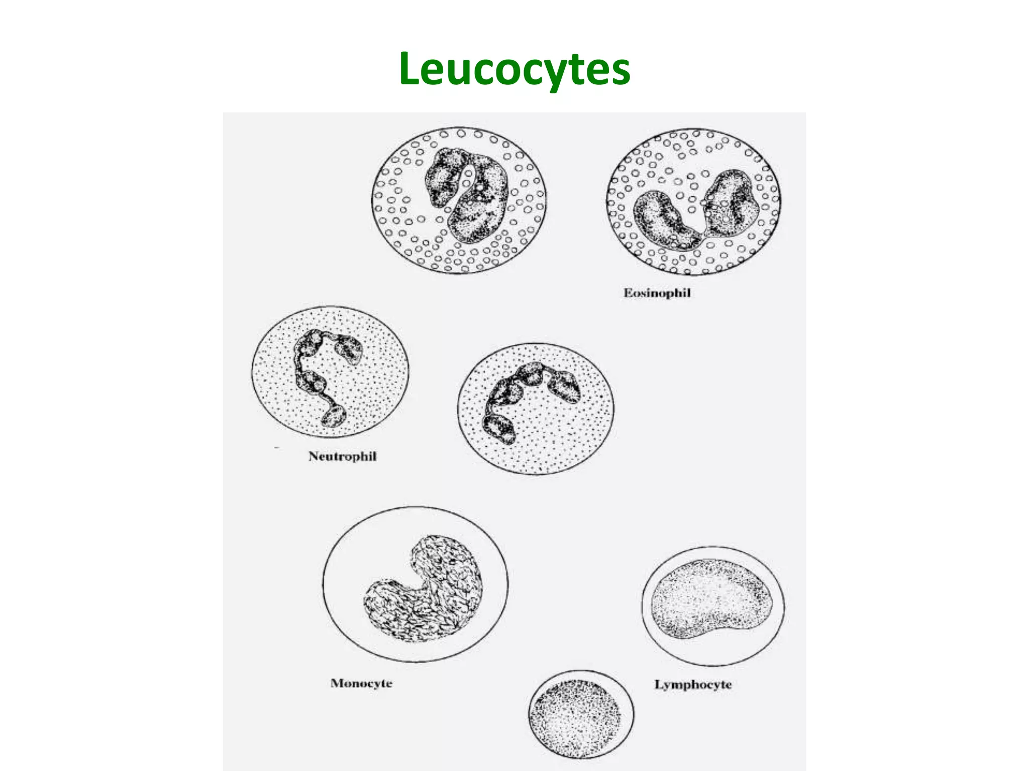
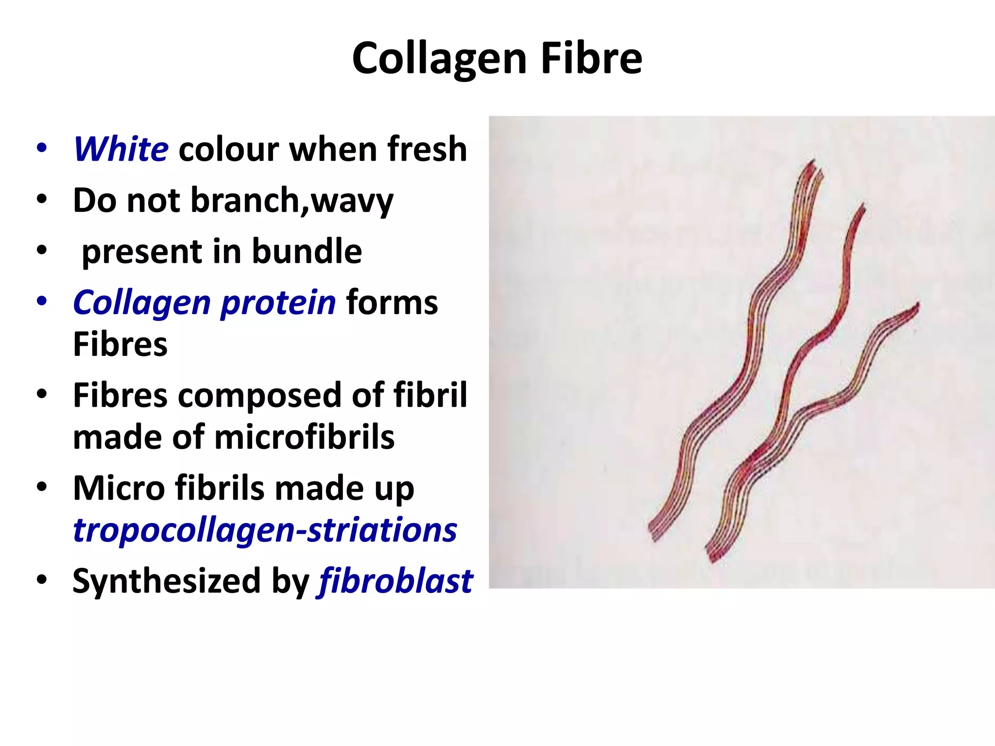
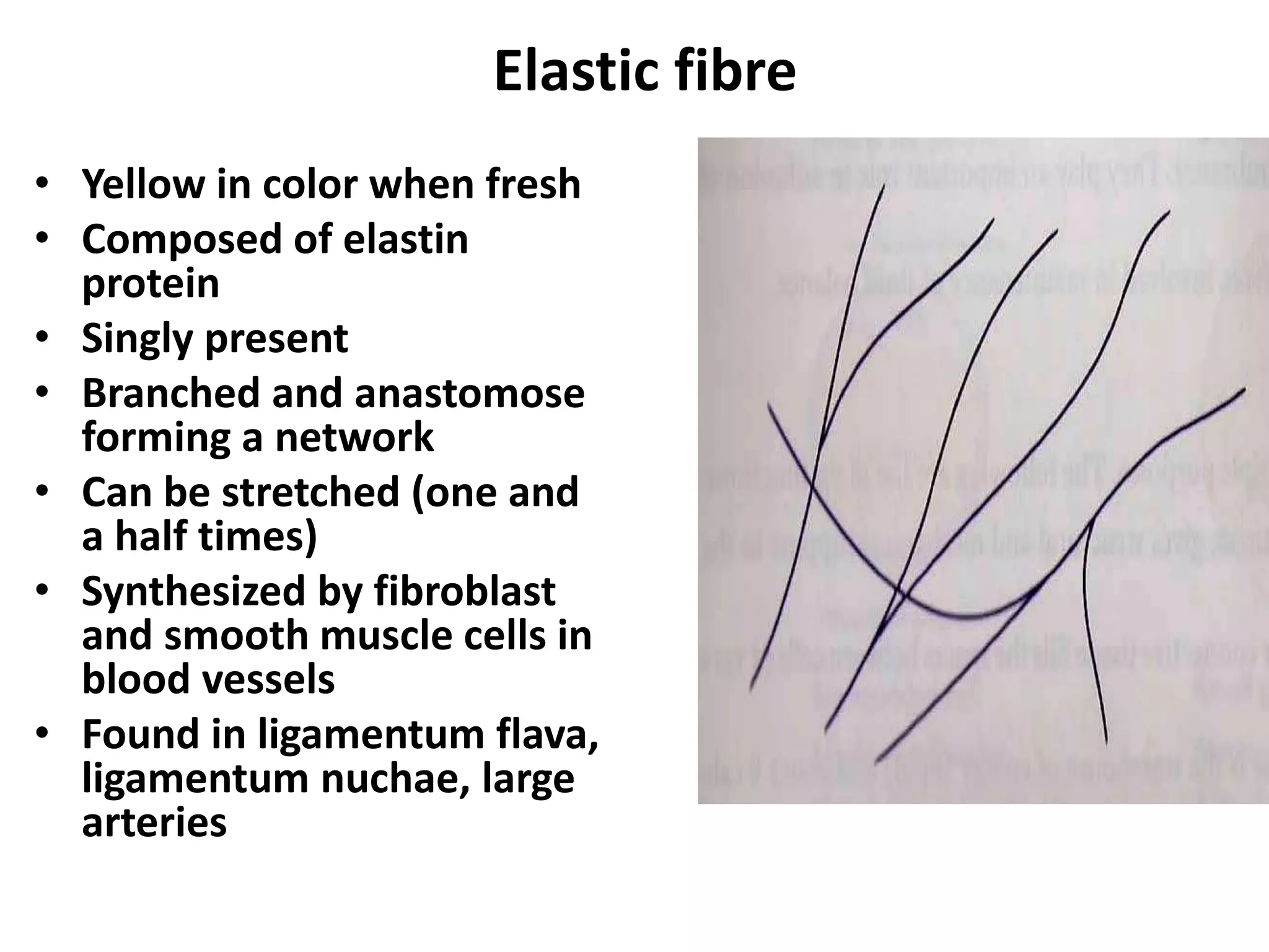
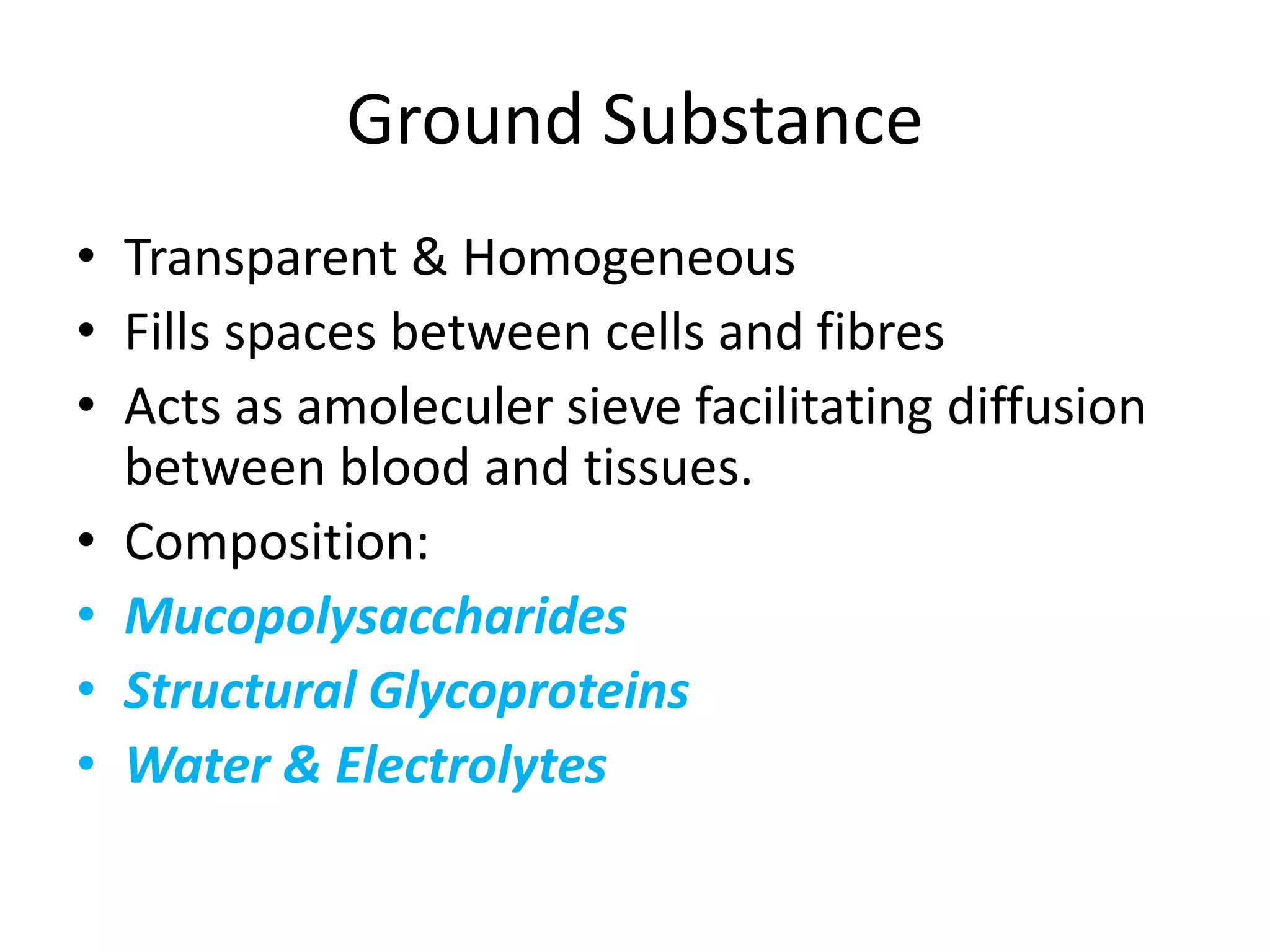
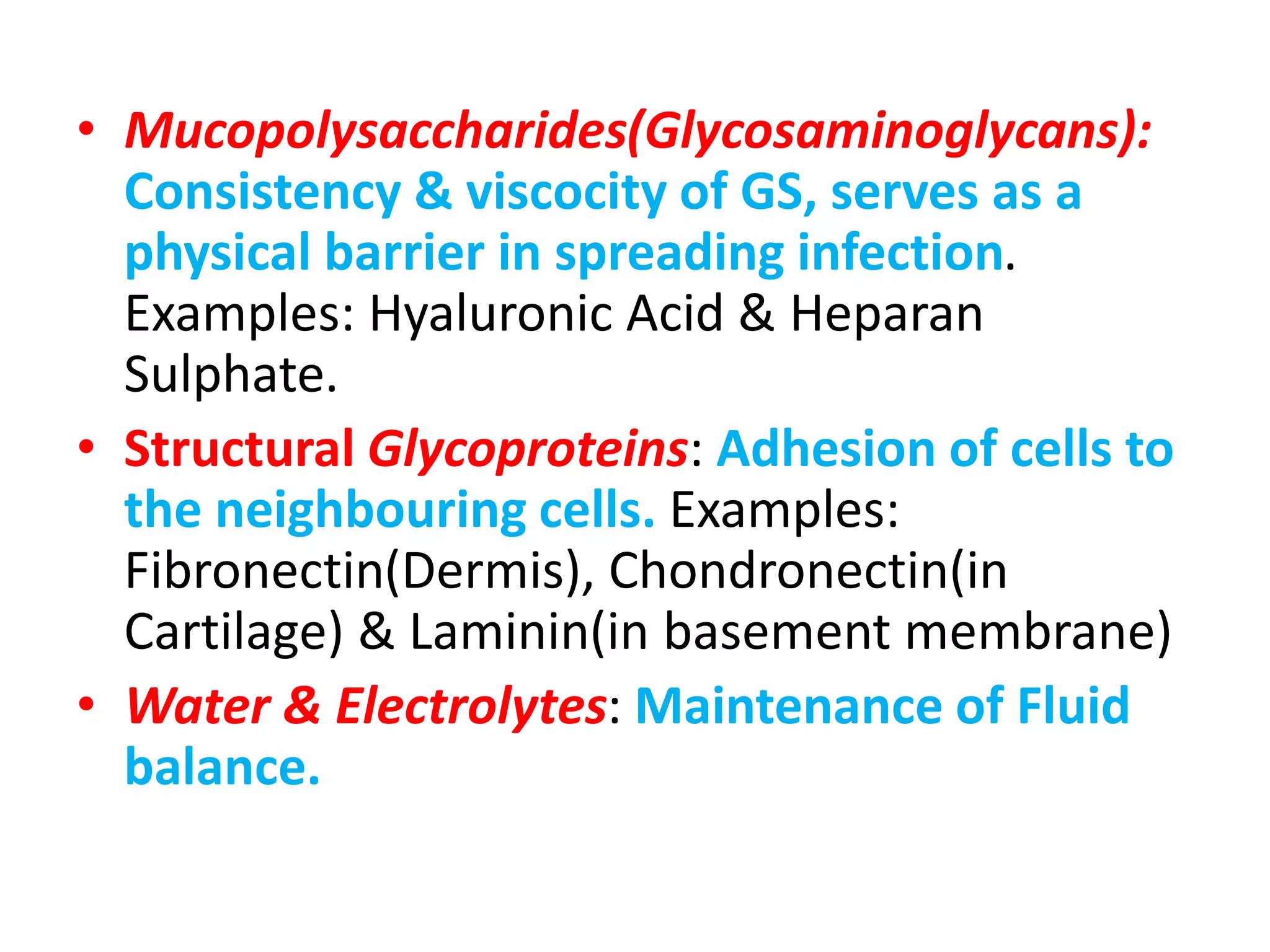
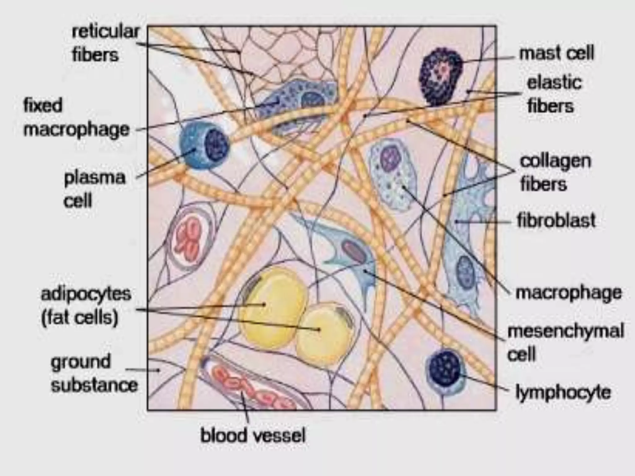
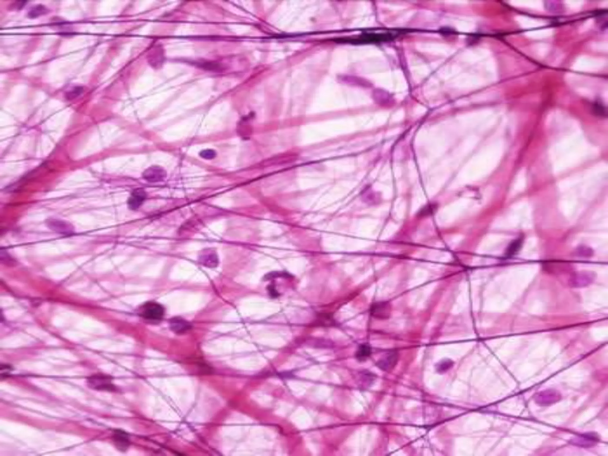
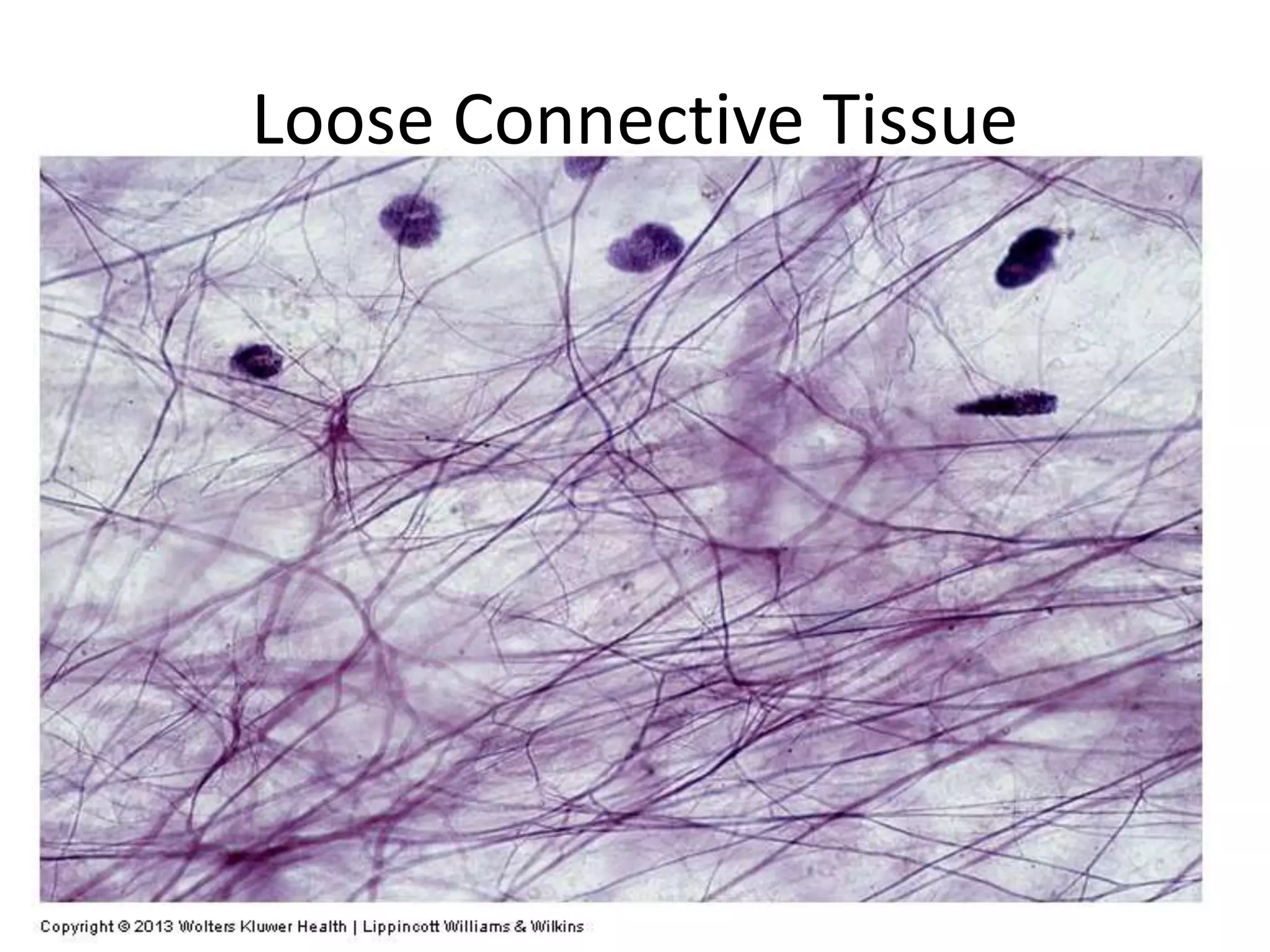
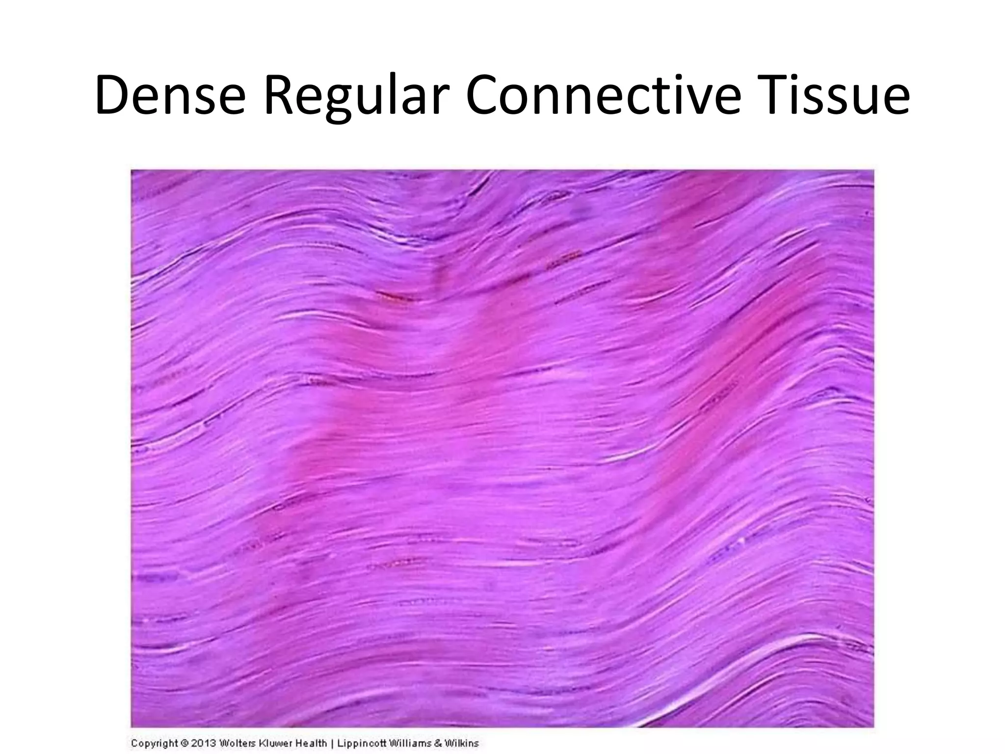
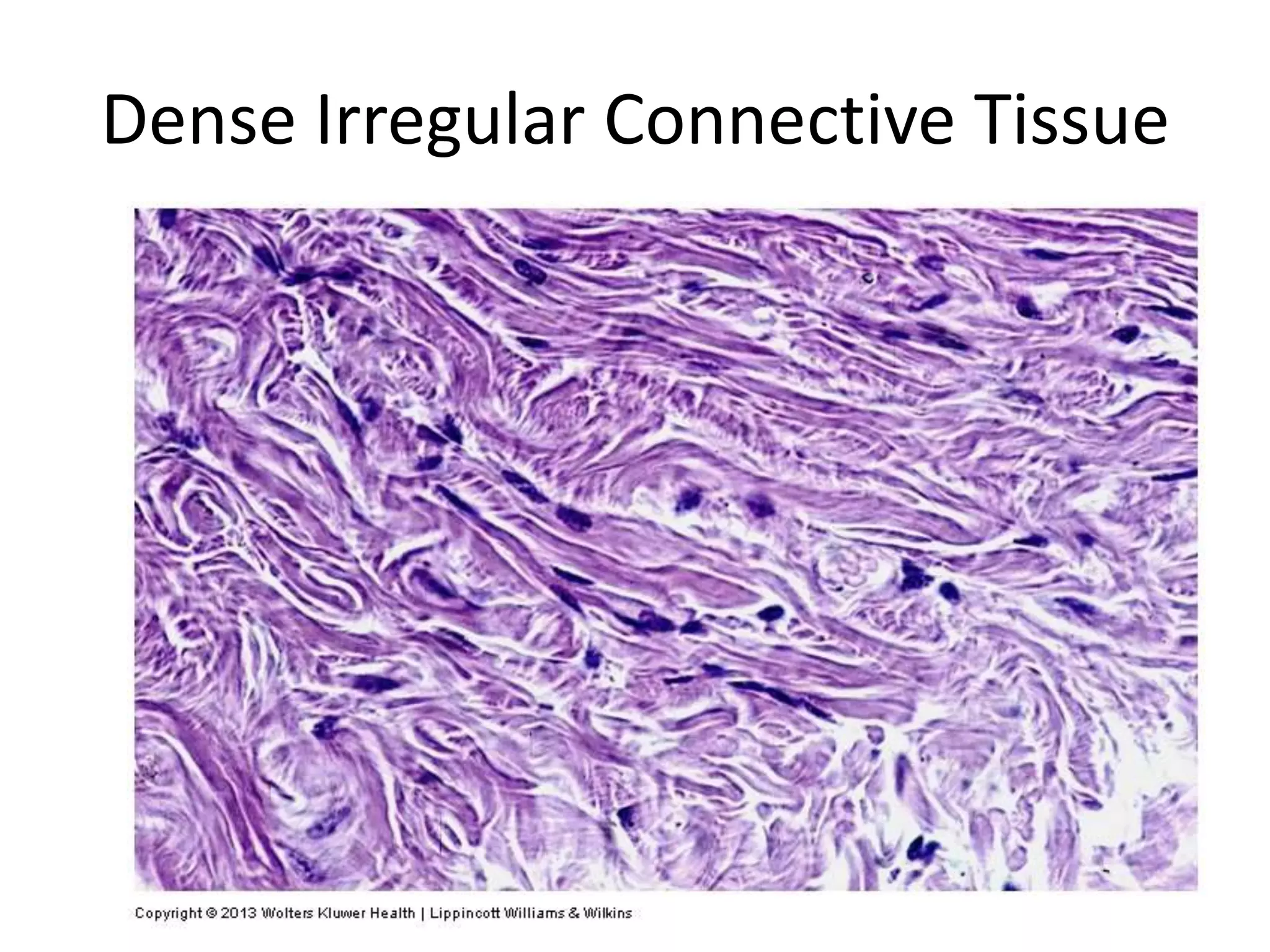
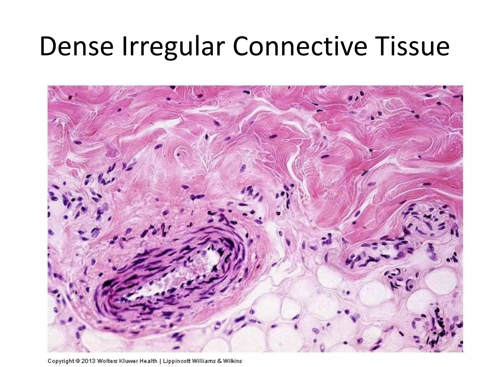
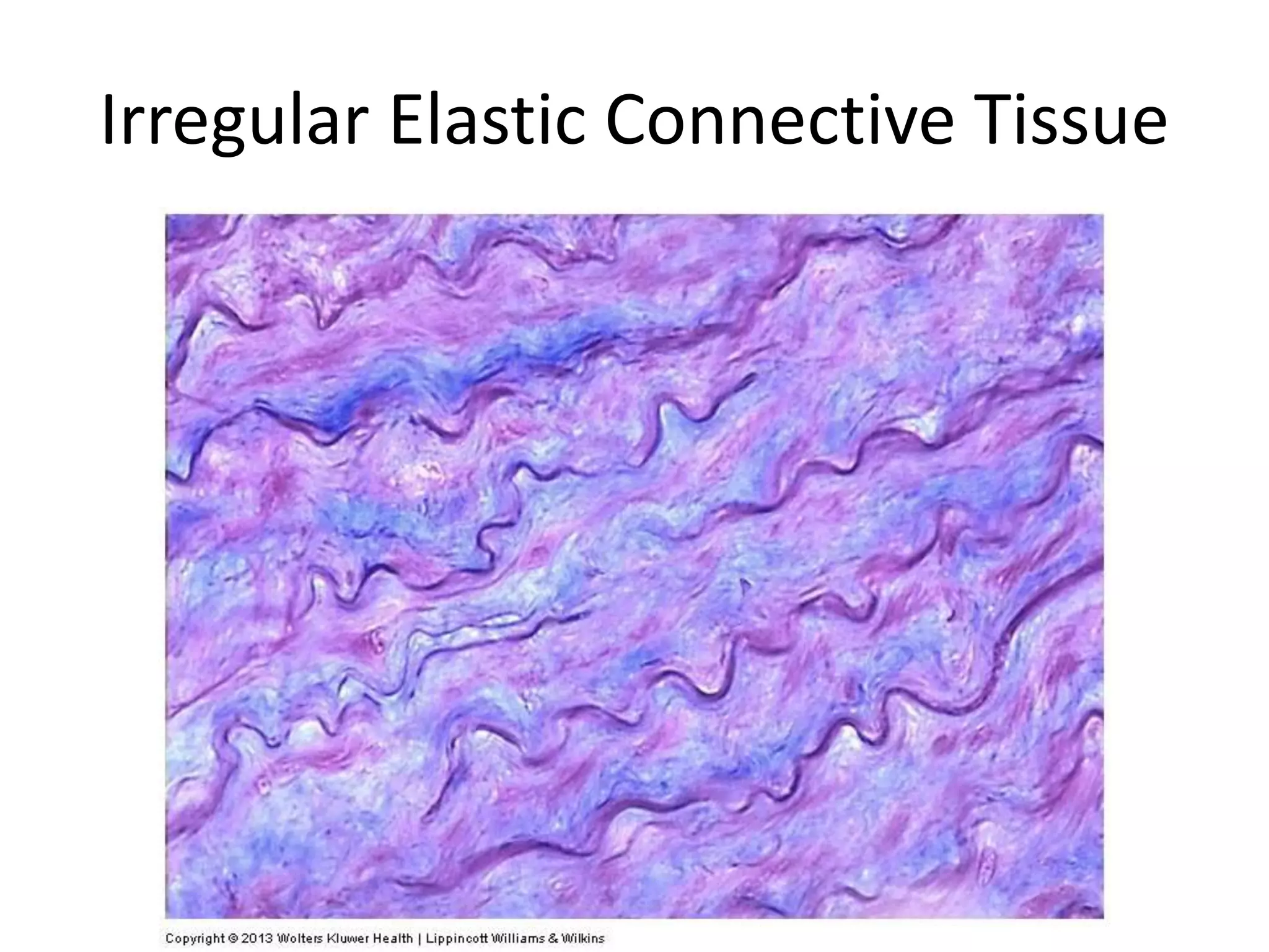
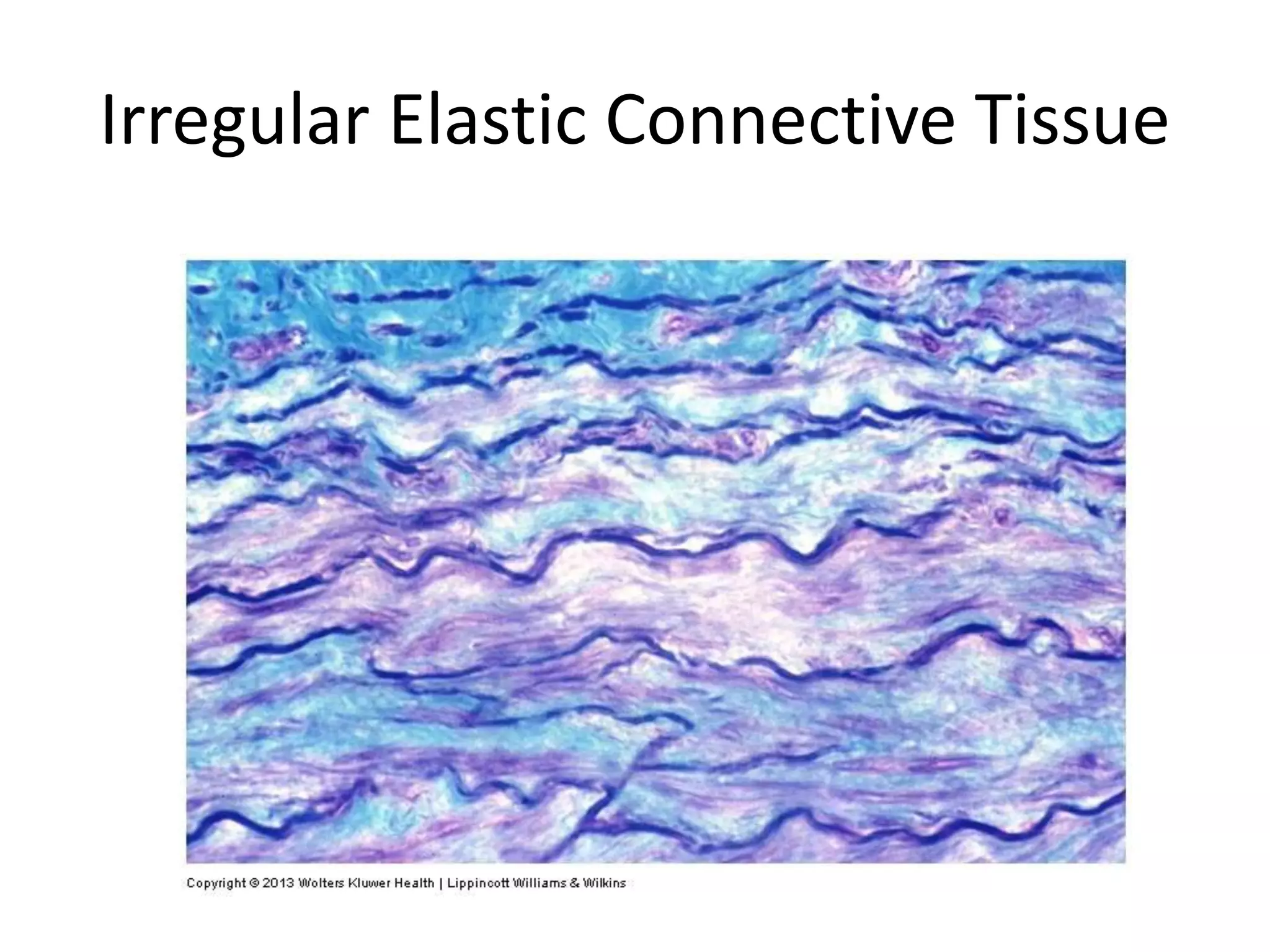
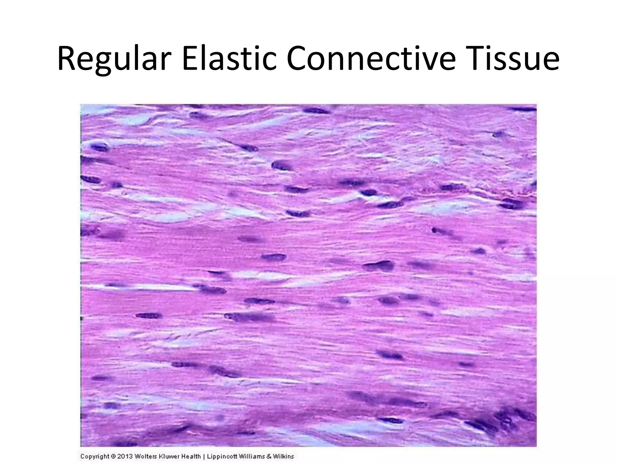
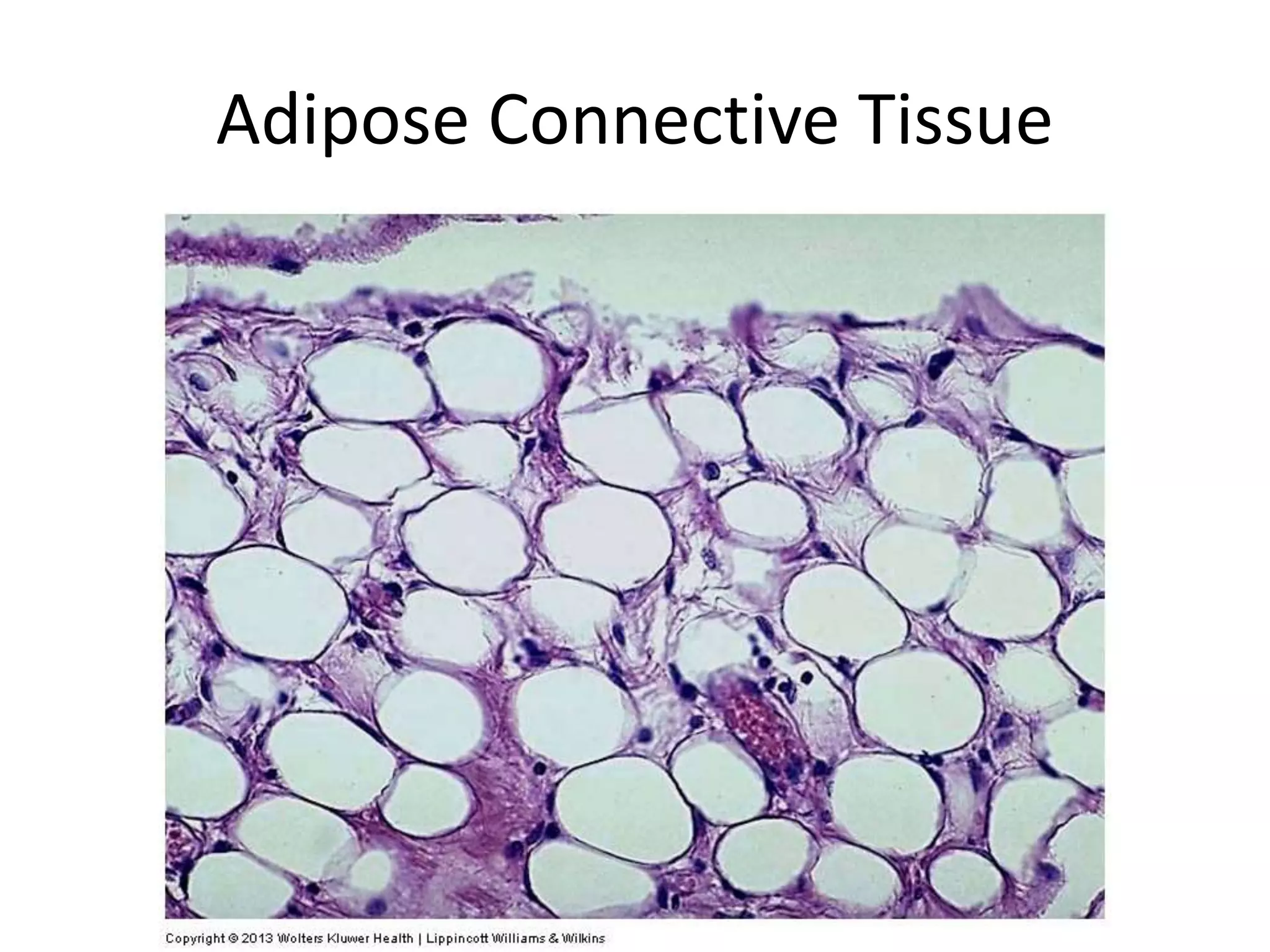
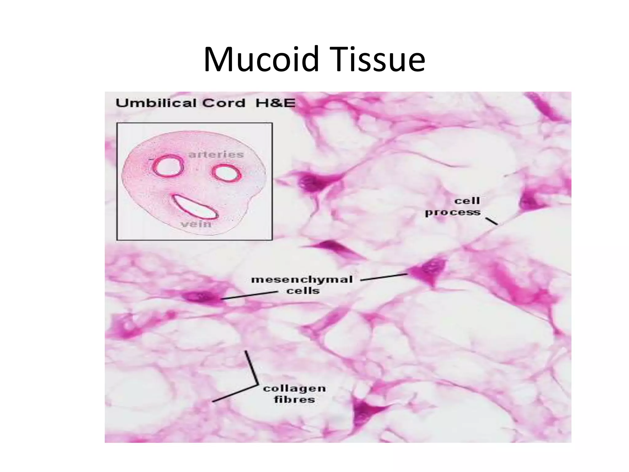
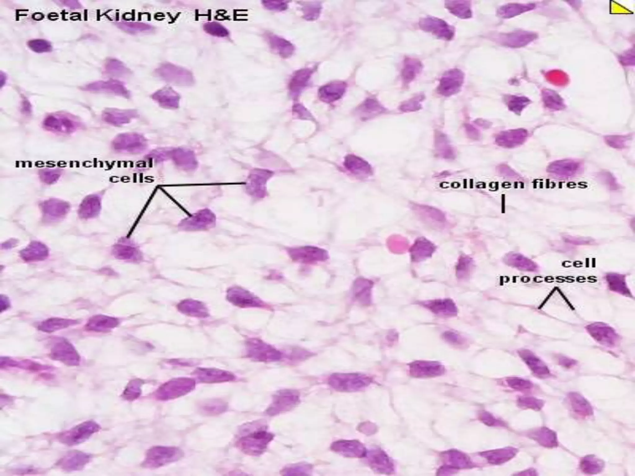
CONNECTIVE TISSUE
CONNECTIVE TISSUE
Connective tissue is one of the basic tissues
which gives structural and metabolic support to
the organ and other tissue of the body.
It connects other tissues.
Functions
• Support Structural & Mechanical
• Packing Fills spaces, Shape to the
organ
• Storage Adipose tissue: energy
Loose areolar CT: water & Electrolytes
• Transport Medium for Nutrients &
Metabolic wastes
• Repair Fibroblasts:matrix and fibres
• Defense Cells: Phagocytosis or
Antibodies
CONNECTIVE TISSUE
• GENERAL FEATURES
• 1. Cells
2. Matrix
-Fibers
-Ground substance
CONNECTIVE TISSUE
Major constituent- Extracellular Matrix
Strength
Cells of Connective Tissue
• A. Fixed cells (intrinsic cells)
1.Fibroblast &
Fibrocytes
2. Mesenchymal cells
3. Adipocyte
4. Fixed macrophages
• B. Free cells (extrinsic
cells/Wandering Cells)
4.Free Macrophage
5. Mast cell
6. Plasma cells
7. Leucocytes
Function
• Fixed Cells: Production &
Maintenance of Extracellular
Matrix.
• Free Cells:Tissue reaction to injury
or invasion of Microorganisms.
Fibroblast
• Most commonly seen
• Fusiform with slender cytoplasmic process
• Large oval nucleus,
• Responsible for fiber production
• Old cells are fibrocyte,
• Contractile Cells are myofibroblast
Fibroblast
Adipocytes
• Store lipid
• Appears as empty
space
• Incapable to division
• Aggregate in adipose
tissue with reticular
fibre
Mesenchymal cells
• Undifferentiated cells
• Stellate in shape,
• Cytoplasmic process,
• Pluripotenant cell
• Near blood vessels as
Advential cell
Macrophages (Histiocytes)
• Free and Fixed type,
• Fixed Cells-
• Irregular Shape
• filopodia process,
• Dark indented eccentric
nucleus,
• Derived from monocyte
• Involved in phagocytosis
• Fused to form giant cell.
• Free Cells- rounded, no
filopodia
Plasma cells
• Oval basophilic cells,
• Eccentric nucleus
• Heterochromatin as cartwheel
nucleus
• Derived from B lymphocyte
• Produces immunoglobulin
• Antibody collected as Russell
body.
• Present in respiratory tract and
gastrointestinal tract
Mast cell
• Round or Fusiform Shaped
• Mostly along blood vessels,
• Metachromatic granules in
cytoplasm,
• Granules have Histamine or
Heparin,
• Look like basophil, so called as
connective tissue basophil .
• Connective tissue mast cell –
heparin granule, present in skin.
• Mucosal mast cell –small,
present in lamina propria of git
and respiratory tract
Leucocytes-
. Granulocytes-
Neutrophils,
Esinophils,
Basophiles
. Agranulocytes
Lymphocytes,
Monocytes
Leucocytes
Collagen Fibre
• White colour when fresh
• Do not branch,wavy
• present in bundle
• Collagen protein forms
Fibres
• Fibres composed of fibril
made of microfibrils
• Micro fibrils made up
tropocollagen-striations
• Synthesized by fibroblast
Collagen
• Tropocollagen is
synthesized by
fibroblasts and released
into extracellular space
where they get
polymerized to form
collagen fibrils
• Collagen on boiling gives
gelatin
• More than 25 types are
present
Collagen is also
synthesized by
• Chondroblasts: in
collagen
• Osteoblasts : in bone
• Smooth Muscle: in
blood vessels
• Odontoblasts: in the
tooth
Types
• Type1-bones ,tendons, dermis etc
• Type2-cartilage
• Type3-reticular fibres
• Type4-basement membrane
• Type5-blood vessels
Synthesis
• Amino acids
• Procollagen
• Three chains
• Tropocollagen
• Collagen
Elastic fibre
• Yellow in color when fresh
• Composed of elastin
protein
• Singly present
• Branched and anastomose
forming a network
• Can be stretched (one and
a half times)
• Synthesized by fibroblast
and smooth muscle cells in
blood vessels
• Found in ligamentum flava,
ligamentum nuchae, large
arteries
Reticular fibre
• Structurally similar to
collagen fibres
• Are very thin Immature
collagen fibre
• Actively branch to form
delicate network therefore
named Reticular
• Form supportive framework
of lymphoid tissue
• Stained black by silver salts
(argyrophillic)
• Composed of Collagen Type III
Ground Substance
• Transparent & Homogeneous
• Fills spaces between cells and fibres
• Acts as amoleculer sieve facilitating diffusion
between blood and tissues.
• Composition:
• Mucopolysaccharides
• Structural Glycoproteins
• Water & Electrolytes
• Mucopolysaccharides(Glycosaminoglycans):
Consistency & viscocity of GS, serves as a
physical barrier in spreading infection.
Examples: Hyaluronic Acid & Heparan
Sulphate.
• Structural Glycoproteins: Adhesion of cells to
the neighbouring cells. Examples:
Fibronectin(Dermis), Chondronectin(in
Cartilage) & Laminin(in basement membrane)
• Water & Electrolytes: Maintenance of Fluid
balance.
Ground substance
• Polysacchrides - hexurate or galectose
• Carbohydrate protein complex (proteoglycanes)-
• 1- mucopolysacchride (glucosaminoglycanes)
• NonSulphates-chondroitin and hyluronic
acid
• Sulphates - chondotinesulphate, heparitine
sulphate, keratohyline
• 2- glycoprotienes- fibronectine(dermis),
chondronectine (cartilage), laminin (b.m)
• water & minerals
Classification
Connective tissue
1.Ordinary connective tissue- Loose connective tissue
-Dense connective tissue
Regular
Irregular
2.connective tissue with special properties
Adipose tissue
Mucoid tissue
Reticular tissue
Pigmented tissue
3.Scleral connective tissue -Bone
-Cartilage
4.Lymphoid and heamopoietc connective tissue
Examples
• Loose areolar connective tissue-
subperitoneal tissue, endomysium, lamina
propria
• Dense collagenous C T
Regular- tendon, ligament, aponeurosis
Irregular-dermis of skin
• Connective T with special properties
Elastic-Ligamentum nuchae
Mucoid/ Embryonic tissue- Wharton’s jelly
Reticular Tissue- Stroma of lymphoid organ
Loose Connective Tissue
Dense Regular Connective Tissue
Dense Irregular Connective Tissue
Irregular Elastic Connective Tissue
Regular Elastic Connective Tissue
Reticular Connective Tissue
Adipose Connective Tissue
Irregular Adipose Connective Tissue
Mucoid Tissue
MCQ
• Plasma Cells are derived from
• 1. Monocytes
• 2. Basophils
• 3.T lymphocytes
• 4. B Lymphocytes
MCQ
• Large number of elastic fibres are present in
• 1. Tendon
• 2. Ligamentum Nuchae
• 3. Basement Membrane
• 4. Aponeurosis
MCQ
The fat cells of Multilocular adipose tissue
(Brown fat) is characterized by the presence of
1.Spherical central nucleus and many lipid
droplets.
2.Flat peripheral nucleus and single lipid
drop
3.Flat central nucleus and single lipid droplet
4.Thin rim of cytoplasm
MCQ
• Which of the following is NOT TRUE about
Collagen
• 1.Constitutes 30% of the dry body weight
• 2.Is synthesized by fibroblasts
• 3.Is composed of Mucopolysaccharides
• 4.Gives gelatin on denaturation

Connective Tissue.pptx

  • 1.
  • 2.
    CONNECTIVE TISSUE Connective tissueis one of the basic tissues which gives structural and metabolic support to the organ and other tissue of the body. It connects other tissues.
  • 3.
    Functions • Support Structural& Mechanical • Packing Fills spaces, Shape to the organ • Storage Adipose tissue: energy Loose areolar CT: water & Electrolytes • Transport Medium for Nutrients & Metabolic wastes • Repair Fibroblasts:matrix and fibres • Defense Cells: Phagocytosis or Antibodies
  • 4.
    CONNECTIVE TISSUE • GENERALFEATURES • 1. Cells 2. Matrix -Fibers -Ground substance
  • 5.
    CONNECTIVE TISSUE Major constituent-Extracellular Matrix Strength
  • 7.
    Cells of ConnectiveTissue • A. Fixed cells (intrinsic cells) 1.Fibroblast & Fibrocytes 2. Mesenchymal cells 3. Adipocyte 4. Fixed macrophages • B. Free cells (extrinsic cells/Wandering Cells) 4.Free Macrophage 5. Mast cell 6. Plasma cells 7. Leucocytes
  • 8.
    Function • Fixed Cells:Production & Maintenance of Extracellular Matrix. • Free Cells:Tissue reaction to injury or invasion of Microorganisms.
  • 9.
    Fibroblast • Most commonlyseen • Fusiform with slender cytoplasmic process • Large oval nucleus, • Responsible for fiber production • Old cells are fibrocyte, • Contractile Cells are myofibroblast
  • 10.
  • 11.
    Adipocytes • Store lipid •Appears as empty space • Incapable to division • Aggregate in adipose tissue with reticular fibre
  • 12.
    Mesenchymal cells • Undifferentiatedcells • Stellate in shape, • Cytoplasmic process, • Pluripotenant cell • Near blood vessels as Advential cell
  • 13.
    Macrophages (Histiocytes) • Freeand Fixed type, • Fixed Cells- • Irregular Shape • filopodia process, • Dark indented eccentric nucleus, • Derived from monocyte • Involved in phagocytosis • Fused to form giant cell. • Free Cells- rounded, no filopodia
  • 14.
    Plasma cells • Ovalbasophilic cells, • Eccentric nucleus • Heterochromatin as cartwheel nucleus • Derived from B lymphocyte • Produces immunoglobulin • Antibody collected as Russell body. • Present in respiratory tract and gastrointestinal tract
  • 16.
    Mast cell • Roundor Fusiform Shaped • Mostly along blood vessels, • Metachromatic granules in cytoplasm, • Granules have Histamine or Heparin, • Look like basophil, so called as connective tissue basophil . • Connective tissue mast cell – heparin granule, present in skin. • Mucosal mast cell –small, present in lamina propria of git and respiratory tract
  • 18.
  • 19.
  • 20.
    Collagen Fibre • Whitecolour when fresh • Do not branch,wavy • present in bundle • Collagen protein forms Fibres • Fibres composed of fibril made of microfibrils • Micro fibrils made up tropocollagen-striations • Synthesized by fibroblast
  • 21.
    Collagen • Tropocollagen is synthesizedby fibroblasts and released into extracellular space where they get polymerized to form collagen fibrils • Collagen on boiling gives gelatin • More than 25 types are present Collagen is also synthesized by • Chondroblasts: in collagen • Osteoblasts : in bone • Smooth Muscle: in blood vessels • Odontoblasts: in the tooth
  • 22.
    Types • Type1-bones ,tendons,dermis etc • Type2-cartilage • Type3-reticular fibres • Type4-basement membrane • Type5-blood vessels
  • 23.
    Synthesis • Amino acids •Procollagen • Three chains • Tropocollagen • Collagen
  • 24.
    Elastic fibre • Yellowin color when fresh • Composed of elastin protein • Singly present • Branched and anastomose forming a network • Can be stretched (one and a half times) • Synthesized by fibroblast and smooth muscle cells in blood vessels • Found in ligamentum flava, ligamentum nuchae, large arteries
  • 25.
    Reticular fibre • Structurallysimilar to collagen fibres • Are very thin Immature collagen fibre • Actively branch to form delicate network therefore named Reticular • Form supportive framework of lymphoid tissue • Stained black by silver salts (argyrophillic) • Composed of Collagen Type III
  • 26.
    Ground Substance • Transparent& Homogeneous • Fills spaces between cells and fibres • Acts as amoleculer sieve facilitating diffusion between blood and tissues. • Composition: • Mucopolysaccharides • Structural Glycoproteins • Water & Electrolytes
  • 27.
    • Mucopolysaccharides(Glycosaminoglycans): Consistency &viscocity of GS, serves as a physical barrier in spreading infection. Examples: Hyaluronic Acid & Heparan Sulphate. • Structural Glycoproteins: Adhesion of cells to the neighbouring cells. Examples: Fibronectin(Dermis), Chondronectin(in Cartilage) & Laminin(in basement membrane) • Water & Electrolytes: Maintenance of Fluid balance.
  • 28.
    Ground substance • Polysacchrides- hexurate or galectose • Carbohydrate protein complex (proteoglycanes)- • 1- mucopolysacchride (glucosaminoglycanes) • NonSulphates-chondroitin and hyluronic acid • Sulphates - chondotinesulphate, heparitine sulphate, keratohyline • 2- glycoprotienes- fibronectine(dermis), chondronectine (cartilage), laminin (b.m) • water & minerals
  • 29.
    Classification Connective tissue 1.Ordinary connectivetissue- Loose connective tissue -Dense connective tissue Regular Irregular 2.connective tissue with special properties Adipose tissue Mucoid tissue Reticular tissue Pigmented tissue 3.Scleral connective tissue -Bone -Cartilage 4.Lymphoid and heamopoietc connective tissue
  • 30.
    Examples • Loose areolarconnective tissue- subperitoneal tissue, endomysium, lamina propria • Dense collagenous C T Regular- tendon, ligament, aponeurosis Irregular-dermis of skin • Connective T with special properties Elastic-Ligamentum nuchae Mucoid/ Embryonic tissue- Wharton’s jelly Reticular Tissue- Stroma of lymphoid organ
  • 33.
  • 34.
  • 35.
  • 36.
  • 37.
  • 38.
  • 39.
  • 40.
  • 41.
  • 42.
  • 43.
  • 44.
  • 45.
  • 46.
  • 48.
  • 50.
    MCQ • Plasma Cellsare derived from • 1. Monocytes • 2. Basophils • 3.T lymphocytes • 4. B Lymphocytes
  • 51.
    MCQ • Large numberof elastic fibres are present in • 1. Tendon • 2. Ligamentum Nuchae • 3. Basement Membrane • 4. Aponeurosis
  • 52.
    MCQ The fat cellsof Multilocular adipose tissue (Brown fat) is characterized by the presence of 1.Spherical central nucleus and many lipid droplets. 2.Flat peripheral nucleus and single lipid drop 3.Flat central nucleus and single lipid droplet 4.Thin rim of cytoplasm
  • 53.
    MCQ • Which ofthe following is NOT TRUE about Collagen • 1.Constitutes 30% of the dry body weight • 2.Is synthesized by fibroblasts • 3.Is composed of Mucopolysaccharides • 4.Gives gelatin on denaturation