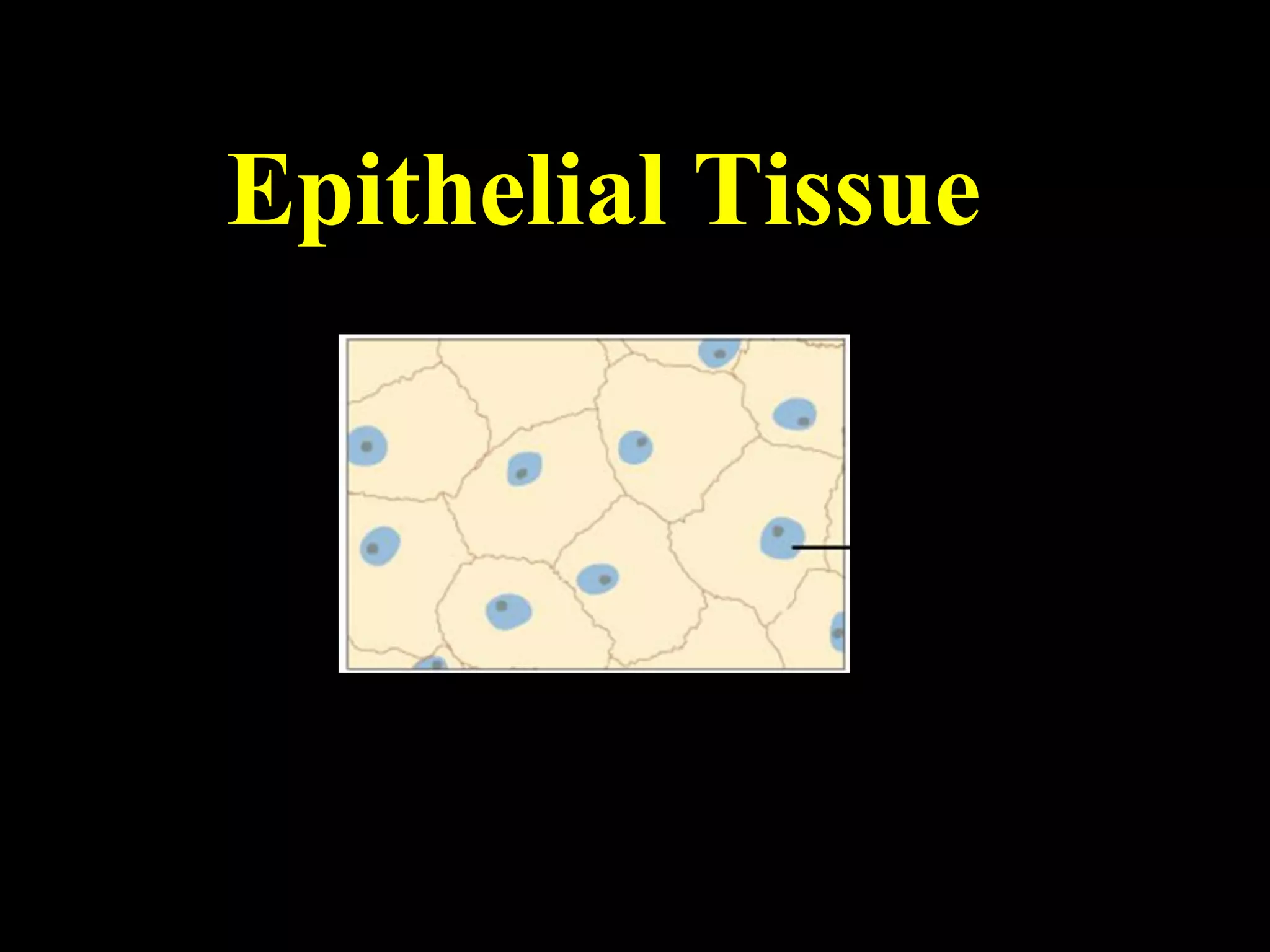
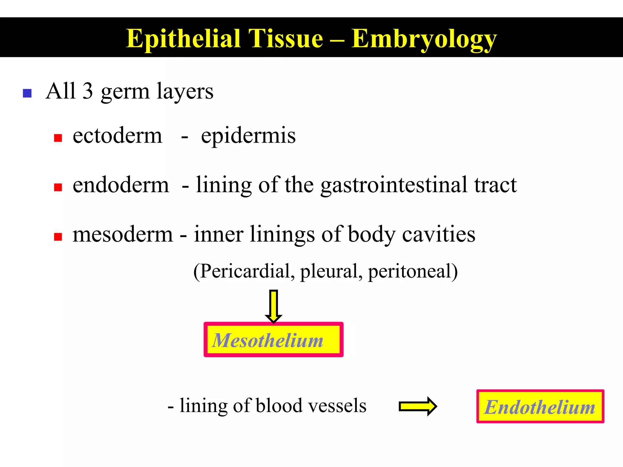
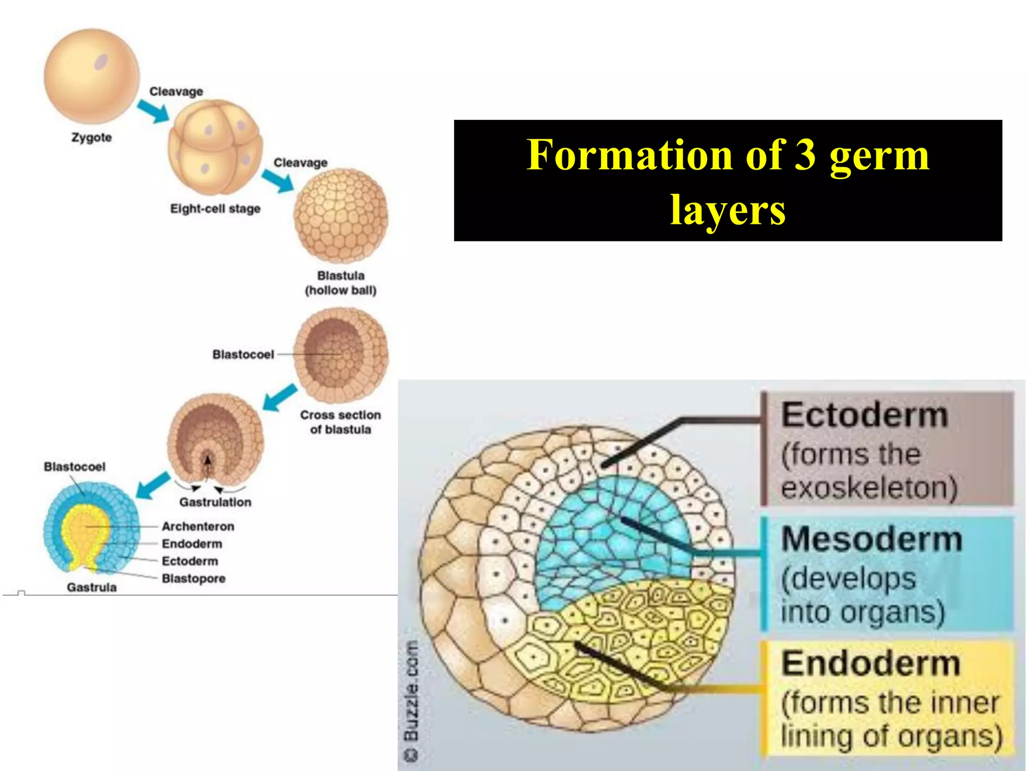
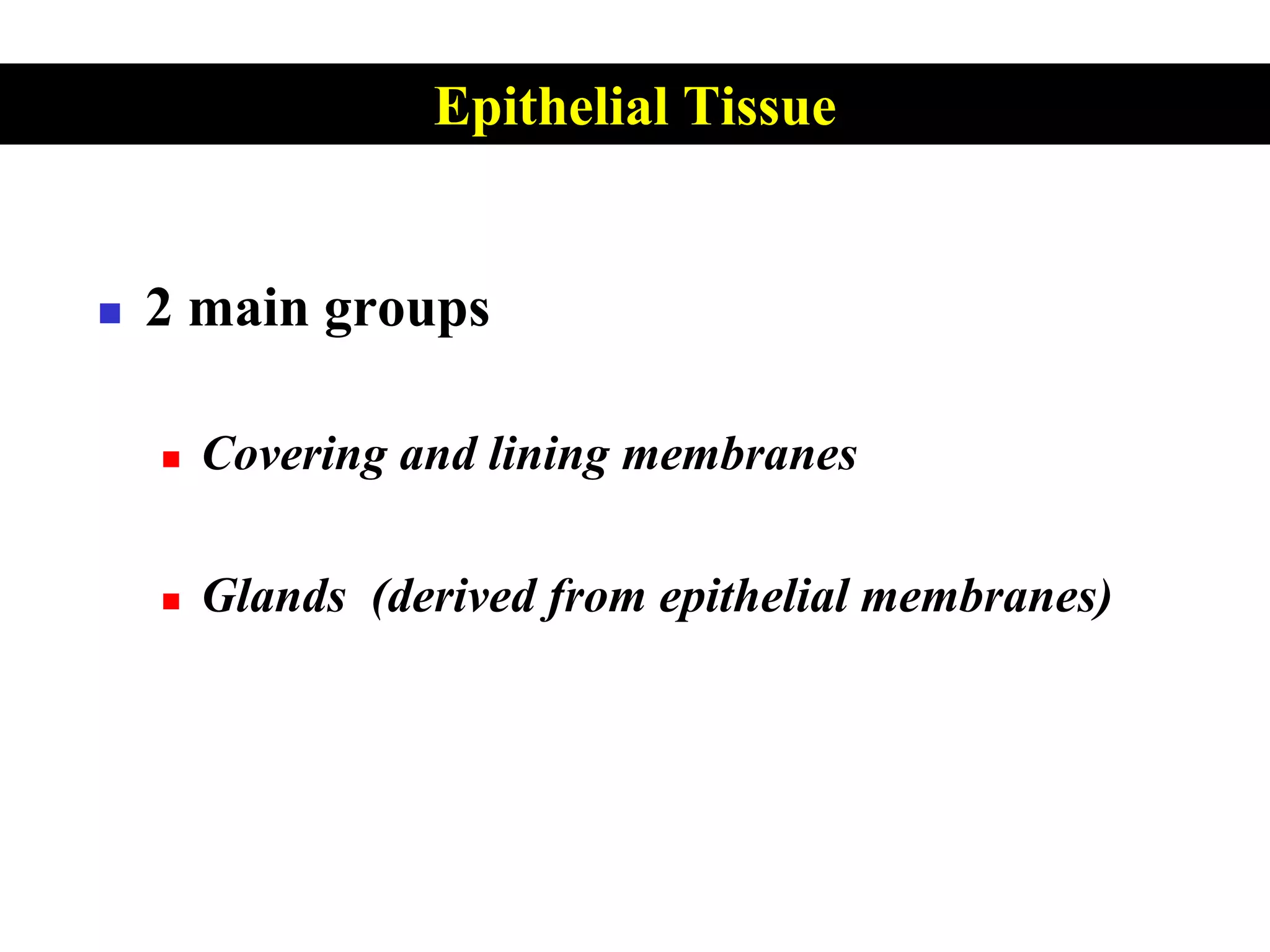
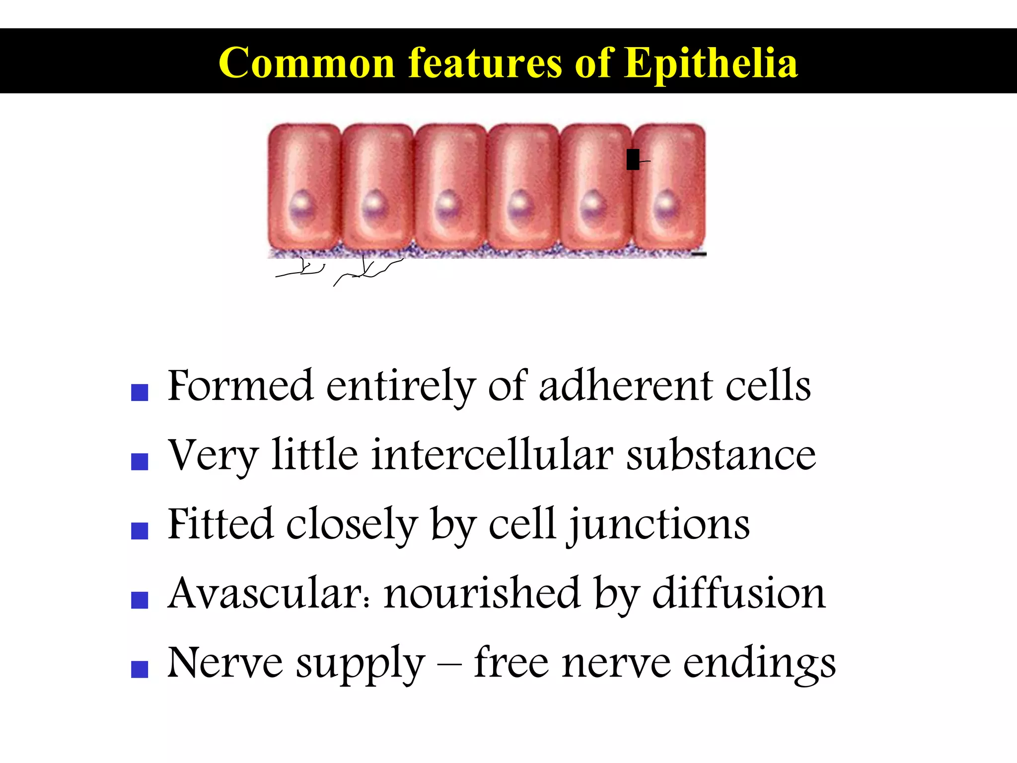
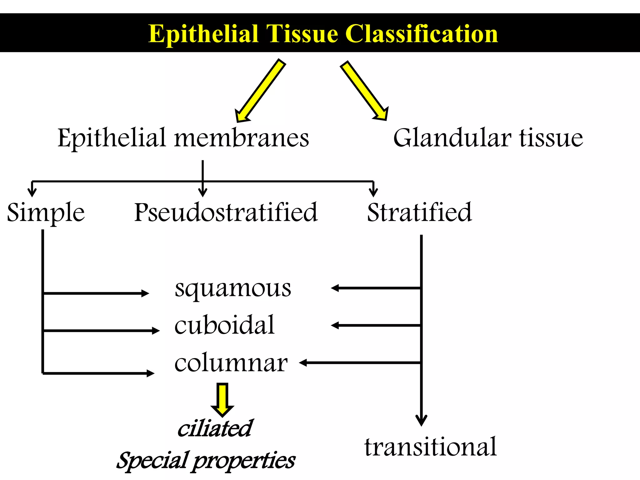
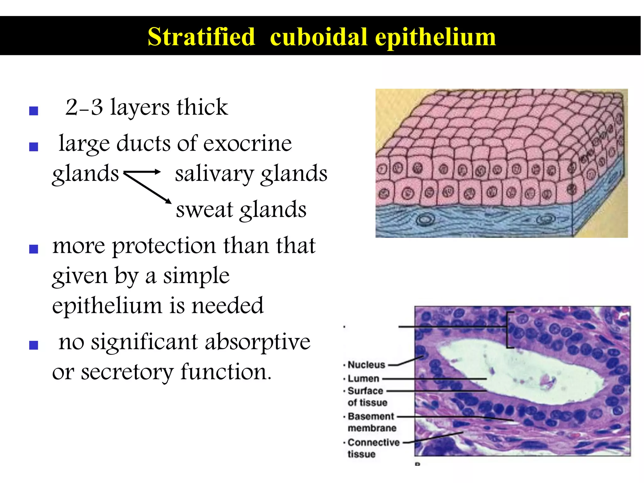
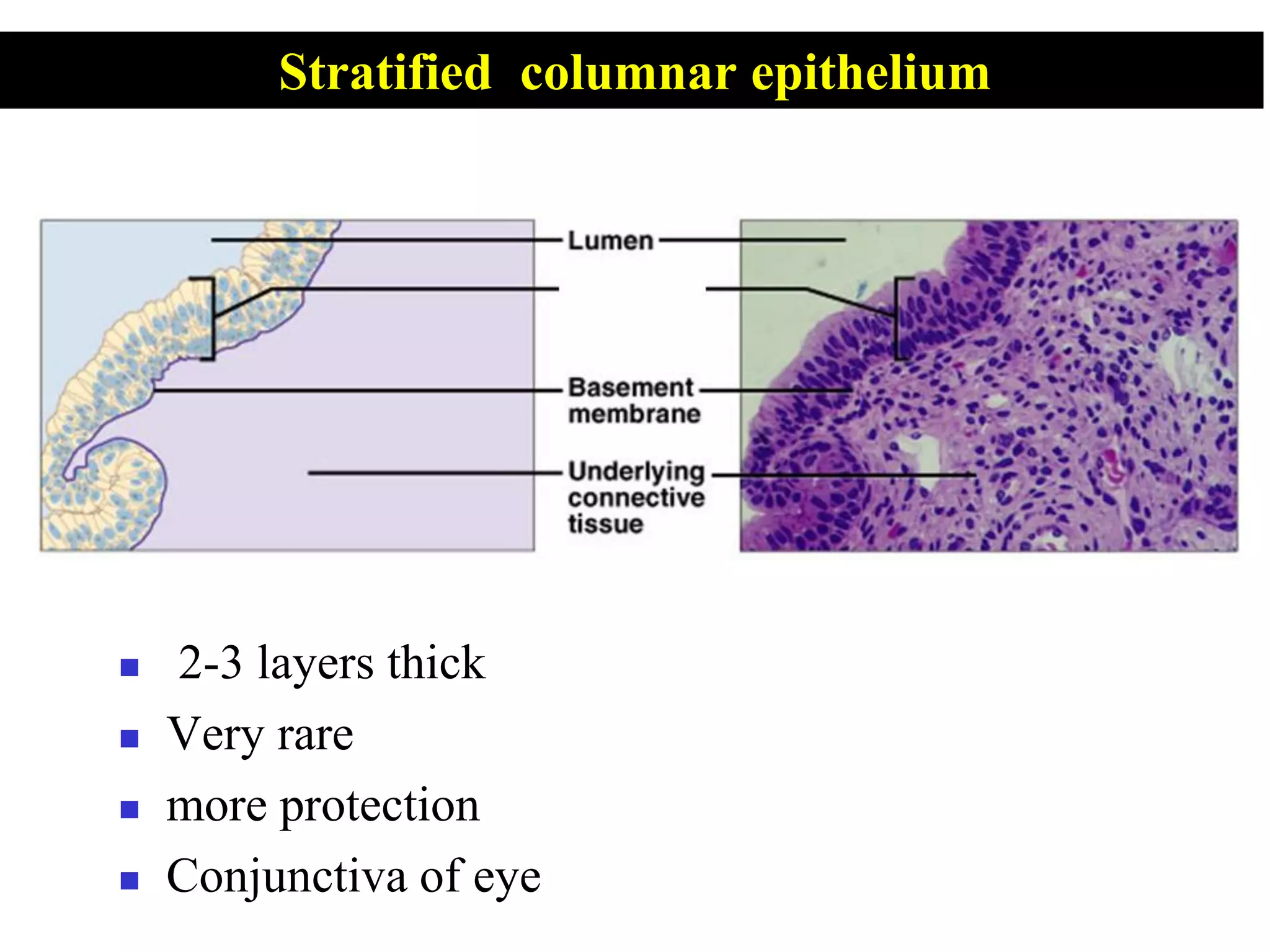
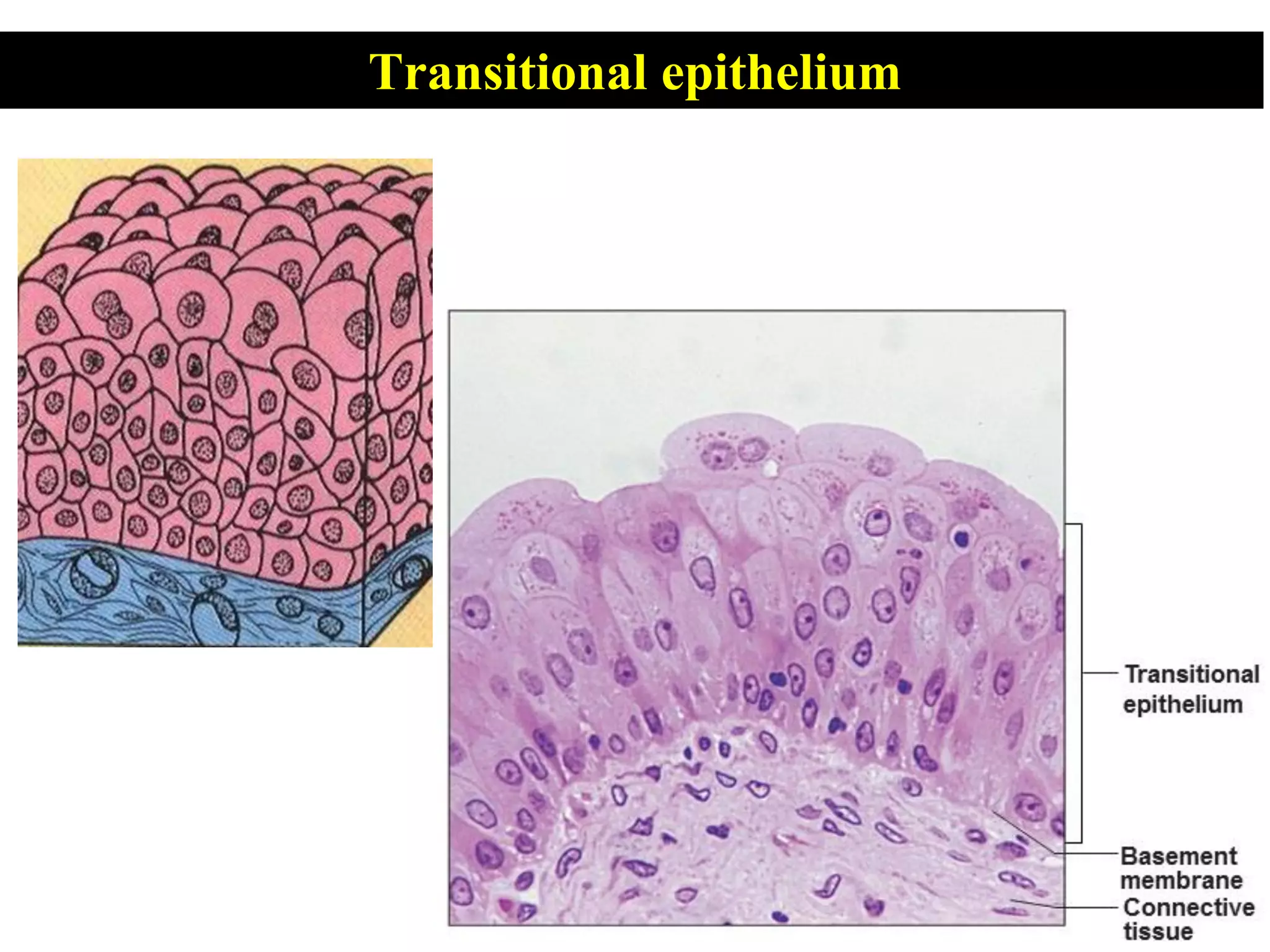
Epithelial Tissue
 Define the term epithelium.
 Classify epithelial tissue.
 State the location of each type of epithelial tissue.
 State the specific function of each type of epithelial tissue.
Objectives
?????
Tissue
 Epithelial
 Connective tissue
 Nervous tissue
 Muscular tissue
Tissue
 Collections of specialized cells
and cell products that perform
a specific function.
• Epithelial tissue
covers exposed surfaces, lines passageways, forms glands.
• Connective tissue
fills internal space, structural support, storage of energy.
• Muscle tissue
contracts for specific movements.
• Neural tissue
carries information from one part of the body to another.
Four Primary Tissue types
 Covers exposed surfaces, lines
passageways & body cavities,
and forms glands.
Epithelial Tissue
 All 3 germ layers
 ectoderm - epidermis
 endoderm - lining of the gastrointestinal tract
 mesoderm - inner linings of body cavities
(Pericardial, pleural, peritoneal)
- lining of blood vessels
Epithelial Tissue – Embryology
Mesothelium
Endothelium
Formation of 3 germ
layers
 2 main groups
 Covering and lining membranes
 Glands (derived from epithelial membranes)
Epithelial Tissue
 Formed entirely of adherent cells
 Very little intercellular substance
 Fitted closely by cell junctions
 Avascular: nourished by diffusion
 Nerve supply – free nerve endings
Common features of Epithelia
apical surface
basal surface
basement membrane
(underlying connective tissue)
lateral surface
apical surfaces
Microvilli cilia
interdigitation
Basal infoldings
Some Characteristics ….
Functional polarity …
Mechanical integrity …..

 Cell shape
 Number of layers
 Type of adherence
 Mitotic activity
 Secretory or absorptive potential
 Adaptation to sensory perception
Epithelia vary in
Squamous
Cuboidal
Columnar
Classification based on cell shape
Stratified
Simple
Apical surface
Basal surface
Apical surface
Basal surface
Classification based on number of cell layers
Cell layers
+
Cell shapes
+
Specializations on apical surface
Epithelial Tissue Classification
based on
Epithelial Tissue Classification
Simple Pseudostratified Stratified
squamous
cuboidal
columnar
Epithelial membranes Glandular tissue
transitional
ciliated
Special properties
• single, thin flat scale like cells
• little cytoplasm
• nucleus bulge in the lumen
Simple squamous (pavement) epithelium
A – cell membrane , B – nucleus , C - cytoplasm
B
A
C
Apical View of Simple Squamous
Cross-Sectional View of Blood Vessel
Simple squamous epithelium
 surfaces involved in passive diffusion
alveoli of lung
blood capillaries endothelium
 lining body cavities mesothelium
 subarachnoid and subdural spaces
 chambers of the eye
Simple squamous epithelium - Locations
 Facilitates the movement of the viscera
(mesothelium)
 active transport by pinocytosis
(mesothelium and endothelium)
 secretion of biologically active molecules
(mesothelium).
Simple squamous epithelium - Functions
 Functions:
secretion
excretion
 Locations:
 Kidney tubules
 Small ducts
 secretory portions of small glands
 ovary surface
 Thyroid
Simple cuboidal epithelium
Simple columnar
epithelium
Simple columnar epithelium
.
 Functions:
Absorption & secretion
 Locations:
Lining of intestine, gallbladder, bronchioles,
stomach
 ciliated - cilia beat to move mucus along the surface.
 cilia are found on the luminal surface of most of
the lining cells
 facilitates transport of the ova in Uterine tube
Simple columnar ciliated epithelium
Cilia
Lumen
Nucleus
Pseudostratified columnar epithelium
 Functions:
Protection; cilia-mediated
transport of particles
rapped in mucus
 Locations:
 Lining of trachea
 Bronchi
 nasal cavity.
Lining of the large airways
Line A - epithelium.
B- Cilia
C - Nuclei
Pseudostratified columnar ciliated epithelium
Epithelial Tissue Classification
Simple Pseudostratified Stratified
squamous
cuboidal
columnar
Epithelial membranes Glandular tissue
transitional
ciliated
Special properties
Stratified squamous epithelium
Non keratinised stratified squamous (wet) epithelium
 Functions:
Protection, secretion;
prevents water loss
 Locations:
 Mouth
 Oesophagus
 Larynx
 Vagina
 Anal canal
 A - epithelium
 B – surface nuclei
Non keratinised stratified squamous (wet) epithelium
 found in parts of the
body exposed to the
atmosphere.
 covered with
waterproof coating -
keratin.
 palms and soles
thick keratin
protects from
abrasion and desiccation.
keratinised stratified squamous (dry) epithelium
Stratified cuboidal epithelium
 2-3 layers thick
 large ducts of exocrine
glands salivary glands
sweat glands
 more protection than that
given by a simple
epithelium is needed
 no significant absorptive
or secretory function.
 2-3 layers thick
 Very rare
 more protection
 Conjunctiva of eye
Stratified columnar epithelium
 found in the urinary tract:
 specialized to stretch (distensibility)
withstand the toxicity of urine.
(Protection)
 made up of 5-6 layers of cells.
 In a relaxed state superficial cells
large rounded pale stained
 luminal surface scalloped outline
 thick plates separated by narrow bands of thin membrane
Transitional epithelium
 Tight junctions between the surface epithelial cells
prevent intercellular diffusion
 In the deeper layers desmosomes are fewer
 Numerous interdigitations and infoldings
 Bladder, ureters, renal calyces
Transitional epithelium
SUMMERY
SUMMERY
SUMMERY
SUMMERY
REFFERENCES
 Junqueira, L.C., Carneiro (1998). Basic
histology. 9 th ed., stamford: Appleton &
lange
 Burkit, H.G, young, B. (1993). Wheaters
functional histology. 4 th ed.,
london:Churchill livingstone
 Singh, I., (2011). Human histology. 6thed.,
New Delhi: Jaypeebrothers.
Epithelial Tissue.pdf

Epithelial Tissue.pdf

  • 1.
  • 2.
     Define theterm epithelium.  Classify epithelial tissue.  State the location of each type of epithelial tissue.  State the specific function of each type of epithelial tissue. Objectives
  • 3.
    ????? Tissue  Epithelial  Connectivetissue  Nervous tissue  Muscular tissue
  • 4.
    Tissue  Collections ofspecialized cells and cell products that perform a specific function.
  • 5.
    • Epithelial tissue coversexposed surfaces, lines passageways, forms glands. • Connective tissue fills internal space, structural support, storage of energy. • Muscle tissue contracts for specific movements. • Neural tissue carries information from one part of the body to another. Four Primary Tissue types
  • 6.
     Covers exposedsurfaces, lines passageways & body cavities, and forms glands. Epithelial Tissue
  • 7.
     All 3germ layers  ectoderm - epidermis  endoderm - lining of the gastrointestinal tract  mesoderm - inner linings of body cavities (Pericardial, pleural, peritoneal) - lining of blood vessels Epithelial Tissue – Embryology Mesothelium Endothelium
  • 8.
    Formation of 3germ layers
  • 10.
     2 maingroups  Covering and lining membranes  Glands (derived from epithelial membranes) Epithelial Tissue
  • 11.
     Formed entirelyof adherent cells  Very little intercellular substance  Fitted closely by cell junctions  Avascular: nourished by diffusion  Nerve supply – free nerve endings Common features of Epithelia
  • 12.
  • 13.
    apical surface basal surface basementmembrane (underlying connective tissue) lateral surface apical surfaces Microvilli cilia interdigitation Basal infoldings Some Characteristics …. Functional polarity … Mechanical integrity ….. 
  • 14.
     Cell shape Number of layers  Type of adherence  Mitotic activity  Secretory or absorptive potential  Adaptation to sensory perception Epithelia vary in
  • 15.
  • 16.
    Stratified Simple Apical surface Basal surface Apicalsurface Basal surface Classification based on number of cell layers
  • 17.
    Cell layers + Cell shapes + Specializationson apical surface Epithelial Tissue Classification based on
  • 18.
    Epithelial Tissue Classification SimplePseudostratified Stratified squamous cuboidal columnar Epithelial membranes Glandular tissue transitional ciliated Special properties
  • 19.
    • single, thinflat scale like cells • little cytoplasm • nucleus bulge in the lumen Simple squamous (pavement) epithelium
  • 20.
    A – cellmembrane , B – nucleus , C - cytoplasm B A C Apical View of Simple Squamous
  • 21.
    Cross-Sectional View ofBlood Vessel Simple squamous epithelium
  • 22.
     surfaces involvedin passive diffusion alveoli of lung blood capillaries endothelium  lining body cavities mesothelium  subarachnoid and subdural spaces  chambers of the eye Simple squamous epithelium - Locations
  • 23.
     Facilitates themovement of the viscera (mesothelium)  active transport by pinocytosis (mesothelium and endothelium)  secretion of biologically active molecules (mesothelium). Simple squamous epithelium - Functions
  • 24.
     Functions: secretion excretion  Locations: Kidney tubules  Small ducts  secretory portions of small glands  ovary surface  Thyroid Simple cuboidal epithelium
  • 25.
  • 26.
    Simple columnar epithelium . Functions: Absorption & secretion  Locations: Lining of intestine, gallbladder, bronchioles, stomach
  • 27.
     ciliated -cilia beat to move mucus along the surface.  cilia are found on the luminal surface of most of the lining cells  facilitates transport of the ova in Uterine tube Simple columnar ciliated epithelium
  • 28.
    Simple columnar ciliatedepithelium Cilia Lumen Nucleus
  • 29.
    Pseudostratified columnar epithelium Functions: Protection; cilia-mediated transport of particles rapped in mucus  Locations:  Lining of trachea  Bronchi  nasal cavity.
  • 30.
    Lining of thelarge airways Line A - epithelium. B- Cilia C - Nuclei Pseudostratified columnar ciliated epithelium
  • 31.
    Epithelial Tissue Classification SimplePseudostratified Stratified squamous cuboidal columnar Epithelial membranes Glandular tissue transitional ciliated Special properties
  • 32.
  • 33.
    Non keratinised stratifiedsquamous (wet) epithelium  Functions: Protection, secretion; prevents water loss  Locations:  Mouth  Oesophagus  Larynx  Vagina  Anal canal
  • 34.
     A -epithelium  B – surface nuclei Non keratinised stratified squamous (wet) epithelium
  • 35.
     found inparts of the body exposed to the atmosphere.  covered with waterproof coating - keratin.  palms and soles thick keratin protects from abrasion and desiccation. keratinised stratified squamous (dry) epithelium
  • 36.
    Stratified cuboidal epithelium 2-3 layers thick  large ducts of exocrine glands salivary glands sweat glands  more protection than that given by a simple epithelium is needed  no significant absorptive or secretory function.
  • 37.
     2-3 layersthick  Very rare  more protection  Conjunctiva of eye Stratified columnar epithelium
  • 38.
     found inthe urinary tract:  specialized to stretch (distensibility) withstand the toxicity of urine. (Protection)  made up of 5-6 layers of cells.  In a relaxed state superficial cells large rounded pale stained  luminal surface scalloped outline  thick plates separated by narrow bands of thin membrane Transitional epithelium
  • 39.
  • 40.
     Tight junctionsbetween the surface epithelial cells prevent intercellular diffusion  In the deeper layers desmosomes are fewer  Numerous interdigitations and infoldings  Bladder, ureters, renal calyces Transitional epithelium
  • 41.
  • 42.
  • 43.
  • 44.
  • 45.
    REFFERENCES  Junqueira, L.C.,Carneiro (1998). Basic histology. 9 th ed., stamford: Appleton & lange  Burkit, H.G, young, B. (1993). Wheaters functional histology. 4 th ed., london:Churchill livingstone  Singh, I., (2011). Human histology. 6thed., New Delhi: Jaypeebrothers.