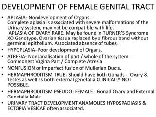
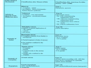
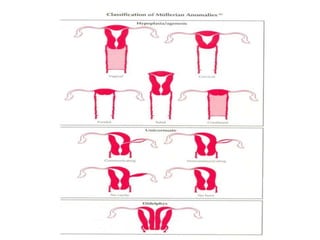
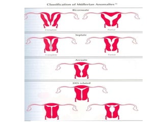
CONGENITAL
MALFORMATIONS

FEMALE GENITAL TRACT
DEVELOPMENT OF FEMALE GENITAL TRACT
• GONADAL DEVELOPMENT IS SEPARATE FROM THAT
  OF THE DUCTS. NORMAL OVARY MAY BE PRESENT
  WITH ABSENT UTERUS & TUBES.
• MULLERIAN & WOLFIAN DUCTS ARE CLOSELY
  RELATED . HENCE ASSOCIATION WITH
  MALFORMATIONS OF THE RENAL TRACT
• SEVERE CONGENITAL ANAMOLIES MAY BE
  ASSOCIATED WITH CHROMOSOMAL
  ABNORMALITIES
DEVELOPMENT OF FEMALE GENITAL TRACT
 EARLY EMBRYO – INTERMEDIATE CELL MASS FORMS
  MEDIAL PART OF UROGENITAL RIDGE FORMS URINARY TRACT
  & GENITAL TRACT ; except Bladder, Urethra,Vagina,Vulva.
 Initially all embryos are undifferentiated. Hence INDIFFERENT
  Gonad . Cells from Coelom migrate to medial part of
  urogenital ridge . Development depends on the XY Genotype
  Chromosomal expression of H-Y Factor which promotes
  Testicular differentiation and secretion of ANTI-MULLERIAN
  FACTOR which causes regression of the Mullerian system
  while the Wolfian Ducts grow in response to Testosterone
  secreted from testes and differentiate into the
  retetubules, Epididymis, Vas, Prostrate, Seminal Vesicles.
 Absence of Y genes, leads to Gonad forming into Uterus and
  since no AMF is secreted so Mullerian ducts differentiate
  while the Wolfian ducts regress in XX Genotype.
DEVELOPMENT OF FEMALE GENITAL TRACT
• MULLERIAN DUCTS ARE SOLID COLUMNS/CORDS OF
  CELLS, which later CANALISES.
• THEY ARE INITIALLY SEPERATE CEPHALIC part but join
  up Caudally and then open into the Urogenital sinus as
  mullerian tubercles.
• Upper part forms Fallopian Tubes, Lower part forms the
  Uterus,Cervix,Upper2/3 of Vagina
• The cavity (lumen) is formed by canalisation.
• SINO-VAGINAL BULBS from the Mesodermal thickening
  forms the lower 1/3 of VAGINA.
• The membrane seperating upper & lower vagina
  disappears.
• WOLFIAN DUCT remnants may form cysts
  Epoopheron, Paraoopheron, Gartner’s Duct
DEVELOPMENT OF FEMALE GENITAL TRACT
• VULVA develops in the area between the coccyx and
  the umbilicus from 2 Genital tubercles.
• The tubercles fuse to form a single tubercle the
  Clitoris. Posteriorly two genital swellings are present
  which form the Labia Majora ; medial to this are 2
  folds which form the Labia minora
DEVELOPMENT OF FEMALE GENITAL TRACT
• APLASIA- Nondevelopment of Organs.
  Complete aplasia is associated with severe malformations of the
  Urinary system, may not be compatible with life.
   APLASIA OF OVARY RARE. May be found in TURNER’S Syndrome
  XO Genotype, Ovarian tissue replaced by a fibrous band without
  germinal epithelium. Associated absence of tubes.
• HYPOPLASIA- Poor development of Organs.
• ATRESIA- Noncanalisation of part / whole of the system.
  Commonest Vagina Part / Complete Atresia
• NONFUSION or Imperfect fusion of Mullerian Ducts.
• HERMAPHRODITISM TRUE- Should have both Gonads - Ovary &
  Testes as well as both external genetalia CLINICALLY NOT
  POSSIBLE.
• HERMAPHRODITISM PSEUDO- FEMALE : Gonad Ovary and External
  Genetalia Male
• URINARY TRACT DEVELOPMENT ANAMOLIES HYPOSPADIASIS &
  ECTOPIA VESICAE often associated.
CONGENITAL MALFORMATIONS
• VAGINAL ATRESIA : Partial / Complete .
  Ovary,Tubes,Uterus are normal. No outlet for OVA
  or Menstrual blood. Blood collects in Vagina.
1.H/o Primary Amenorrhoea Cryptomenorrhoea.
2.Internal collection of blood
   - Pain and discomfort
   - Haematocolpos
   - Haematrometra
   - Haematosalpinx
   - Haematoperitoneum
3.Urinary Retention
CONGENITAL MALFORMATIONS
• IMPERFORATE VAGINA (HYMEN)
Bluish discoloration of hymen
Rx Drainage (i)Excision of Hymen(ii)Canalisation of
  Vagina(iii)Allow blood to drain by gravity
Do not place drain or press abdomen. PV after a
  month on followup . Antibiotics in case of infection
CONGENITAL MALFORMATIONS
• FUSION DEFECTS
1.UTERUS DIADELPHIS :2 Uterii, 2 Cervix, 2 Vaginas
2.UTERUS BICORNUATE , BICOLLIS :2 Uterii, 2
  Cervix, septate vagina
3.UTERUS BICORNUATE , UNICOLLIS : 2 Uterii, Septate
  Cervix & Vagina
4.SEPTATE / SUBSEPTATE UTERUS
5.SEPTATE VAGINA
6.ARCUATE UTERUS : Fundus not well developed
7.UNICORNUATE : One side Mullerian duct develops
8.RUDIMENTARY HORN : One horn develops normally
  other does not develop. Types – Communicating/
  Noncommunicating
9.UNILATERAL HAEMATOCOLPOS - Cryptomenorrhoea
CONGENITAL MALFORMATIONS
• CLINICAL FEATURES :-
1.Symptomless – accidental discovery on USG/Exam
2.Menstrual problems – Menorrhagia
3.Unilateral dysmenorrhoea – abnormal contraction of one horn
   of uterus
4.Coital difficulty – Pain or Bleeding (septate vagina)
5.Obstetrical Problems –
Infertility (coitus in rudimentary half of vagina)
Ectopic Pregnancy (rudimentary horn)
Habitual Abortions / Premature Labour
Malpresentations – Transverse lie / Breech presentation in
   Septate Uterus & Bicornuate/Unicornuate Uterus
During Labour – Dysfunctional Labour, Obstructed Labour
   (Rudimentary horn), Postpartum Haemorrhage /Retained
   Placenta
CONGENITAL MALFORMATIONS
 DIAGNOSIS :
H/O Clinical Features
OBSTETRICAL HISTORY
CLINICAL EXAM
MAY MISTAKE TWO CORNU FOR FIBROID UTERUS
 INVESTIGATIONS :
ULTRASONOGRAPHY
SALINE SONOGRAPHY
HYSTEROSALPINGOGRAPHY
INTRAVENOUS PYELOGRRAPHY / UROGRAPHY
DIAGNOSTIC LAPROSCOPIC
COMPUTERISED TOMOGRAPHY
MAGNETIC RESONANCE IMAGING
POSITIVE EMISSION TOMOGRAPHY
CONGENITAL MALFORMATIONS
• PROBLEMS ON UNDIAGNOSED CASES
1. DYSMENORRHOEA – dilatation done on one side
   other side produces pain
2. INTRODUCTION OF IUCD – IUCD placed in opposite
   horn, pregnancy develops in undetected horn
3. CAESAREAN Section due to nondilatation of Cervix
4. MTP / MR Suction Evacuation – Pregnancy may
   continue as procedure done in opposite horn
5. CARCINOMA CERVIX may be missed
CONGENITAL MALFORMATIONS
• TREATMENT OPERATIVE
UNIFICATION OF HORNS – METROPLASTY
  May be done by Abdominal Route STRASSMAN’S
  Hysteroscopic Resection of septae
EXCISION OF VAGINAL SEPTA
EXCISION OF RUDIMENTARY HORN to prevent risk
  of Ectopic Pregnancy

Congenital malformations rs

  • 1.
  • 2.
    DEVELOPMENT OF FEMALEGENITAL TRACT • GONADAL DEVELOPMENT IS SEPARATE FROM THAT OF THE DUCTS. NORMAL OVARY MAY BE PRESENT WITH ABSENT UTERUS & TUBES. • MULLERIAN & WOLFIAN DUCTS ARE CLOSELY RELATED . HENCE ASSOCIATION WITH MALFORMATIONS OF THE RENAL TRACT • SEVERE CONGENITAL ANAMOLIES MAY BE ASSOCIATED WITH CHROMOSOMAL ABNORMALITIES
  • 3.
    DEVELOPMENT OF FEMALEGENITAL TRACT  EARLY EMBRYO – INTERMEDIATE CELL MASS FORMS MEDIAL PART OF UROGENITAL RIDGE FORMS URINARY TRACT & GENITAL TRACT ; except Bladder, Urethra,Vagina,Vulva.  Initially all embryos are undifferentiated. Hence INDIFFERENT Gonad . Cells from Coelom migrate to medial part of urogenital ridge . Development depends on the XY Genotype Chromosomal expression of H-Y Factor which promotes Testicular differentiation and secretion of ANTI-MULLERIAN FACTOR which causes regression of the Mullerian system while the Wolfian Ducts grow in response to Testosterone secreted from testes and differentiate into the retetubules, Epididymis, Vas, Prostrate, Seminal Vesicles.  Absence of Y genes, leads to Gonad forming into Uterus and since no AMF is secreted so Mullerian ducts differentiate while the Wolfian ducts regress in XX Genotype.
  • 4.
    DEVELOPMENT OF FEMALEGENITAL TRACT • MULLERIAN DUCTS ARE SOLID COLUMNS/CORDS OF CELLS, which later CANALISES. • THEY ARE INITIALLY SEPERATE CEPHALIC part but join up Caudally and then open into the Urogenital sinus as mullerian tubercles. • Upper part forms Fallopian Tubes, Lower part forms the Uterus,Cervix,Upper2/3 of Vagina • The cavity (lumen) is formed by canalisation. • SINO-VAGINAL BULBS from the Mesodermal thickening forms the lower 1/3 of VAGINA. • The membrane seperating upper & lower vagina disappears. • WOLFIAN DUCT remnants may form cysts Epoopheron, Paraoopheron, Gartner’s Duct
  • 5.
    DEVELOPMENT OF FEMALEGENITAL TRACT • VULVA develops in the area between the coccyx and the umbilicus from 2 Genital tubercles. • The tubercles fuse to form a single tubercle the Clitoris. Posteriorly two genital swellings are present which form the Labia Majora ; medial to this are 2 folds which form the Labia minora
  • 6.
    DEVELOPMENT OF FEMALEGENITAL TRACT • APLASIA- Nondevelopment of Organs. Complete aplasia is associated with severe malformations of the Urinary system, may not be compatible with life. APLASIA OF OVARY RARE. May be found in TURNER’S Syndrome XO Genotype, Ovarian tissue replaced by a fibrous band without germinal epithelium. Associated absence of tubes. • HYPOPLASIA- Poor development of Organs. • ATRESIA- Noncanalisation of part / whole of the system. Commonest Vagina Part / Complete Atresia • NONFUSION or Imperfect fusion of Mullerian Ducts. • HERMAPHRODITISM TRUE- Should have both Gonads - Ovary & Testes as well as both external genetalia CLINICALLY NOT POSSIBLE. • HERMAPHRODITISM PSEUDO- FEMALE : Gonad Ovary and External Genetalia Male • URINARY TRACT DEVELOPMENT ANAMOLIES HYPOSPADIASIS & ECTOPIA VESICAE often associated.
  • 11.
    CONGENITAL MALFORMATIONS • VAGINALATRESIA : Partial / Complete . Ovary,Tubes,Uterus are normal. No outlet for OVA or Menstrual blood. Blood collects in Vagina. 1.H/o Primary Amenorrhoea Cryptomenorrhoea. 2.Internal collection of blood - Pain and discomfort - Haematocolpos - Haematrometra - Haematosalpinx - Haematoperitoneum 3.Urinary Retention
  • 12.
    CONGENITAL MALFORMATIONS • IMPERFORATEVAGINA (HYMEN) Bluish discoloration of hymen Rx Drainage (i)Excision of Hymen(ii)Canalisation of Vagina(iii)Allow blood to drain by gravity Do not place drain or press abdomen. PV after a month on followup . Antibiotics in case of infection
  • 13.
    CONGENITAL MALFORMATIONS • FUSIONDEFECTS 1.UTERUS DIADELPHIS :2 Uterii, 2 Cervix, 2 Vaginas 2.UTERUS BICORNUATE , BICOLLIS :2 Uterii, 2 Cervix, septate vagina 3.UTERUS BICORNUATE , UNICOLLIS : 2 Uterii, Septate Cervix & Vagina 4.SEPTATE / SUBSEPTATE UTERUS 5.SEPTATE VAGINA 6.ARCUATE UTERUS : Fundus not well developed 7.UNICORNUATE : One side Mullerian duct develops 8.RUDIMENTARY HORN : One horn develops normally other does not develop. Types – Communicating/ Noncommunicating 9.UNILATERAL HAEMATOCOLPOS - Cryptomenorrhoea
  • 14.
    CONGENITAL MALFORMATIONS • CLINICALFEATURES :- 1.Symptomless – accidental discovery on USG/Exam 2.Menstrual problems – Menorrhagia 3.Unilateral dysmenorrhoea – abnormal contraction of one horn of uterus 4.Coital difficulty – Pain or Bleeding (septate vagina) 5.Obstetrical Problems – Infertility (coitus in rudimentary half of vagina) Ectopic Pregnancy (rudimentary horn) Habitual Abortions / Premature Labour Malpresentations – Transverse lie / Breech presentation in Septate Uterus & Bicornuate/Unicornuate Uterus During Labour – Dysfunctional Labour, Obstructed Labour (Rudimentary horn), Postpartum Haemorrhage /Retained Placenta
  • 15.
    CONGENITAL MALFORMATIONS  DIAGNOSIS: H/O Clinical Features OBSTETRICAL HISTORY CLINICAL EXAM MAY MISTAKE TWO CORNU FOR FIBROID UTERUS  INVESTIGATIONS : ULTRASONOGRAPHY SALINE SONOGRAPHY HYSTEROSALPINGOGRAPHY INTRAVENOUS PYELOGRRAPHY / UROGRAPHY DIAGNOSTIC LAPROSCOPIC COMPUTERISED TOMOGRAPHY MAGNETIC RESONANCE IMAGING POSITIVE EMISSION TOMOGRAPHY
  • 16.
    CONGENITAL MALFORMATIONS • PROBLEMSON UNDIAGNOSED CASES 1. DYSMENORRHOEA – dilatation done on one side other side produces pain 2. INTRODUCTION OF IUCD – IUCD placed in opposite horn, pregnancy develops in undetected horn 3. CAESAREAN Section due to nondilatation of Cervix 4. MTP / MR Suction Evacuation – Pregnancy may continue as procedure done in opposite horn 5. CARCINOMA CERVIX may be missed
  • 17.
    CONGENITAL MALFORMATIONS • TREATMENTOPERATIVE UNIFICATION OF HORNS – METROPLASTY May be done by Abdominal Route STRASSMAN’S Hysteroscopic Resection of septae EXCISION OF VAGINAL SEPTA EXCISION OF RUDIMENTARY HORN to prevent risk of Ectopic Pregnancy