L'uso delle protesi LRE nelle patologie degenerative e post-traumatiche del g...Alberto Mantovani
Summary. “Use of the LRE prosthesis for degenerative and inflammatory conditions of the elbow”. The authors
present their experience in the use of the LRE Biomet® prosthesis for lateral unicompartmental joint replacement
for the treatment of arthritis as well as fractures of the lateral condyle of the humerus. This is a series
of 7 patients, 3 of whom were women. Their ages ranged from 46 to 73 years.In 2 cases, only the humeral
component was inserted as the articular surface of the head of the radius was intact. The follow-up
ranged from 6-36 months. The results were evaluated using the Mayo Elbow Performance Score (MEPS).
The final score was excellent in 3 patients, good in 3 cases and there was one fair result. The best scores were
noted in the patients in whom both components were used and at longer periods of follow-up. The indications
and advantages of this implant will be discussed.The consistent good results and absence of complications
appear to validate the use of this prosthesis and should open the door to future expansion of indications
for partial joint replacement in the elbow.
INTRODUZIONE
Il trattamento delle fratture del radio distale ha subito un’ importante evoluzione dopo l’introduzione delle placche a stabilità angolare, con vantaggio sulla precoce mobilizzazione del polso e delle dita. Questi sistemi di sintesi però sono abbastanza invasivi, sia che si usi un accesso chirurgico volare che dorsale al radio distale. Per questo è stato ideato da Orbay (1) il chiodo–placca dorsale (Dorsal Nail Plate) DNP (Hand Innovations Depuy), che può essere applicato con un piccolo accesso chirurgico dorsale e minore manipolazione dei tendini estensori rispetto alle placche dorsali.
Il DNP è un mezzo di sintesi che si può definire misto (chiodo-placca). Esso è costituito infatti da una parte a forma di chiodo che viene inserita intramidollare nel radio prossimale e una parte a forma di placca che viene avvitata con viti o pins a stabilità angolare come una mensola dorsale sul radio distale fratturato (fig. 1).
Il DNP visto di profilo ha una forma “a baionetta”. La sua porzione intramidollare a forma di chiodo presenta 3 fori che servono a bloccarlo con tre viti monocorticali alla diafisi del radio. La sua porzione extramidollare, a forma di placca, presenta 4 fori filettati in cui si bloccano viti o pins a stabilità angolare. In questa estremità del DNP vi è anche un foro in cui si avvita il suo manipolo di posizionamento.
TRAPEZIECTOMIA E TENOARTROPLASTICA CON ½ FCR “ANNODATO” (relazione SICM 2013)Alberto Mantovani
Relazione al "51° Congresso Nazionale della Società Italiana di Chirurgia della Mano" Rimini 03 - 05 ottobre 2013
RIASSUNTO
Scopo: Lo scopo di questo studio è di presentare e valutare in modo retrospettivo una tecnica di trattamento dell’artrosi trapezio metacarpale (TM) utilizzata dal 1995. Si tratta di un intervento di trapeziectomia, ligamentoplastica e interposizione tendinea con metà del tendine flexor carpi radialis (FCR), che viene prima bloccato in un tunnel osseo nella base del primo metacarpo e poi interposto e più volte annodato nel nuovo spazio scafo metacarpale, fino a colmarlo stabilmente. Dimostriamo inoltre che il muscolo abductor digastricus (ABD.D.) è una costante anatomica utilizzabile per la via chirurgica di accesso al trapezio.
Materiali e metodi: Sono stati valutati 78 pazienti operati dal 1995 al 2012, da 51 a 86 anni, di cui 72 donne e 6 uomini, con un follow up da 1 a 17 anni. Per la valutazione clinica abbiamo usato il Mayo Wrist Score (MWS) “adattato” per il pollice. La distanza scafo metacarpale è stata misurata all’RX di controllo del follow up ed è stata definita: invariata o ridotta di ± ½ confrontandola con l’RX postoperatorio.
Risultati: I risultati clinici con il MWS adattato al pollice sono stati: ottimi in 53 casi, buoni in 15 casi, discreti in 6 casi e negativi in 4 casi: 2 di questi sono stati rioperati dopo 1 e 2 anni rispettivamente, per dolore persistente. Il tempo medio di ripresa funzionale per il lavoro è stato di 2 mesi (range da 1 a 6 mesi) . La distanza scafo metacarpale è risultata costantemente diminuita ma non ha mai superato il valore di – ½ rispetto all’RX post operatorio e senza correlazione con il risultato clinico. Non sono state registrate complicanze.
Discussione: La tecnica descritta è risultata efficace e riproducibile. Associando la stabilizzazione metacarpale mediante plastica legamentosa con metà del tendine FCR e l’interposizione e annodamento dello stesso tendine nel nuovo spazio scafo metacarpale, si può ridurre il tempo di immobilizzazione post operatoria e anticipare il recupero funzionale del pollice. Si tratta di una tenoartroplastica biologica ottenuta solo con strutture del paziente e non richiede perciò costi aggiuntivi di altri materiali. Viene anche segnalato l’interesse chirurgico della via di accesso anteriore al trapezio, sezionando costantemente il tendine del muscolo ABD.D..
Artrolisi del gomito a cielo aperto nella rigidità post traumaticaAlberto Mantovani
ARTROLISI A CIELO APERTO NELLA RIGIDITA’ POST TRAUMATICA DEL GOMITO: ASPETTI CONSOLIDATI E INNOVAZIONI.
SCOPO DEL LAVORO: si vuole confermare con questo lavoro il ruolo dell’artrolisi a cielo aperto nelle rigidità post traumatiche del gomito secondo le indicazioni consolidate in letteratura. Ma, in assenza di ossificazioni eterotopiche o malunion omero-ulnari, si mette in evidenza anche la possibilità di eseguire la resezione del solo fascio posteriore del legamento collaterale mediale per recuperare la flessione del gomito e di trattare con protesi laterale di rivestimento l’artrosi o la perdità di sostanza ossea del condilo omerale laterale.
MATERIALI E METODI: sono stati esaminati 8 pazienti operati di artrolisi a cielo aperto del gomito per rigidità post traumatica con follow up da 1 a 5 anni. Si sono registrati prima e dopo l’intervento il grado di rigidità secondo Morrey, il dolore con scala visuale analogica, il grado di artrosi secondo Retting & Hastings e l’instabilità con il table top relocation test. In tutti i casi si è praticata la resezione del fascio posteriore del legamento collaterale mediale e in 4 casi si è applicata una protesi laterale di rivestimento: 2 casi per artrosi e 2 casi per perdita di sostanza ossea.
RISULTATI: il miglioramento è stato molto significativo in tutti i casi operati e non si sono registrate né instabilità del gomito operato né altre complicanze.
CONCLUSIONI: il release del legamento collaterale mediale e l’applicazione della protesi laterale di rivestimento sono due gesti chirurgici innovativi e aggiuntivi nell’intervento di artrolisi del gomito per rigidità post traumatica. Si suggerisce di utilizzare la protesi laterale di rivestimento sia per l’artrosi post traumatica che per i difetti ossei del condilo omerale laterale. La resezione del solo fascio posteriore obliquo del legamento collaterale mediale consente un recupero costante della flessione senza creare instabilità.
Trattamento dell'artrosi trapezio-metacarpale con 1/2 FCR "annodato" (2014)Alberto Mantovani
TRATTAMENTO DELL’ARTROSI TRAPEZIO-METACARPALE
CON TRAPEZIECTOMIA, LIGAMENTOPLASTICA E
INTERPOSIZIONE DI METÀ TENDINE FLEXOR CARPI
RADIALIS “ANNODATO”
Alberto Mantovani, Carmen Girardelli, Michele Trevisan, Daniele Carletti, Marco Cassini
UOC Ortopedia e Traumatologia, ULSS n. 21 Regione Veneto, Ospedale Mater Salutis, Legnago (VR)
Pinning laterale bloccato del radio distale: tecnica di LegnagoAlberto Mantovani
CORSO ANNUALE S.E.R.T.O.T. 2013, Piacenza
Fratture di polso
TRATTAMENTO CHIRURGICO MINI INVASIVO
Pinning laterale bloccato del radio distale: Tecnica di Legnago
Osteosintesi percutanea del radio distale: Tecnica di Legnago (2013)Alberto Mantovani
SUMMARY
Purpose: We have developed and used a system of percutaneous fixation of unstable distal radius fractures (DRF)
using 4 Kirschner (K) wires. These wires are passed from the lateral side of the radius and connected among themselves
using a clamp. We call this the “Legnago technique” and the objective of this study is to standardize the
method and make it safe and easily reproducible. Methods: 27 patients aged from 45 to 102, 3 men and 24 women,
were treated using this technique. The indications were strictly limited to type A2 and A3 of the AO classification,
excluding the A3.3. These were usually emergency procedures, performed under local anaesthesia and under
image intensifier control. We recommend a small incision at the entry point of each K wire and blunt dissection up
to the bone in order to avoid impalement of vessels, tendons or nerves.We follow a standard sequence of passing four
K wires, starting with a 2 mm K wire from the radial styloid into the medullary canal of the radius. This is inserted
dorsal to the tendons of the first extensor compartment. The K wire was mounted on a Jacob’s chuck handle and
was pre-bent at its leading end to around 30 degrees. This helps to control the direction of the wire within the bone
and, also, helps in achieving the reduction. The subsequent three wires of diameter 1.8 mm are passed using a motorised
drill from the lateral aspect of the lower end of the radius across the fracture site to engage the opposite cortex.
Finally, each of the wires is bent adequately in a convergent direction along the axis of the wrist on the lateral side
and held together with the help of a clamp. Results: Each patient was evaluated according to MayoWrist Score criteria,
with a follow-up ranging from 4-26 months.We noted 17 excellent results, 7 good and 3 satisfactory. Radiological
consolidation of the fracture was achieved in each patient, at an average delay of 40 days. Union occurred
with no change in the radiological parameters achieved by the operation. The complications included three cases of
superficial infection around the K wires and a partial lesion of the superficial radial nerve. The patients regained
complete autonomy in the use of the affected upper limb for activities of daily living within a week from the operation.
None of the patients underwent supervised physiotherapy. Conclusions: The Legnago technique of percutaneous
fixation of the DRF has proved efficacious in the treatment of unstable extra-articular fractures. The particular
arrangement of insertion of the K-wires and their connection using an external fixator clamp allowed early
active mobilisation of the wrist without plaster support. This concurs with recent experimental demonstrations according
to which the biomechanical stability of the percutaneous fixation of the DRF with externally connected
crossing K wires is superimposable to that obtained by volar locked plates. RivChirMano 2012; 3: 339-349
IL TRATTAMENTO DELL’ARTROSI ISOLATA SCAFO-TRAPEZIO-TRAPEZOIDE CON TENODESI DE...Alberto Mantovani
SUMMARY
The authors report their experience in the treatment of isolated STT arthritis using a novel technique and propose a new clinico-radiological classification for the condition in four stages. In STT arthritis, the joint is rendered unstable due to the rupture of the capitotrapezium and volar scaphotrapezial ligaments. The objective of the operation is to stabilise this joint and, thus, eliminate the pain and the tenosynovitis that occurs due to the instability.
MATERIAL AND METHODOS. The technique involves deepening of the gutter for the flexor carpi radialis in the trapezium, excision of the distal part of the scaphoid and anchorage of the FCR in its fibro-osseous canal.
The hand is immobilised in a plaster for 2-3 weeks depending upon the condition of the tenosynovium and the tendon noted during surgery.
This technique has been utilised in 11 patients between 1995 and 2004 and the results have been reviewed.
RESULTS. Complete and permanent relief of pain was obtained in each of the 11 patients within an average period of 6 months. The follow-up ranged from 1-9 years. There was no loss of range of flexion nor extension as compared to the pre-operative conditions. In fact, we only noted an improvement in the mobility or it remained unchanged.
The scapho-lunate angle, too, was not affected following surgery. The increased angle noted in two patients before surgery, too, remained intact.
CONCLUSIONS. This technique of FCR tenodesis and minimal bony resection of the distal end of the scaphoid is a simple and efficient method that offers consistent results in the treatment of STT arthritis. It probably owes its success to the fact that it tackles the cause of pain i.e. the instability due to the ruptured capitotrapezium and volar scaphotrapezial ligaments.
La pseudoartrosi congenita dell’apofisiunciforme dell’osso uncinato: descrizi...Alberto Mantovani
SUMMARY
Congenital pseudarthrosis of the hook of the hamate has been described in the literature as "os hamuli proprium." However, its existence has never been correlated with other pathological conditions. We report two cases in which this congenital anomaly became symptomatic due to two different disorders related to it. In the first case, the congenital pseudarthrosis of the hook of the hamate produced a pathological rupture of the flexor profundus tendon to the little finger in a 54 years old farmer. In the second case, it resulted in carpal tunnel syndrome in a 35 years old lady who was employed as mechanic worker. The first patient was treated in the emergency and the flexor digitorum superficialis tendon to the ring finger was sutured to the distal stump of the ruptured flexor profundus tendon to the little finger. The second patient was advised surgery for decompression of the median nerve but she has refused to undergo the operation. In our opinion, the pathological symptoms in both patients (ruptured flexor profundus tendon and carpal tunnel syndrome) were directly related to the congenital pseudarthrosis of the hook of the hamate. We have described the diagnostic protocol adopted in the two patients and the radiological study performed in the other members of their families. The latter study revealed an asymptomatic congenital pseudarthrosis of the hook of the hamate in a sister of the first patient. We have also discussed the differential diagnoses and the other possible modalities of treatment that could be utilized in these and other cases.
Utilità della valutazione con ETE (Ecocardiogramma Transesofageo) e ricostruzione 3D durante le procedure di Chiusura Percutanea con device di PFO (Patent Foramen Ovalis) complicati: Anatomia del difetto
il dottor Marco Spoliti illustra come curare nel modo migliore con Protesi la fratture di spalla di persone anziane, frattura omero prossimale, protesi inversa
Similar to CONDROSARCOMA DELLA TESTA OMERALE. RESEZIONE E ENDOPROTESI (20)
ortopedico dott Marco Spoliti Protesi nelle fratture della spalla dell'anzian...
CONDROSARCOMA DELLA TESTA OMERALE. RESEZIONE E ENDOPROTESI
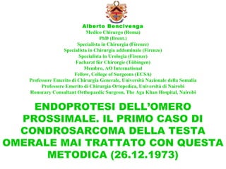
1. Alberto Bencivenga
Medico Chirurgo (Roma)
PhD (Brent.)
Specialista in Chirurgia (Firenze)
Specialista in Chirurgia addominale (Firenze)
Specialista in Urologia (Firenze)
Facharzt für Chirurgie (Tübingen)
Membro, AO International
Fellow, College of Surgeons (ECSA)
Professore Emerito di Chirurgia Generale, Università Nazionale della Somalia
Professore Emerito di Chirurgia Ortopedica, Università di Nairobi
Honorary Consultant Orthopaedic Surgeon, The Aga Khan Hospital, Nairobi
ENDOPROTESI DELL’OMERO
PROSSIMALE. IL PRIMO CASO DI
CONDROSARCOMA DELLA TESTA
OMERALE MAI TRATTATO CON QUESTA
METODICA (26.12.1973)
2. Asha A., età 17 anni
.
Storia di rapido
ingrossamento della
spalla sinistra in pochi
mesi. Episodi febbrili
irregolari ed inspiegati
recentemente.
Aspetto clinico della
lesione.
24. IL CAPO LUNGO DEL BICIPITE È STATO PRESERVATO IL PIÙ LUNGO
POSSIBILE ALLO SCOPO DI OTTENERE UNA STRUTTURA A CUI
SUTURARE IL SISTEMA CAPSULO-LEGAMENTOSO GLENO-OMERALE
25. Il tendine diviso a Y viene
passato nei fori centrali dal
basso per poi passare le due
metà del tendine nei fori
laterali, prima di intesserle alla
Bunnel atraverso il resto del
tendine. Con ciò si otterrà
un’affidabile struttura a cui
poter suturare il complesso
capsulo-legamentoso per incre-
mentare la stabilità articolare
finale.
38. RADIOGRAFIA POST-OPE-
RATORIA.
DA NOTARE IL PICCOLO
CALIBRO DELLA DIAFISI
OMERALE CHE, INIZIAL-
MENTE, PREOCCUPÒ TAN-
TO NOI QUANTO ROBERT
MATHYS SEN., CHE CI CO-
STRUÌ LA PROTESI E GLI
ALESATORI NECESSARI.
DA NOTARE ANCHE LA
PERFETTA CONGRUENZA
ARTICOLARE.
39. RADIOGRAMMI DOPO 8 MESI. DA NOTARE OSSO
NEOFORMATO CHE FISSA BIOLOGICAMENTE LO STELO
DELLA PROTESI.
40. 11 MESI DOPO L’INTERVENTO. L’OSSO NEOFORMATO
COMINCIA A PRODURRE UN CANALE “MIDOLLARE”
SOTTO LA BASE DELLA PROTESI!
41. DOPO 7 ANNI, SI È ULTIMATA LA FORMAZIONE DI UN
CANALE ENDOMIDOLLARE CIRCOLARE ATTORNO ALLO
STELO DELLA PROTESI (
*)
**
RADIOGRAMMA
SUBITO DOPO
L’INTERVENTO
RADIOGRAMMA
DOPO 7 ANNI E 4
MESI
42. UNO STUDIO DELLA CON-
GRUENZA DELL’ARTICOLA-
ZIONE SCAPOLO-OMERALE
SOTTO CARICO (PAZIENTE
APPOGGIATA CON LE MANI
SULLE PARALLELE E CON I
PIEDI SOLLEVATI).