SlideShare a Scribd company logo
COURSE OF COMMON PERONEAL NERVE
FOOT DROP
AND
it's management
Dr. Manas Kanti Sarkar.
2nd yr. PGT
BMCH
ANATOMY OF
COMMON
PERRONEAL NERVE
LUMBO SACRAL PLEXUS [VENTRAL RAMI OF
(L1 –S3) IN LOWER LIMB.]
SCIATIC NERVE(WIDEST 2 cm in diameter )
1)TIBIAL –VENTRAL BRANCH (L4-S3)
2)COMMON PERONEAL-DORSAL BR.(L4-
S2)
SUPERFICIAL DEEP
BOTH HAVETERMINATING MEDIAL LATERAL
DIVISIN
COURSE
Smaller terminal br. Of sciatic nerve conveys fibres from dorsal branch of ventral rami.
IN POPLITTEAL FOSSA
Nv. enter the fossa beneath long head of biceps femoris and slopes down laterally along
the medial margin of biceps tendon.
At the lateral angle of the popliteal fossa it crosses superficial to plantaris, lat. Head of
gastrocnemius and on reaching the back it rests on the fleshy sheet of soleus(where it can
be rolled against bone)
Finally nerve curves forward on the lateral side of neck of fibula deep to peroneus longus
tendon divided into 2 terminal branches- i) superficial & ii)deep.
BRANCHES- A) CUTANEOS- *LATERAL CUTANEOU S NERVE OF CALF.
*SURAL COMMUNICATING.
B)ARTICULAR- 3 branches
*SUPERIOR LATERAL GENICULAR
*INFERIOR LATERAL GENICULAR
* RECURRENT GENICULAR.
*** SUPERFICIAL PERONEAL NERVE***
• ARISE FROM-
Superficial division of common peroneal nerve on the lat.
Side of neck of fibula.
 COURSE-
Initially deep to peroneus longus & passes downward
between peroneus longus and brevis(supplies both muscles)
&pierce the deep fascia behind the ant. Inter muscular
septum at the jn. b/n upper two third and lower third and
divide into med. & lat. Br above the ankle joint & appear on
the dorsum of foot.
 BRANCHES – Muscular (peroneus longus and Brevis)
cutaneous
***DEEP PERONEAL NERVE***
• COURSE-
After winding round the neck of fibula pierces the ant inter
muscular septum appear in the ant extensor compartment
Deep to EDL & superficial to IOM.
$In prox. third –intervenes TA
EDL.
$In middle third TA
. EHL
$In distal third EHL& EDL
BRANCHES OF DEEP PERONEAL
NERVE:
MUSCULAR: All four muscles of ant crural
compartment-
1. TIBIALISANT.
2. EHL
3. EDL
4. PERONEUSTERTIUS.
ARTICULR: ANKLE JOINT
NERVE SUPPLY OF DORSUM OF FOOT
FOOT DROP-
 PARALYSIS OF MUSCLES OF EXTENSOR AND PERONEAL COMPARTMENT CAUSES
PLANTIFLEXION AND INVERSION OF FOOT ALONG WITH CERTAIN DEGREE OF
FLATTENING OF LONGITUDINAL ARCH, WHICH IS NORMALLY MAINTAINED SOME
EXTENT BYTIBIALIS ANTERIOR AND PERONEUS LONGUS.
 FOOT BEING ELEVATED TO ALLOW THE DROPPED TOESTO CLEAR THE GROUND,SO
THE PATIENTWALKS IN A HIGH STEPPING GAIT.
Causes
Injury to the peroneal nerve
1. Fracture head ,neck of fibula.
2. prolonged external pressure as application of tight plaster cast.
3. severe varus injuries of knee.( injury of postero lateral corner of knee)
4. surgical procedure like – HTO.
KNEE PROSTHESIS.
5. knee dislocation.
6.weight loss.
7. sports specific nerve entrapment in runners.
 SCIATIC NERVE INJURY CAUSING COMMON PERONEAL NERVE PALSY
1. fracture dislocation of hip.
2.penetrating injury in the proximal buttock.
3.shaft femur fracture.
4.Hip exposure.
Neurological conditions that can contribute to foot drop include:
• stroke
• multiple sclerosis (MS)
• cerebral palsy
• Charcot-Marie-Tooth disease
Conditions that cause the muscles to progressively weaken or
deteriorate may cause foot drop:
• muscular dystrophy
• amyotrophic lateral sclerosis (Lou Gehrig’s disease)
• polio
Rupture of AnteriorTibialis
Compartment Syndrome
Diabetes
Alcohol Abuse
Vulnerability of Peroneal Nerve
• Anatomy around the neck of fibula implicated as the main
reason for susceptibility nerve injury – fibular tunnel formed by
peroneus longus blending with crural fascia anteriorly &
posteriorly with fibular neck and soleus muscle. Dynamic
contraction of these muscles causes repeatative stretching of
nv.
• Funiculi of the peroneal nerve - larger and less connective tissue
• Fewer autonomic fibers, so in any injury, motor and sensory
fibers bear the brunt of the trauma.
• More superficial course, especially at the fibular neck
MUSCLES in MAINTAING
NEUTRAL POSITION OF FOOT.
• DORSIFLEXORS
TIBIALISANTERIOR
EXTENSOR HALLUCIS LONGUS
EXTENSOR DIGITORUM LONGUS
PERONEUSTERTIUS
• EVERTORS
PERONEUS LONGUS
PERONEUS BREVIS
SYMPTOMS
• Difficulty in lifting the foot.
• Pain, weakness numbness along the distribution of nerve.
SIGN
• INS- EQUINOUS DEFORMITY , CLAWING OFTOES ,
WASTING OF MUSCLES.
• MOTOR- INABILITYTO DORSIFLEXTHE FOOT AND
TOES
• SENSORY LOSS- LATERAL ASPECT OF LEG AND
DORSUM OF FOOT.
FOOT DROP
• Drop foot SW: Greater flexion at the knee to
accommodate the inability to dorsiflex - stair climbing
movement.
• Drop foot IC: Instead of normal heel-toe foot strike, foot
may either slap the ground or the entire foot may be
planted on the ground all at once.
• Drop footTC:Terminal contact is quite different -
inability to support their body weight – walker can be
used
IMAGING
• X-Ray :
Post-Traumatic - tibia/fibula and ankle - any bony injury.
Anatomic dysfunction (eg. Charcot joint)
• High resolution Ultrasonography :
Helpful in diagnosing masses around peroneal nerve
Additional advantage of allowing for dynmic
examination with muscle contraction, joint motion
and palpation of tender area.
• Magnetic Resonance: to detect Tumor or a
compressive mass lesion to the peroneal nerve.
Electromyogram
• This study can confirm the type of neuropathy, establish
the site of the lesion, estimate extent of injury, and
provide a prognosis.
• Sequential studies are useful to monitor recovery of
acute lesions.
TREATMENT
• Depends on the underlying cause.
• If cause is successfully treated foot drop may improve or
even disappear.
• Medical treatment - Painful Paresthesia
• Sympathetic block
• Amitriptyline
• Nortriptyline
• Pregabalin
PhysicalTherapy
• Exercises that strengthen the leg muscles
• Maintain the range of motion in knee and ankle
• Improve gait problems associated with foot drop.
Nerve Stimulation
Stimulating the nerve (peroneal nerve) improves foot drop
especially if it caused by a stroke.
SURGICAL REPAIR
• When nerve insult is the cause - restore the nerve continuity
– by Autogenous interfascicular nerve grafting is the
preferred technique for bridging the gap.
MINIMUM GAP CAN BE SUTURED- SCIATIC NERVE 15CM & CPN 12CM.
INDICATION-
If failing to show any clinical and electrophysiological
evidence of recovery 3 months or more after the injury:
CRITICAL LIMIT OF DELAY-SCIATIC NOT >(12-15)&CPN NOT>12
MONTHS.
In the mean time patient is put on a foot drop splint or srung caliper to improve
the gait.
AFTER CARE- Hip spica 6 wks (suture removal after 10 days)
Hinged knee brace 6wks
physiotherapy and exercises.
• WHEN THE NERVE DOES NOT RECOVERAND
FUNCTION IS SUFFICICINTLY IMPAIRED
1.BARR procedure(1947);
Tendon tibialis posterior is freed from its insertion and
transferred through IOM and insertion of tendon on medial
cuneiform on the dorsum of foot.
it is necessary to stabilize the hind foot by triple
arthrodesis.
advantage
Provide active dorsiflexion of ankle joint and improve gait.
2.SRINIVASAN et al (1968)
Distal end ofTibialis posterior tendon is splitted into 2
halves one half inserting to EHL other half to EDL &
peroneus brevis with ankle held in ankle held in 10 degree of
dorsiflexion knee in 30 degree flexion .
they also recommend TENDO ACHILLIS lengthening by Z
plasty.
Walking commenced at 6 weeks. After cast removal.
• Neurotendinous transposition
• Lateral head of gastrocnemius is transposed to the
tendons of the anterior muscle group with simultaneous
transposition of the proximal end of deep peroneal nerve.
• The nerve is sutured to the motor nerve of the
gartrocnemius.
N.B.-
• AFTERTENDONTRANSFER Cast and Non-
Weight Bearing ambulation for 6 weeks
• PHYSIOTHERAPY
To correct gait abnormalities
• CHRONICAND CONTRACTURECASES
Achilles tendon lengthening
• In patients whom foot drop is due to neurologic and anatomic factors
(polio,Charcot joint ) - Arthrodesis
• Subtalar Stabilizing procedure orTriple Arthrodesis can be done.
COMPLICATIONS
• Surgical procedure- wound infection may occur.
• Nerve graft failure
• In tendon transfer procedures- recurrent deformity
• In arthrodesis or fusion procedures- pseudoarthrosis,
delayed union, or nonunion.
Thank you

More Related Content

What's hot

Dislocation of patella
Dislocation of patellaDislocation of patella
Dislocation of patella
orthoprince
 
Principles of management of volkmann’s contracture
Principles of management of volkmann’s contracturePrinciples of management of volkmann’s contracture
Principles of management of volkmann’s contracture
Soliudeen Arojuraye
 
Brachial plexus injury
Brachial plexus injuryBrachial plexus injury
Brachial plexus injury
Paudel Sushil
 
Extensor apparatus hand
Extensor apparatus hand Extensor apparatus hand
Extensor apparatus hand
orthoprince
 
Median nerve injuries
Median nerve injuriesMedian nerve injuries
Median nerve injuries
Nabil Khalil
 

What's hot (20)

Dislocation of patella
Dislocation of patellaDislocation of patella
Dislocation of patella
 
Achilles tendon for presentation
Achilles tendon for presentation Achilles tendon for presentation
Achilles tendon for presentation
 
Foot drop
Foot dropFoot drop
Foot drop
 
Bennett , rolando , tendon injuries
Bennett , rolando , tendon injuriesBennett , rolando , tendon injuries
Bennett , rolando , tendon injuries
 
Radial nerve palsy
Radial nerve palsyRadial nerve palsy
Radial nerve palsy
 
Ulnar nerve injury PPT
Ulnar nerve injury PPTUlnar nerve injury PPT
Ulnar nerve injury PPT
 
Bicipital tendonitis
Bicipital tendonitisBicipital tendonitis
Bicipital tendonitis
 
Piriformis Syndrome
Piriformis Syndrome Piriformis Syndrome
Piriformis Syndrome
 
Peripheral nerve injuries
Peripheral nerve injuriesPeripheral nerve injuries
Peripheral nerve injuries
 
Principles of management of volkmann’s contracture
Principles of management of volkmann’s contracturePrinciples of management of volkmann’s contracture
Principles of management of volkmann’s contracture
 
Brachial plexus injury
Brachial plexus injuryBrachial plexus injury
Brachial plexus injury
 
Radial nerve injury
Radial nerve injuryRadial nerve injury
Radial nerve injury
 
Coxa vara
Coxa varaCoxa vara
Coxa vara
 
Extensor apparatus hand
Extensor apparatus hand Extensor apparatus hand
Extensor apparatus hand
 
Volksmann contracture
Volksmann contracture Volksmann contracture
Volksmann contracture
 
Compressive neuropathies of upper limb
Compressive neuropathies of upper limbCompressive neuropathies of upper limb
Compressive neuropathies of upper limb
 
Meniscus injury
Meniscus injuryMeniscus injury
Meniscus injury
 
Mallet finger
Mallet fingerMallet finger
Mallet finger
 
Median nerve injuries
Median nerve injuriesMedian nerve injuries
Median nerve injuries
 
Pes cavus
Pes cavusPes cavus
Pes cavus
 

Similar to Foot drop

INJECTION PALSY PRESENTING WITH FOOT DROP
INJECTION PALSY PRESENTING WITH FOOT DROPINJECTION PALSY PRESENTING WITH FOOT DROP
INJECTION PALSY PRESENTING WITH FOOT DROP
Peace Samuel
 

Similar to Foot drop (20)

WRIST DROP,FINGERDROP, by parvathy new.pptx
WRIST DROP,FINGERDROP, by parvathy new.pptxWRIST DROP,FINGERDROP, by parvathy new.pptx
WRIST DROP,FINGERDROP, by parvathy new.pptx
 
ANATOMY OF SCIATIC NERVE AND FOOT DROP
ANATOMY OF SCIATIC NERVE AND FOOT DROPANATOMY OF SCIATIC NERVE AND FOOT DROP
ANATOMY OF SCIATIC NERVE AND FOOT DROP
 
Muscle tone munish G B PANT DELHI
Muscle tone munish G B PANT DELHIMuscle tone munish G B PANT DELHI
Muscle tone munish G B PANT DELHI
 
Regional anaesthesia lower limb blocks
Regional anaesthesia lower limb blocksRegional anaesthesia lower limb blocks
Regional anaesthesia lower limb blocks
 
Tone new
Tone newTone new
Tone new
 
Lateral & posterior compartment of leg
Lateral & posterior compartment of leg   Lateral & posterior compartment of leg
Lateral & posterior compartment of leg
 
INJECTION PALSY PRESENTING WITH FOOT DROP
INJECTION PALSY PRESENTING WITH FOOT DROPINJECTION PALSY PRESENTING WITH FOOT DROP
INJECTION PALSY PRESENTING WITH FOOT DROP
 
Radial nerve palsy
Radial nerve palsyRadial nerve palsy
Radial nerve palsy
 
Peripheral Nerve Injury
Peripheral Nerve InjuryPeripheral Nerve Injury
Peripheral Nerve Injury
 
USMLE MSK L006 Lower 04 Muscles of leg anatomy medical .pdf
USMLE   MSK L006 Lower 04 Muscles of leg anatomy medical .pdfUSMLE   MSK L006 Lower 04 Muscles of leg anatomy medical .pdf
USMLE MSK L006 Lower 04 Muscles of leg anatomy medical .pdf
 
Knee CME 1.pptx
Knee CME 1.pptxKnee CME 1.pptx
Knee CME 1.pptx
 
805_Front-of-Forearm.pptx
805_Front-of-Forearm.pptx805_Front-of-Forearm.pptx
805_Front-of-Forearm.pptx
 
Bicep Femoris Tendinitis
Bicep Femoris TendinitisBicep Femoris Tendinitis
Bicep Femoris Tendinitis
 
FLEXOR COMPARTMENT of forearm (1).pptx
FLEXOR COMPARTMENT  of  forearm (1).pptxFLEXOR COMPARTMENT  of  forearm (1).pptx
FLEXOR COMPARTMENT of forearm (1).pptx
 
8. COMMON PERONEAL NERVE.pdf
8. COMMON PERONEAL NERVE.pdf8. COMMON PERONEAL NERVE.pdf
8. COMMON PERONEAL NERVE.pdf
 
Muscles_of_mastication.pptx
Muscles_of_mastication.pptxMuscles_of_mastication.pptx
Muscles_of_mastication.pptx
 
Tarsal tunnel syndrome complete review
Tarsal tunnel syndrome complete reviewTarsal tunnel syndrome complete review
Tarsal tunnel syndrome complete review
 
lower limb nerve injury.pptx
lower limb nerve injury.pptxlower limb nerve injury.pptx
lower limb nerve injury.pptx
 
Foot drop
Foot dropFoot drop
Foot drop
 
Sciatic Nerve Anatomy
Sciatic Nerve AnatomySciatic Nerve Anatomy
Sciatic Nerve Anatomy
 

Recently uploaded

plant breeding methods in asexually or clonally propagated crops
plant breeding methods in asexually or clonally propagated cropsplant breeding methods in asexually or clonally propagated crops
plant breeding methods in asexually or clonally propagated crops
parmarsneha2
 
The Roman Empire A Historical Colossus.pdf
The Roman Empire A Historical Colossus.pdfThe Roman Empire A Historical Colossus.pdf
The Roman Empire A Historical Colossus.pdf
kaushalkr1407
 
Accounting and finance exit exam 2016 E.C.pdf
Accounting and finance exit exam 2016 E.C.pdfAccounting and finance exit exam 2016 E.C.pdf
Accounting and finance exit exam 2016 E.C.pdf
YibeltalNibretu
 

Recently uploaded (20)

The approach at University of Liverpool.pptx
The approach at University of Liverpool.pptxThe approach at University of Liverpool.pptx
The approach at University of Liverpool.pptx
 
The Challenger.pdf DNHS Official Publication
The Challenger.pdf DNHS Official PublicationThe Challenger.pdf DNHS Official Publication
The Challenger.pdf DNHS Official Publication
 
plant breeding methods in asexually or clonally propagated crops
plant breeding methods in asexually or clonally propagated cropsplant breeding methods in asexually or clonally propagated crops
plant breeding methods in asexually or clonally propagated crops
 
INU_CAPSTONEDESIGN_비밀번호486_업로드용 발표자료.pdf
INU_CAPSTONEDESIGN_비밀번호486_업로드용 발표자료.pdfINU_CAPSTONEDESIGN_비밀번호486_업로드용 발표자료.pdf
INU_CAPSTONEDESIGN_비밀번호486_업로드용 발표자료.pdf
 
Synthetic Fiber Construction in lab .pptx
Synthetic Fiber Construction in lab .pptxSynthetic Fiber Construction in lab .pptx
Synthetic Fiber Construction in lab .pptx
 
1.4 modern child centered education - mahatma gandhi-2.pptx
1.4 modern child centered education - mahatma gandhi-2.pptx1.4 modern child centered education - mahatma gandhi-2.pptx
1.4 modern child centered education - mahatma gandhi-2.pptx
 
MARUTI SUZUKI- A Successful Joint Venture in India.pptx
MARUTI SUZUKI- A Successful Joint Venture in India.pptxMARUTI SUZUKI- A Successful Joint Venture in India.pptx
MARUTI SUZUKI- A Successful Joint Venture in India.pptx
 
Danh sách HSG Bộ môn cấp trường - Cấp THPT.pdf
Danh sách HSG Bộ môn cấp trường - Cấp THPT.pdfDanh sách HSG Bộ môn cấp trường - Cấp THPT.pdf
Danh sách HSG Bộ môn cấp trường - Cấp THPT.pdf
 
The Roman Empire A Historical Colossus.pdf
The Roman Empire A Historical Colossus.pdfThe Roman Empire A Historical Colossus.pdf
The Roman Empire A Historical Colossus.pdf
 
Matatag-Curriculum and the 21st Century Skills Presentation.pptx
Matatag-Curriculum and the 21st Century Skills Presentation.pptxMatatag-Curriculum and the 21st Century Skills Presentation.pptx
Matatag-Curriculum and the 21st Century Skills Presentation.pptx
 
GIÁO ÁN DẠY THÊM (KẾ HOẠCH BÀI BUỔI 2) - TIẾNG ANH 8 GLOBAL SUCCESS (2 CỘT) N...
GIÁO ÁN DẠY THÊM (KẾ HOẠCH BÀI BUỔI 2) - TIẾNG ANH 8 GLOBAL SUCCESS (2 CỘT) N...GIÁO ÁN DẠY THÊM (KẾ HOẠCH BÀI BUỔI 2) - TIẾNG ANH 8 GLOBAL SUCCESS (2 CỘT) N...
GIÁO ÁN DẠY THÊM (KẾ HOẠCH BÀI BUỔI 2) - TIẾNG ANH 8 GLOBAL SUCCESS (2 CỘT) N...
 
Sectors of the Indian Economy - Class 10 Study Notes pdf
Sectors of the Indian Economy - Class 10 Study Notes pdfSectors of the Indian Economy - Class 10 Study Notes pdf
Sectors of the Indian Economy - Class 10 Study Notes pdf
 
Students, digital devices and success - Andreas Schleicher - 27 May 2024..pptx
Students, digital devices and success - Andreas Schleicher - 27 May 2024..pptxStudents, digital devices and success - Andreas Schleicher - 27 May 2024..pptx
Students, digital devices and success - Andreas Schleicher - 27 May 2024..pptx
 
Salient features of Environment protection Act 1986.pptx
Salient features of Environment protection Act 1986.pptxSalient features of Environment protection Act 1986.pptx
Salient features of Environment protection Act 1986.pptx
 
NLC-2024-Orientation-for-RO-SDO (1).pptx
NLC-2024-Orientation-for-RO-SDO (1).pptxNLC-2024-Orientation-for-RO-SDO (1).pptx
NLC-2024-Orientation-for-RO-SDO (1).pptx
 
NCERT Solutions Power Sharing Class 10 Notes pdf
NCERT Solutions Power Sharing Class 10 Notes pdfNCERT Solutions Power Sharing Class 10 Notes pdf
NCERT Solutions Power Sharing Class 10 Notes pdf
 
Instructions for Submissions thorugh G- Classroom.pptx
Instructions for Submissions thorugh G- Classroom.pptxInstructions for Submissions thorugh G- Classroom.pptx
Instructions for Submissions thorugh G- Classroom.pptx
 
Jose-Rizal-and-Philippine-Nationalism-National-Symbol-2.pptx
Jose-Rizal-and-Philippine-Nationalism-National-Symbol-2.pptxJose-Rizal-and-Philippine-Nationalism-National-Symbol-2.pptx
Jose-Rizal-and-Philippine-Nationalism-National-Symbol-2.pptx
 
B.ed spl. HI pdusu exam paper-2023-24.pdf
B.ed spl. HI pdusu exam paper-2023-24.pdfB.ed spl. HI pdusu exam paper-2023-24.pdf
B.ed spl. HI pdusu exam paper-2023-24.pdf
 
Accounting and finance exit exam 2016 E.C.pdf
Accounting and finance exit exam 2016 E.C.pdfAccounting and finance exit exam 2016 E.C.pdf
Accounting and finance exit exam 2016 E.C.pdf
 

Foot drop

  • 1. COURSE OF COMMON PERONEAL NERVE FOOT DROP AND it's management Dr. Manas Kanti Sarkar. 2nd yr. PGT BMCH
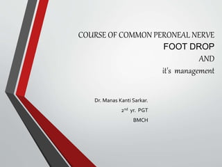
  • 2. ANATOMY OF COMMON PERRONEAL NERVE LUMBO SACRAL PLEXUS [VENTRAL RAMI OF (L1 –S3) IN LOWER LIMB.] SCIATIC NERVE(WIDEST 2 cm in diameter ) 1)TIBIAL –VENTRAL BRANCH (L4-S3) 2)COMMON PERONEAL-DORSAL BR.(L4- S2) SUPERFICIAL DEEP BOTH HAVETERMINATING MEDIAL LATERAL DIVISIN
  • 3. COURSE Smaller terminal br. Of sciatic nerve conveys fibres from dorsal branch of ventral rami. IN POPLITTEAL FOSSA Nv. enter the fossa beneath long head of biceps femoris and slopes down laterally along the medial margin of biceps tendon. At the lateral angle of the popliteal fossa it crosses superficial to plantaris, lat. Head of gastrocnemius and on reaching the back it rests on the fleshy sheet of soleus(where it can be rolled against bone) Finally nerve curves forward on the lateral side of neck of fibula deep to peroneus longus tendon divided into 2 terminal branches- i) superficial & ii)deep. BRANCHES- A) CUTANEOS- *LATERAL CUTANEOU S NERVE OF CALF. *SURAL COMMUNICATING. B)ARTICULAR- 3 branches *SUPERIOR LATERAL GENICULAR *INFERIOR LATERAL GENICULAR * RECURRENT GENICULAR.
  • 4. *** SUPERFICIAL PERONEAL NERVE*** • ARISE FROM- Superficial division of common peroneal nerve on the lat. Side of neck of fibula.  COURSE- Initially deep to peroneus longus & passes downward between peroneus longus and brevis(supplies both muscles) &pierce the deep fascia behind the ant. Inter muscular septum at the jn. b/n upper two third and lower third and divide into med. & lat. Br above the ankle joint & appear on the dorsum of foot.  BRANCHES – Muscular (peroneus longus and Brevis) cutaneous
  • 5. ***DEEP PERONEAL NERVE*** • COURSE- After winding round the neck of fibula pierces the ant inter muscular septum appear in the ant extensor compartment Deep to EDL & superficial to IOM. $In prox. third –intervenes TA EDL. $In middle third TA . EHL $In distal third EHL& EDL
  • 6. BRANCHES OF DEEP PERONEAL NERVE: MUSCULAR: All four muscles of ant crural compartment- 1. TIBIALISANT. 2. EHL 3. EDL 4. PERONEUSTERTIUS. ARTICULR: ANKLE JOINT
  • 7. NERVE SUPPLY OF DORSUM OF FOOT
  • 8. FOOT DROP-  PARALYSIS OF MUSCLES OF EXTENSOR AND PERONEAL COMPARTMENT CAUSES PLANTIFLEXION AND INVERSION OF FOOT ALONG WITH CERTAIN DEGREE OF FLATTENING OF LONGITUDINAL ARCH, WHICH IS NORMALLY MAINTAINED SOME EXTENT BYTIBIALIS ANTERIOR AND PERONEUS LONGUS.  FOOT BEING ELEVATED TO ALLOW THE DROPPED TOESTO CLEAR THE GROUND,SO THE PATIENTWALKS IN A HIGH STEPPING GAIT.
  • 9. Causes Injury to the peroneal nerve 1. Fracture head ,neck of fibula. 2. prolonged external pressure as application of tight plaster cast. 3. severe varus injuries of knee.( injury of postero lateral corner of knee) 4. surgical procedure like – HTO. KNEE PROSTHESIS. 5. knee dislocation. 6.weight loss. 7. sports specific nerve entrapment in runners.  SCIATIC NERVE INJURY CAUSING COMMON PERONEAL NERVE PALSY 1. fracture dislocation of hip. 2.penetrating injury in the proximal buttock. 3.shaft femur fracture. 4.Hip exposure.
  • 10. Neurological conditions that can contribute to foot drop include: • stroke • multiple sclerosis (MS) • cerebral palsy • Charcot-Marie-Tooth disease Conditions that cause the muscles to progressively weaken or deteriorate may cause foot drop: • muscular dystrophy • amyotrophic lateral sclerosis (Lou Gehrig’s disease) • polio
  • 11. Rupture of AnteriorTibialis Compartment Syndrome Diabetes Alcohol Abuse
  • 12. Vulnerability of Peroneal Nerve • Anatomy around the neck of fibula implicated as the main reason for susceptibility nerve injury – fibular tunnel formed by peroneus longus blending with crural fascia anteriorly & posteriorly with fibular neck and soleus muscle. Dynamic contraction of these muscles causes repeatative stretching of nv. • Funiculi of the peroneal nerve - larger and less connective tissue • Fewer autonomic fibers, so in any injury, motor and sensory fibers bear the brunt of the trauma. • More superficial course, especially at the fibular neck
  • 13. MUSCLES in MAINTAING NEUTRAL POSITION OF FOOT. • DORSIFLEXORS TIBIALISANTERIOR EXTENSOR HALLUCIS LONGUS EXTENSOR DIGITORUM LONGUS PERONEUSTERTIUS • EVERTORS PERONEUS LONGUS PERONEUS BREVIS
  • 14. SYMPTOMS • Difficulty in lifting the foot. • Pain, weakness numbness along the distribution of nerve.
  • 15. SIGN • INS- EQUINOUS DEFORMITY , CLAWING OFTOES , WASTING OF MUSCLES. • MOTOR- INABILITYTO DORSIFLEXTHE FOOT AND TOES • SENSORY LOSS- LATERAL ASPECT OF LEG AND DORSUM OF FOOT.
  • 16.
  • 17.
  • 18. FOOT DROP • Drop foot SW: Greater flexion at the knee to accommodate the inability to dorsiflex - stair climbing movement. • Drop foot IC: Instead of normal heel-toe foot strike, foot may either slap the ground or the entire foot may be planted on the ground all at once. • Drop footTC:Terminal contact is quite different - inability to support their body weight – walker can be used
  • 19.
  • 20.
  • 21. IMAGING • X-Ray : Post-Traumatic - tibia/fibula and ankle - any bony injury. Anatomic dysfunction (eg. Charcot joint) • High resolution Ultrasonography : Helpful in diagnosing masses around peroneal nerve Additional advantage of allowing for dynmic examination with muscle contraction, joint motion and palpation of tender area. • Magnetic Resonance: to detect Tumor or a compressive mass lesion to the peroneal nerve.
  • 22. Electromyogram • This study can confirm the type of neuropathy, establish the site of the lesion, estimate extent of injury, and provide a prognosis. • Sequential studies are useful to monitor recovery of acute lesions.
  • 23. TREATMENT • Depends on the underlying cause. • If cause is successfully treated foot drop may improve or even disappear. • Medical treatment - Painful Paresthesia • Sympathetic block • Amitriptyline • Nortriptyline • Pregabalin
  • 24. PhysicalTherapy • Exercises that strengthen the leg muscles • Maintain the range of motion in knee and ankle • Improve gait problems associated with foot drop.
  • 25. Nerve Stimulation Stimulating the nerve (peroneal nerve) improves foot drop especially if it caused by a stroke.
  • 26. SURGICAL REPAIR • When nerve insult is the cause - restore the nerve continuity – by Autogenous interfascicular nerve grafting is the preferred technique for bridging the gap. MINIMUM GAP CAN BE SUTURED- SCIATIC NERVE 15CM & CPN 12CM. INDICATION- If failing to show any clinical and electrophysiological evidence of recovery 3 months or more after the injury: CRITICAL LIMIT OF DELAY-SCIATIC NOT >(12-15)&CPN NOT>12 MONTHS. In the mean time patient is put on a foot drop splint or srung caliper to improve the gait. AFTER CARE- Hip spica 6 wks (suture removal after 10 days) Hinged knee brace 6wks physiotherapy and exercises.
  • 27. • WHEN THE NERVE DOES NOT RECOVERAND FUNCTION IS SUFFICICINTLY IMPAIRED 1.BARR procedure(1947); Tendon tibialis posterior is freed from its insertion and transferred through IOM and insertion of tendon on medial cuneiform on the dorsum of foot. it is necessary to stabilize the hind foot by triple arthrodesis. advantage Provide active dorsiflexion of ankle joint and improve gait.
  • 28. 2.SRINIVASAN et al (1968) Distal end ofTibialis posterior tendon is splitted into 2 halves one half inserting to EHL other half to EDL & peroneus brevis with ankle held in ankle held in 10 degree of dorsiflexion knee in 30 degree flexion . they also recommend TENDO ACHILLIS lengthening by Z plasty. Walking commenced at 6 weeks. After cast removal.
  • 29. • Neurotendinous transposition • Lateral head of gastrocnemius is transposed to the tendons of the anterior muscle group with simultaneous transposition of the proximal end of deep peroneal nerve. • The nerve is sutured to the motor nerve of the gartrocnemius.
  • 30. N.B.- • AFTERTENDONTRANSFER Cast and Non- Weight Bearing ambulation for 6 weeks • PHYSIOTHERAPY To correct gait abnormalities • CHRONICAND CONTRACTURECASES Achilles tendon lengthening • In patients whom foot drop is due to neurologic and anatomic factors (polio,Charcot joint ) - Arthrodesis • Subtalar Stabilizing procedure orTriple Arthrodesis can be done.
  • 31. COMPLICATIONS • Surgical procedure- wound infection may occur. • Nerve graft failure • In tendon transfer procedures- recurrent deformity • In arthrodesis or fusion procedures- pseudoarthrosis, delayed union, or nonunion.