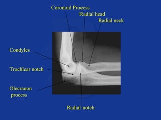
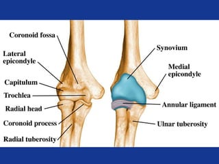
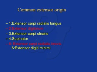
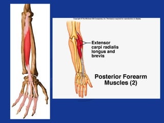
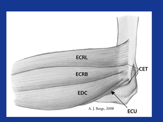
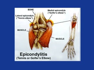
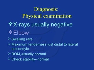
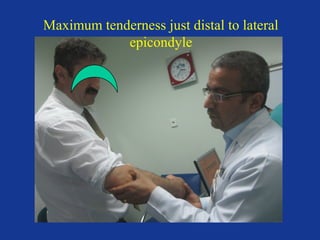
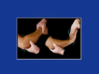
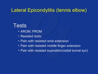
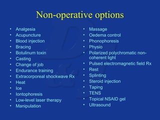
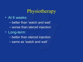
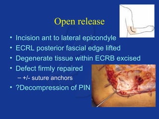
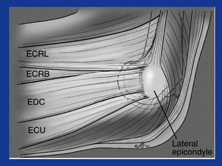
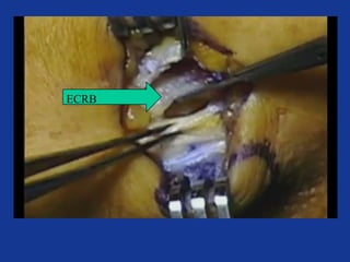
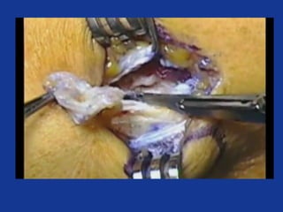
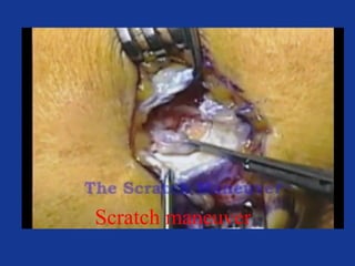
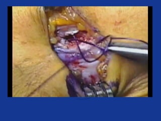
Epicondylitis
:Subject of the presentation 
Lateral Epicondylitis 
PPrreeppaarreedd bbyy:: DDrr.. RRaasseekkhh MMSS oorrtthhoo 
KKaabbuull aaffgghhaanniissttaann 
Date :19/04/2013
Elbow Anatomy 
• Elbow joint is made of 
– 3 bones 
– 3 joints 
– One capsule 
– Hinge joint 
– Flexion(145) and extension(0-5)
(Diarthrosis)- freely moveable
• Planar Joint 
• Hinge Joint 
• Pivot Joint 
• Saddle Joint 
• Ball & Socket Joint 
• Condyloid or Ellipsoid Joint
• Convex surface of bone fits in concave surface of 
2nd bone 
• Unixlateral like a door hinge 
• Examples: 
- Knee, elbow, ankle, interphalangeal joints 
• Movements produced: 
- flexion 
- extension 
- hyperextension
• Rounded surface of bone articulates with 
the ring formed by the 2nd bone & ligament 
• Monoaxial since it only allows rotation 
around longitudinal axis 
• Examples: 
- proximal radioulnar joint 
- supination 
- pronation 
- atlanto-axial joint 
- Turning head side to side “no”
Medial Collateral Ligament (MCL) 
Anterior 
Medial 
Collateral 
Ligament 
Posterior 
Medial 
Collateral 
Ligament 
Resists valgus 
forces 
Limits extension 
Transverse ligament
© 2007 McGraw-Hill Higher Education. All rights reserved.
Lateral Collateral 
Resists varus stress 
Weaker than MCL 
Tensed in flexion and 
extention
Secondary Stabilizers
Lateral epicondyle 
Capitulum 
Proximal radioulnar 
jt. 
Radial head 
Radial neck 
Radial tuberosity 
Olecranon fossa 
Medial 
epicondyle 
Trochlea 
Coronoid 
process
Coronoid Process 
Radial head 
Radial neck 
Condyles 
Trochlear notch 
Olecranon 
process 
Radial notch
Common extensor origin 
– 1:Extensor carpi radialis longus 
– 2:Extensor digitorum 
– 3:Extensor carpi ulnaris 
– 4:Supinator 
– 5: Extensor carpi radialis brevis 
6:Extensor digiti minimi
Epicondylitis
LATERAL EPICONDYLITIS
TENNIS ELBOW
Definition 
• • “A pathologic condition of the common 
• extensor muscles at their origin on the 
• lateral humeral epicondyle. Epicondylitis 
• suggests an inflammation at one of the 
• epicondyles of the elbow.”
Lateral Epicondylitis (tennis elbow) 
• Pathology 
– 30 – 50 years old 
– Repetitive micro-trauma 
– Chronic tear in the origin of the extensor 
carpi radialis brevis
Lateral Epicondylitis (tennis elbow) 
• Mechanism of Injury 
– Overuse syndrome caused by repeated 
forceful wrist and finger movements 
– Tennis players 
– Prolonged and rapid activities
Risk factors 
• Obesity 
• Repetitive movements 
• Forceful activities 
• Manual labour
Etiology 
• Extrinsic factors 
• Repetitive movements 
• Forceful activities 
• Manual labour 
• Intrinsic factors 
• Anatomical factors 
• Age related factors 
• Systemic factors 
Tendon degeneration 
Decreased vascularity 
Decreased healing
Common Complaints 
• Diffuse pain 
• • Morning stiffness 
• • Occasional night pain 
• • Dropping of objects/ weak grip strength 
• • Pain w/ palpation of lat. epicondyle 
• • Pain w/active or resisted extension 
• • Pain w/ grasping objects with the effected 
hand
Symptoms 
Lateral Arm / elbow /forearm pain 
 Increased with use(holding/picking up items) 
 If popping / clicking present, consider 
problem within joint(loose bodies, 
osteochondral lesions )
Acute vs. Chronic 
• Tendonitis 
• Localized edema 
• Inflammation of wrist 
extensor tendons 
• Microtearing 
• Tendinosis 
• Decreased edema 
• Non-inflammatory 
• Localized fibrosis 
• Collagen necrosis 
• Fibroblastic 
hyperplasia
Acute vs. Chronic
Diagnosis: 
Physical examination 
X-rays usually negative 
Elbow 
Swelling rare 
Maximum tenderness just distal to lateral 
epicondyle 
ROM,-usually normal 
Check stability--normal
Maximum tenderness just distal to lateral 
epicondyle
Lateral Epicondylitis (tennis elbow) 
Tests 
• AROM; PROM 
• Resisted tests: 
• Pain with resisted wrist extension 
• Pain with resisted middle finger extension 
• Pain with resisted supination(radial tunnel syn)
Special Tests 
• Cozen’s Sign 
– Elbow flexed; Forearm pronated 
– Wrist extension and radial deviation against 
resistance 
– Positive when pain at lateral epicondyle 
• Mill’s Test 
– While palpating the lateral epicondyle 
– The examiner pronates the patient’s forearm, flexes 
the wrist, and extends the elbow 
– Positive when pain at lateral epicondyle or lack of 
full elbow extension
Special Tests 
• Grip Strength Measures 
• Middle Finger Test 
– Resistance just distal to PIP joint of the 
middle finger with forearm in pronation 
– Positive in tennis elbow with pain at lateral 
epicondyle
Differential diagnosis of ‘Tennis 
Elbow’ 
• C6/7 radiculopathy 
• Radial tunnel syndrome 
(10%) 
• Distal biceps tendon degeneration 
• Radiocapitellar arthritis 
• Capsular infolding 
• Posterolateral instability
C6/7 radiculopathy
50 
Spurling sign 
• . Axial compression of 
the spine and rotation 
to the ipsilateral side of 
symptoms reproduces 
or worsens cervical 
radiculopathy. 
• Pain on the side of 
rotation is usually 
indicative of foraminal 
stenosis and nerve root 
irritation.
Radial Tunnel Syndrome 
• Compression of 
radial nerve under 
extensors in forearm 
• Deep, lateral 
forearm pain, often 
at night 
• No sensory 
component 
• Often confused 
with lateral 
epicondylitis (they 
co-exist 5% of the 
time) pain is more 
distal
Radial Tunnel Syndrome: Diagnosis 
• Extended middle finger 
test 
• Pain with resisted 
supination 
• Electrodiagnostic tests 
not helpful 
• Injection of local 
anesthetic into radial 
tunnel completely 
relieves symptoms and 
is diagnostic
Radial tunnel syndrome
Distal biceps tendon 
degeneration
Radiocapitellar arthritis
Management 
• Key points 
• It is a self limiting condition – no-one ever has it 
forever. 
• 90% of people are better after 1 year. 
• Physiotherapy, activity modification and simple 
exercises will control the symptoms in most people. 
• Injections are reserved for very resistant cases. 
• An operation is only considered as a last resort.
Management 
• Non-operative 
– successful in 95% 
• Operative 
– only after failed non-operative Rx 
– usually successful
Non-operative options 
• Analgesia 
• Acupuncture 
• Blood injection 
• Bracing 
• Botulinum toxin 
• Casting 
• Change of job 
• Endurance training 
• Extracorporeal shockwave Rx 
• Heat 
• Ice 
• Iontophoresis 
• Low-level laser therapy 
• Manipulation 
• Massage 
• Oedema control 
• Phonophoresis 
• Physio 
• Polarized polychromatic non-coherent 
light 
• Pulsed electromagnetic field Rx 
• Rest 
• Splinting 
• Steroid injection 
• Taping 
• TENS 
• Topical NSAID gel 
• Ultrasound
Physiotherapy 
• At 6 weeks: 
– better than ‘watch and wait’ 
– worse than steroid injection 
• Long-term: 
– better than steroid injection 
– same as ‘watch and wait’
Brace / elbow clasp 
• Between 12 and 24 weeks: 
– Pain reduction 
– Improved functionality 
– Improved pain-free grip strength 
• No better at 12 months
Physical Therapy 
• Ultrasound 
– Limited low quality evidence 
– Used as an adjunct; not independently 
• Pulsed ultrasound to break up scar 
tissue, promote healing, and increase 
blood flow in the area
Manual Techniques 
• Deep Transverse Friction Massage 
– No benefit when combined with concurrent 
physiotherapy modalities when compared 
to control group 
• Manipulation of the Wrist 
– Scaphoid Thrust Manipulation 
• Cervicothoracic Spine Manipulation 
– Non-thrust manipulation and traction of 
cervical spine 
– Lateral Cervical Glide Technique
Steroid injection 
• Good short-term relief for 6 weeks 
• Poorer outcome in the longer term than 
– watch and wait 
– physio
Injections 
• Short-term benefits (2-6 weeks) 
• Greater perception of benefits (pain 
reduction, global improvement, grip 
strength) but did not persist long term 
• Several studies have found that oral 
NSAIDS and PT have greater benefits 
than corticosteroid injections at both 6 
weeks and 6 month follow-ups
Platelet Rich Plasma 
Therapy 
• A 2006 study looking at the treatment of lateral 
epicondylitis with platelet rich plasma therapy 
– Over 90% of the patients were completely satisfied 
with their results and did not opt for surgery in the 
weeks and months following a single treatment 
• Eight weeks after the treatment, the platelet-rich 
plasma patients noted 60% improvement in their visual 
analog pain scores versus 16% improvement in control 
patients (P =.001).
►PRP Application Technique 
Withdraw peripheral blood 
Place blood in canister 
Centrifuge
►PRP Application Technique 
Remove PPP 
Shake vigorously for 30 seconds 
PPllaatteelleett PPoooorr PPllaassmmaa 
((PPPPPP)) 
PPllaatteelleett RRiicchh PPllaassmmaa 
((PPRRPP)) 
PPaacckkeedd RReedd BBlloooodd CCeellllss
PRP: Contraindications 
• Thrombocytopenia 
• Anticoagulation therapy 
• Active infection 
• Tumor 
• Metastatic disease 
• Pregnancy 
(Hall et al, JAAOS 2009)
Predictors of poor outcome 
• Manual labour 
• High physical strain at work 
• High level of baseline pain 
• Lower social class
Operative options 
• Open release 
• Arthroscopic release 
• Percutaneous release 
• Suture anchor repair 
• Microtenotomy 
• Anconeus transposition 
• Radiofrequency probe
Open release 
• Incision ant to lateral epicondyle 
• ECRL posterior fascial edge lifted 
• Degenerate tissue within ECRB excised 
• Defect firmly repaired 
– +/- suture anchors 
• ?Decompression of PIN
Open release 
• Excellent / good 75 – 91% 
• Poor / failed 2 – 11% 
• 80 – 95% return to normal activity in 4/12
Surgery
ECRL 
EDC 
L. Cond
ECRB
Scratch maneuver
Lateral Epicondylar Release 
Return to Work Protocol 
• Week 0 – 1: off work 
• Week 1 – 4: one-handed work 
• Week 4 – 12: light duty work 
• Week 12: regular duty work
Arthroscopy 
• 70% satisfactory to excellent 
• 473 cases 
– 4 deep infection 
– 33 prolonged drainage 
– 12 transient nerve palsies 
Arthroscopic tennis elbow release. Kalainov D et al. Techniques in Hand and Upper Extremity 
Surgery. 2007;11(1):2-7 
• Arthroscopy leaves residual tendinopathy 
– Gross and histological 
– Results in poorer outcomes 
Lateral Epicondylitis: In Vivo Assessment of Arthroscopic Debridement and Correlation With 
Patient Outcomes. Cummins CA. Am J Sports Med Sep 2006, 34(9):1486
Conclusions 
•  Nirschl Mini techniques less risk, lower costs, best 
success 
•  Tendinosis surgery is not a release operation 
•  Tendinosis surgery is resection of pain producing 
tissue 
•  Direct vision clearly identifies pathological tissue 
•  No harm to normal tissue – rapid rehab 
•  Can do combined procedures (medial and lateral) 
when indicated
Management summery 
• Activity modification,stretching,tennis elbow strap 
and cock up wrist splint 
• NSAIDs 
• Therapy (Iontophoresis) 
• Corticosteroid injection 
• Offer PRP injection in some individuals 
• Surgery: 
 Open technique 
 Arthroscopic technique when intra-articular pathology suspected 
or when more rapid recovery needed 
 Perform concomitant radial tunnel decompression in patients 
with both conditions
Questions?
Thanks for your kind 
attention

Tennis elbow(le)

  • 1.
  • 2.
    :Subject of thepresentation Lateral Epicondylitis PPrreeppaarreedd bbyy:: DDrr.. RRaasseekkhh MMSS oorrtthhoo KKaabbuull aaffgghhaanniissttaann Date :19/04/2013
  • 3.
    Elbow Anatomy •Elbow joint is made of – 3 bones – 3 joints – One capsule – Hinge joint – Flexion(145) and extension(0-5)
  • 4.
  • 5.
    • Planar Joint • Hinge Joint • Pivot Joint • Saddle Joint • Ball & Socket Joint • Condyloid or Ellipsoid Joint
  • 7.
    • Convex surfaceof bone fits in concave surface of 2nd bone • Unixlateral like a door hinge • Examples: - Knee, elbow, ankle, interphalangeal joints • Movements produced: - flexion - extension - hyperextension
  • 8.
    • Rounded surfaceof bone articulates with the ring formed by the 2nd bone & ligament • Monoaxial since it only allows rotation around longitudinal axis • Examples: - proximal radioulnar joint - supination - pronation - atlanto-axial joint - Turning head side to side “no”
  • 11.
    Medial Collateral Ligament(MCL) Anterior Medial Collateral Ligament Posterior Medial Collateral Ligament Resists valgus forces Limits extension Transverse ligament
  • 13.
    © 2007 McGraw-HillHigher Education. All rights reserved.
  • 15.
    Lateral Collateral Resistsvarus stress Weaker than MCL Tensed in flexion and extention
  • 18.
  • 21.
    Lateral epicondyle Capitulum Proximal radioulnar jt. Radial head Radial neck Radial tuberosity Olecranon fossa Medial epicondyle Trochlea Coronoid process
  • 22.
    Coronoid Process Radialhead Radial neck Condyles Trochlear notch Olecranon process Radial notch
  • 24.
    Common extensor origin – 1:Extensor carpi radialis longus – 2:Extensor digitorum – 3:Extensor carpi ulnaris – 4:Supinator – 5: Extensor carpi radialis brevis 6:Extensor digiti minimi
  • 27.
  • 28.
  • 29.
  • 31.
    Definition • •“A pathologic condition of the common • extensor muscles at their origin on the • lateral humeral epicondyle. Epicondylitis • suggests an inflammation at one of the • epicondyles of the elbow.”
  • 33.
    Lateral Epicondylitis (tenniselbow) • Pathology – 30 – 50 years old – Repetitive micro-trauma – Chronic tear in the origin of the extensor carpi radialis brevis
  • 34.
    Lateral Epicondylitis (tenniselbow) • Mechanism of Injury – Overuse syndrome caused by repeated forceful wrist and finger movements – Tennis players – Prolonged and rapid activities
  • 35.
    Risk factors •Obesity • Repetitive movements • Forceful activities • Manual labour
  • 36.
    Etiology • Extrinsicfactors • Repetitive movements • Forceful activities • Manual labour • Intrinsic factors • Anatomical factors • Age related factors • Systemic factors Tendon degeneration Decreased vascularity Decreased healing
  • 37.
    Common Complaints •Diffuse pain • • Morning stiffness • • Occasional night pain • • Dropping of objects/ weak grip strength • • Pain w/ palpation of lat. epicondyle • • Pain w/active or resisted extension • • Pain w/ grasping objects with the effected hand
  • 38.
    Symptoms Lateral Arm/ elbow /forearm pain  Increased with use(holding/picking up items)  If popping / clicking present, consider problem within joint(loose bodies, osteochondral lesions )
  • 39.
    Acute vs. Chronic • Tendonitis • Localized edema • Inflammation of wrist extensor tendons • Microtearing • Tendinosis • Decreased edema • Non-inflammatory • Localized fibrosis • Collagen necrosis • Fibroblastic hyperplasia
  • 40.
  • 41.
    Diagnosis: Physical examination X-rays usually negative Elbow Swelling rare Maximum tenderness just distal to lateral epicondyle ROM,-usually normal Check stability--normal
  • 42.
    Maximum tenderness justdistal to lateral epicondyle
  • 44.
    Lateral Epicondylitis (tenniselbow) Tests • AROM; PROM • Resisted tests: • Pain with resisted wrist extension • Pain with resisted middle finger extension • Pain with resisted supination(radial tunnel syn)
  • 45.
    Special Tests •Cozen’s Sign – Elbow flexed; Forearm pronated – Wrist extension and radial deviation against resistance – Positive when pain at lateral epicondyle • Mill’s Test – While palpating the lateral epicondyle – The examiner pronates the patient’s forearm, flexes the wrist, and extends the elbow – Positive when pain at lateral epicondyle or lack of full elbow extension
  • 47.
    Special Tests •Grip Strength Measures • Middle Finger Test – Resistance just distal to PIP joint of the middle finger with forearm in pronation – Positive in tennis elbow with pain at lateral epicondyle
  • 48.
    Differential diagnosis of‘Tennis Elbow’ • C6/7 radiculopathy • Radial tunnel syndrome (10%) • Distal biceps tendon degeneration • Radiocapitellar arthritis • Capsular infolding • Posterolateral instability
  • 49.
  • 50.
    50 Spurling sign • . Axial compression of the spine and rotation to the ipsilateral side of symptoms reproduces or worsens cervical radiculopathy. • Pain on the side of rotation is usually indicative of foraminal stenosis and nerve root irritation.
  • 51.
    Radial Tunnel Syndrome • Compression of radial nerve under extensors in forearm • Deep, lateral forearm pain, often at night • No sensory component • Often confused with lateral epicondylitis (they co-exist 5% of the time) pain is more distal
  • 52.
    Radial Tunnel Syndrome:Diagnosis • Extended middle finger test • Pain with resisted supination • Electrodiagnostic tests not helpful • Injection of local anesthetic into radial tunnel completely relieves symptoms and is diagnostic
  • 56.
  • 57.
    Distal biceps tendon degeneration
  • 58.
  • 59.
    Management • Keypoints • It is a self limiting condition – no-one ever has it forever. • 90% of people are better after 1 year. • Physiotherapy, activity modification and simple exercises will control the symptoms in most people. • Injections are reserved for very resistant cases. • An operation is only considered as a last resort.
  • 60.
    Management • Non-operative – successful in 95% • Operative – only after failed non-operative Rx – usually successful
  • 64.
    Non-operative options •Analgesia • Acupuncture • Blood injection • Bracing • Botulinum toxin • Casting • Change of job • Endurance training • Extracorporeal shockwave Rx • Heat • Ice • Iontophoresis • Low-level laser therapy • Manipulation • Massage • Oedema control • Phonophoresis • Physio • Polarized polychromatic non-coherent light • Pulsed electromagnetic field Rx • Rest • Splinting • Steroid injection • Taping • TENS • Topical NSAID gel • Ultrasound
  • 65.
    Physiotherapy • At6 weeks: – better than ‘watch and wait’ – worse than steroid injection • Long-term: – better than steroid injection – same as ‘watch and wait’
  • 67.
    Brace / elbowclasp • Between 12 and 24 weeks: – Pain reduction – Improved functionality – Improved pain-free grip strength • No better at 12 months
  • 68.
    Physical Therapy •Ultrasound – Limited low quality evidence – Used as an adjunct; not independently • Pulsed ultrasound to break up scar tissue, promote healing, and increase blood flow in the area
  • 69.
    Manual Techniques •Deep Transverse Friction Massage – No benefit when combined with concurrent physiotherapy modalities when compared to control group • Manipulation of the Wrist – Scaphoid Thrust Manipulation • Cervicothoracic Spine Manipulation – Non-thrust manipulation and traction of cervical spine – Lateral Cervical Glide Technique
  • 70.
    Steroid injection •Good short-term relief for 6 weeks • Poorer outcome in the longer term than – watch and wait – physio
  • 71.
    Injections • Short-termbenefits (2-6 weeks) • Greater perception of benefits (pain reduction, global improvement, grip strength) but did not persist long term • Several studies have found that oral NSAIDS and PT have greater benefits than corticosteroid injections at both 6 weeks and 6 month follow-ups
  • 72.
    Platelet Rich Plasma Therapy • A 2006 study looking at the treatment of lateral epicondylitis with platelet rich plasma therapy – Over 90% of the patients were completely satisfied with their results and did not opt for surgery in the weeks and months following a single treatment • Eight weeks after the treatment, the platelet-rich plasma patients noted 60% improvement in their visual analog pain scores versus 16% improvement in control patients (P =.001).
  • 73.
    ►PRP Application Technique Withdraw peripheral blood Place blood in canister Centrifuge
  • 74.
    ►PRP Application Technique Remove PPP Shake vigorously for 30 seconds PPllaatteelleett PPoooorr PPllaassmmaa ((PPPPPP)) PPllaatteelleett RRiicchh PPllaassmmaa ((PPRRPP)) PPaacckkeedd RReedd BBlloooodd CCeellllss
  • 75.
    PRP: Contraindications •Thrombocytopenia • Anticoagulation therapy • Active infection • Tumor • Metastatic disease • Pregnancy (Hall et al, JAAOS 2009)
  • 77.
    Predictors of pooroutcome • Manual labour • High physical strain at work • High level of baseline pain • Lower social class
  • 78.
    Operative options •Open release • Arthroscopic release • Percutaneous release • Suture anchor repair • Microtenotomy • Anconeus transposition • Radiofrequency probe
  • 79.
    Open release •Incision ant to lateral epicondyle • ECRL posterior fascial edge lifted • Degenerate tissue within ECRB excised • Defect firmly repaired – +/- suture anchors • ?Decompression of PIN
  • 80.
    Open release •Excellent / good 75 – 91% • Poor / failed 2 – 11% • 80 – 95% return to normal activity in 4/12
  • 81.
  • 94.
  • 95.
  • 97.
  • 100.
    Lateral Epicondylar Release Return to Work Protocol • Week 0 – 1: off work • Week 1 – 4: one-handed work • Week 4 – 12: light duty work • Week 12: regular duty work
  • 108.
    Arthroscopy • 70%satisfactory to excellent • 473 cases – 4 deep infection – 33 prolonged drainage – 12 transient nerve palsies Arthroscopic tennis elbow release. Kalainov D et al. Techniques in Hand and Upper Extremity Surgery. 2007;11(1):2-7 • Arthroscopy leaves residual tendinopathy – Gross and histological – Results in poorer outcomes Lateral Epicondylitis: In Vivo Assessment of Arthroscopic Debridement and Correlation With Patient Outcomes. Cummins CA. Am J Sports Med Sep 2006, 34(9):1486
  • 111.
    Conclusions • Nirschl Mini techniques less risk, lower costs, best success •  Tendinosis surgery is not a release operation •  Tendinosis surgery is resection of pain producing tissue •  Direct vision clearly identifies pathological tissue •  No harm to normal tissue – rapid rehab •  Can do combined procedures (medial and lateral) when indicated
  • 112.
    Management summery •Activity modification,stretching,tennis elbow strap and cock up wrist splint • NSAIDs • Therapy (Iontophoresis) • Corticosteroid injection • Offer PRP injection in some individuals • Surgery:  Open technique  Arthroscopic technique when intra-articular pathology suspected or when more rapid recovery needed  Perform concomitant radial tunnel decompression in patients with both conditions
  • 113.
  • 114.
    Thanks for yourkind attention

Editor's Notes

  • #40 Depending on the severity and number of small tendon injuries that build up, the ECRB may not be able to fully heal. Nirschl defined four stages of lateral epicondylitis, showing the introduction of permanent damage beginning at Stage 2. The stages are: 1. Inflammatory changes that are reversible 2. Nonreversible pathologic changes to origin of the ECRB muscle 3. Rupture of ECRB muscle origin 4. Secondary changes such as fibrosis or calcification
  • #41 Fedorczyk
  • #46 Sensitivity and Specificity have not been determined
  • #68 63 patients randomised
  • #69 US: Has thermal and mechanical effects on target tissue leading to increased metabolism, circulation, extensibility of connective tissue, and tissue regeneration Provides modest pain reduction over 1-3 months Combining US with deep transverse friction massage or corticosteroids is no better than US alone Lundeberg et al. reported no significant difference in healing between groups; no difference between pulsed/continuous Binder et al. reported significantly enhanced recovery in US group vs. placebo group D’Vaz et al. found that very low intensity pulsed US using a home unit device to promote fracture healing was equivalent to placebo in reducing pain *different parameters and stages of healing CAT- used 6 studies Low quality RCT  2b on Sackett level of evidence scale
  • #70 DTFM: Thought to realign abnormal collagen fiber structure, break up adhesions and scar tissue, and increase healing Insufficient Evidence Johnson et al. Brosseau et al. – low sample size, lack of studies available, inconsistent pain scales, combination of several modalities Wrist Manip. (Struijs et al.) 31 patients with symptoms ranging from 6 wks-6 mos.; 9 treatments over 6-week pd. Random assignment  manipulation group & therapy group Manipulation group: wrist extended dorsally and scaphoid is manipulated ventrally Therapy group: pulsed US, friction massage, HEP Treatment group exhibited significantly less pain (VAS) but success rates of 2 groups were not significantly different Cervicothoracic Spine Manip. (Gunn and Milbrandt) 50 patients, 5.3 weeks, non randomized study, low level of evidence Many patients had unsuccessful treatment in past including surgery Results = 86% patients reported a good or satisfactory improvement following treatment (persisted at 6 month f/u) Similar Study (Cleland et al.) 10 patients, randomly assigned, 10 treatments over 6 weeks Results = showed improvements in pain-free grip force and DASH but not on pain rating scale Lateral Cervical Glide (Elvey/Vicenzino et al.) Patient is supine and with head supported and involved UE placed in neurodynamic test position purported to preferentially stress or load the radial nerve; therapist applies lateral cervical glide toward contra-lateral side of symptoms Results = Initially, improved ROM, reduced pain and increased pain threshold F/u Study, significant improvements in pressure pain threshold and increase in pain-free grip force *Long term f/u is needed
  • #72 Corticosteroid Injections: Uncertain long-term effectiveness and advantages over other conservative treatments For short term relief, injections may be more effective than oral NSAIDS in decreasing pain and increasing function but for long-term relief, oral NSAIDS and PT have greater benefits Johnson et al.
  • #73 Mishra et al. 2006 Cohort Study 140 patients; all initially given a standardized physical therapy protocol and a variety of other nonoperative treatments All patients were considering surgery Eight weeks after the treatment, the platelet-rich plasma patients noted 60% improvement in their visual analog pain scores versus 16% improvement in control patients Final follow-up (range 12-38 months), the PRP patients reported 93% reduction in pain compared with before the treatment and did not opt for surgery following their treatment 60% of pts withdrew from study or sought other treatment after 8-wk period
  • #82 Johnson et al.