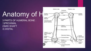
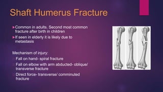
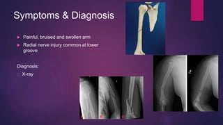
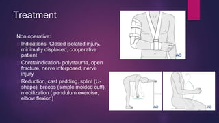
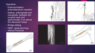
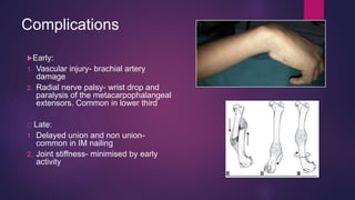
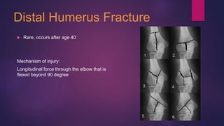
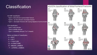
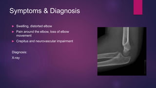
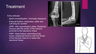
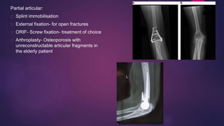
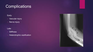
Fractures of the
humerus
BY: SUNNY SHARON MARIA & NIVEDITA
PANT
Anatomy of Humerus
3 PARTS OF HUMERAL BONE :
1)PROXIMAL
2)MID SHAFT
3) DISTAL
Shaft Humerus Fracture
Common in adults. Second most common
fracture after birth in children
If seen in elderly it is likely due to
metastasis
Mechanism of injury:
Fall on hand- spiral fracture
Fall on elbow with arm abducted- oblique/
transverse fracture
Direct force- transverse/ comminuted
fracture
Symptoms & Diagnosis
 Painful, bruised and swollen arm
 Radial nerve injury common at lower
groove
Diagnosis:
X-ray
Treatment
Non operative:
Indications- Closed isolated injury,
minimally displaced, cooperative
patient
Contraindication- polytrauma, open
fracture, nerve interposed, nerve
injury
Reduction, cast padding, splint (U-
shape), braces (simple molded cuff),
mobilization ( pendulum exercise,
elbow flexion)
Operative:
External fixation-
anterolateral pin insertion
Nailing- anterograde and
retrograde- between the
surgical neck and
approximally 5 cm above
the olecranon fossa
Bridge plating
ORIF- spiral and long
oblique fractures
Complications
Early:
1. Vascular injury- brachial artery
damage
2. Radial nerve palsy- wrist drop and
paralysis of the metacarpophalangeal
extensors. Common in lower third
Late:
1. Delayed union and non union-
common in IM nailing
2. Joint stiffness- minimised by early
activity
Distal Humerus Fracture
 Rare, occurs after age 40
Mechanism of injury:
Longitudinal force through the elbow that is
flexed beyond 90 degree
Classification
AO-ASIF classification
Type A- extra-articular supracondylar fracture
Type B – an intra-articular unicondylar fracture
Type C – bicondylar fractures with varying degrees of communication
OTA classification:
Type A- extra articular
Type B- partially articular
Type C- completely articular (T or Y shaped)
Mehne and Matta A-F classification:
A – HIGHT
B – LOW T
C – Y FRACTURE
D – H FRACTURE
E – MEDIAL LAMBDA
F – LATERAL LAMBDA
Symptoms & Diagnosis
 Swelling, distorted elbow
 Pain around the elbow, loss of elbow
movement
 Crepitus and neurovascular impairment
Diagnosis:
X-ray
Treatment
Extra articular:
Splint immobilisation- minimally displaced
External fixation- temporary, collar and
cuff sling for comfort
ORIF- with neutralisation plate- Displaced
fracture with the center of the fracture
proximal to the olecranon fossa
ORIF- Plate fixation with/without lag
screw- Displaced fracture with the center
of the fracture close to or below the
olecranon fossa
Partial articular:
Splint immobilisation
External fixation- for open fractures
ORIF- Screw fixation- treatment of choice
Arthroplasty- Osteoporosis with
unreconstructable articular fragments in
the elderly patient
Complete articular:
Splint immobilisation
External fixation
ORIF- plate fixation- Treatment of
choice
Arthroplasty- Osteoporosis or
very low transverse fracture
pattern with unreconstructable
articular fragments in the elderly
patient
Complications
Early:
Vascular injury
Nerve injury
Late:
Stiffness
Heterotrophic ossification
Supracondylar fracture
 Commonest fracture in children.
 Uncommon after the physes have
closed.
 Also called Malgaigne’s fracture.
 The humerus breaks just above the
condyles.
 The distal fragment may be displaced
either posteriorly or anteriorly.
 Common in boys.
 Types- flexion & extension (90%).
MECHANISM OF INJURY
 Fall on the outstretched hand with forearm in pronation.
 The distal fragment is pushed backwards and twisted inwards.
 Posterior angulations or displacement suggests a hyperextension injury (common).
 Anterior displacement is due to direct fall on the point of elbow with joint in flexion (rare)
CLASSIFICATION
 Type I – an undisplaced fracture.
 Type II – an angulated fracture with posterior cortex still in continuity.
IIA – a less severe injury with the distal fragment merely angulated.
IIB – a severe injury; the fragment is both angulated and malrotated.
 Type III – a completely displaced fracture.
CLINICAL FEATURES
• History of fall
• Pain in the elbow.
• Elbow is swollen and tender.
• S-deformity of the elbow (posterior)
• Bony landmarks are abnormal.
• Both active and passive movements of the elbow are decreased.
• Assessment of the neurovascular status.
• Relationships between the tip of the olecranon and the epicondyle are normally
aligned.
INVESTIGATIONS
• X ray
seen clearly in lateral view.
Fat pad sign in undisplaced fracture.
Posteriorly displaced - fracture line runs obliquely downwards and forwards and distal
fragment is tilted backwards or shifted backwards.
Anteriorly displaced – fracture line runs downwards and backwards
and distal fragment is tilted forwards.
TREATMENT
UNDISPLACED
The elbow is immobilized at 90 degrees and neutral rotation for 3 weeks.
MILD POSTERIORLY ANGULATED
Reduction under anaesthesia.
If the reduction is unstable, the fracture should be fixed with percutaneous
K_x0002_wires.
Immobilized for 3 weeks
ANGULATED AND MALROTATED OR POSTERIORLY DISPLACED
Usually associated with severe swelling, often unstable, risk of neurovascular injury.
Reduced under general anaesthesiaand then held with percutaneous K_x0002_wires.
Open reduction –
i. When fracture cannot be reduced closed;
ii. An open fracture;
iii. A fracture associated with vascular damage.
iv. Interposition of the biceps
Complications
EARLY-
Vascular injury- brachial artery
Nerve injury- anterior interosseousn,>median n.>radial n.> ulnar n.
Volkman’s ischemia & compartment syndrome
LATE-
Malunion- uncorrected sideways tilt and rotation may lead to varus or
valgus deformity.
Elbow stiffness
Myositis ossificans.
Tardy ulnar nerve palsy
Nonunion – least common
Medial condyle & epicondyle fractures
 Injuries to the medial aspect of the distal humerus in young children can
range from an avulsion fracture of the medial epicondyle to a much more
serious Salter-Harris type IV fracture of the medial condyle.
 Medial condyle fractures involve a fracture line that extends through and
separates the medial metaphysis and epicondyle from the rest of the
humerus; by definition, the fracture line must involve the trochlear
articular surface.
 Medial condyle fractures must be distinguished from medial epicondyle
fractures that involve the medial column but are extra-articular The
distinction between these two types of fractures is key to the selection of
appropriate treatment. Both fracture patterns may be difficult to diagnose
in young children, especially before the secondary ossification centers
have formed
 Medial condyle fracture
The patient usually presents with a recent history of a significant fall on an outstretched hand or directly on the apex of the
flexed elbow. The elbow may be severely painful after this injury. Swelling, deformity, and loss of function of the elbow may
be present. Palpable crepitus may be present over the medial condyle. Elbow motion may be decreased as a consequence
of swelling and pain. The patient often holds the elbow fixed at approximately 90° of flexion. The patient may present with
medial dislocation of the forearm, referred to as a fracture dislocation.
Distal neurovascular changes may occur, especially in the ulnar nerve distribution. Other injuries may be present that are
easier to detect, such as elbow dislocation or fracture of the radial head or olecranon, which may distract the physician from
making the diagnosis of medial condyle fracture. A high index of suspicion for this type of injury concurrent with other elbow
injuries can ensure timely diagnosis and treatment.
 Medial epicondyle fracture
The presentation of a patient with a medial epicondyle fracture does not differ significantly from that of a patient with a
medial condyle fracture, as described above. A through physical examination should include a valgus stress test to assess
for instability of the anterior oblique band of the ulnar collateral ligament . The test is performed with the patient supine and
the arm abducted 90º. The shoulder and arm are externally rotated 90º, with the elbow flexed at least 15º to unlock the
olecranon. Valgus stress is then placed through the elbow to assess for ligamentous instability
Imaging Studies
Diagnosis is usually based on standard anteroposterior (AP) and lateral radiographs of the affected
elbow
Arthrography may be used to determine the extent of a fracture and to help distinguish an epicondyle
fracture from a condyle fracture.
Magnetic resonance imaging (MRI) may be used to evaluate soft-tissue injury and may be helpful in
evaluating cartilaginous injury.
Treatment
 Salter-Harris type IV medial condyle fractures with 2 mm or more of displacement usually must be treated by means of open
reduction with internal fixation (ORIF). A displaced medial condyle fragment or instability of the fragment with closed reduction is
an indication for open reduction with rigid internal fixation. Accurate apposition of the fracture surfaces is important to reduce the
risk of growth-plate disturbance and to prevent loss of motion due to articular incongruence. Nondisplaced medial condyle
fractures can be treated without surgery.
 For nondisplaced or minimally displaced medial epicondyle fractures, nonoperative management is the procedure of choice.
More controversy exists with displacement of 5-15 mm. Similar functional results have been reported with operative and
nonoperative surgical management. Long-term functional assessment has demonstrated similar results even with radiographic
nonunion being apparent on most of the fractures treated nonoperatively.
 Irreducible incarceration of the medial epicondyle fragment and open fracture are indications for operative management. Excision
of the comminuted medial epicondyle fragment has been associated with less beneficial results. Other controversial relative
surgical indications include complete ulnar nerve dysfunction after an injury or reduction attempt and valgus instability in high-
demand athletes.
 If the patient is unable to tolerate a long surgical procedure because of polytrauma, closed reduction and cast immobilization with
90° of flexion is an option
Avulsion of lateral epicondyle
 This fracture type is classified by the
AO/OTA as 13A1.1.
 This is an avulsion fracture of the lateral
epicondyle. By definition, there is no
involvement of the joint (capitellum).
 The fracture is the equivalent of an
avulsion of the humeral attachment of the
lateral collateral ligament.
 Typically, this injury occurs in young
patients.
 The fragment may become displaced into
the lateral elbow joint cavity.
Avulsion of medial epicondyle
 This fracture type is classified by the
AO/OTA as 13A1.2.
 These fractures represent avulsion of the
humeral origin of the medial collateral
ligament and common flexor origin,
resulting from a valgus force to the elbow
 Medial epicondyle fractures are more
common in skeletally immature patients.
In rare cases, the fragment is incarcerated in the ulnohumeral joint.
Incarcerated fragment
treatment
 Splint immobilization
 ORIF - Screw fixation
Main indications Any significant
displacement, incarceration in joint,
or varus or posterolateral rotatory
instability of the elbow
humerus fracture

humerus fracture

  • 1.
    Fractures of the humerus BY:SUNNY SHARON MARIA & NIVEDITA PANT
  • 2.
    Anatomy of Humerus 3PARTS OF HUMERAL BONE : 1)PROXIMAL 2)MID SHAFT 3) DISTAL
  • 3.
    Shaft Humerus Fracture Commonin adults. Second most common fracture after birth in children If seen in elderly it is likely due to metastasis Mechanism of injury: Fall on hand- spiral fracture Fall on elbow with arm abducted- oblique/ transverse fracture Direct force- transverse/ comminuted fracture
  • 4.
    Symptoms & Diagnosis Painful, bruised and swollen arm  Radial nerve injury common at lower groove Diagnosis: X-ray
  • 5.
    Treatment Non operative: Indications- Closedisolated injury, minimally displaced, cooperative patient Contraindication- polytrauma, open fracture, nerve interposed, nerve injury Reduction, cast padding, splint (U- shape), braces (simple molded cuff), mobilization ( pendulum exercise, elbow flexion)
  • 6.
    Operative: External fixation- anterolateral pininsertion Nailing- anterograde and retrograde- between the surgical neck and approximally 5 cm above the olecranon fossa Bridge plating ORIF- spiral and long oblique fractures
  • 7.
    Complications Early: 1. Vascular injury-brachial artery damage 2. Radial nerve palsy- wrist drop and paralysis of the metacarpophalangeal extensors. Common in lower third Late: 1. Delayed union and non union- common in IM nailing 2. Joint stiffness- minimised by early activity
  • 8.
    Distal Humerus Fracture Rare, occurs after age 40 Mechanism of injury: Longitudinal force through the elbow that is flexed beyond 90 degree
  • 9.
    Classification AO-ASIF classification Type A-extra-articular supracondylar fracture Type B – an intra-articular unicondylar fracture Type C – bicondylar fractures with varying degrees of communication OTA classification: Type A- extra articular Type B- partially articular Type C- completely articular (T or Y shaped) Mehne and Matta A-F classification: A – HIGHT B – LOW T C – Y FRACTURE D – H FRACTURE E – MEDIAL LAMBDA F – LATERAL LAMBDA
  • 10.
    Symptoms & Diagnosis Swelling, distorted elbow  Pain around the elbow, loss of elbow movement  Crepitus and neurovascular impairment Diagnosis: X-ray
  • 11.
    Treatment Extra articular: Splint immobilisation-minimally displaced External fixation- temporary, collar and cuff sling for comfort ORIF- with neutralisation plate- Displaced fracture with the center of the fracture proximal to the olecranon fossa ORIF- Plate fixation with/without lag screw- Displaced fracture with the center of the fracture close to or below the olecranon fossa
  • 12.
    Partial articular: Splint immobilisation Externalfixation- for open fractures ORIF- Screw fixation- treatment of choice Arthroplasty- Osteoporosis with unreconstructable articular fragments in the elderly patient
  • 13.
    Complete articular: Splint immobilisation Externalfixation ORIF- plate fixation- Treatment of choice Arthroplasty- Osteoporosis or very low transverse fracture pattern with unreconstructable articular fragments in the elderly patient
  • 14.
  • 15.
    Supracondylar fracture  Commonestfracture in children.  Uncommon after the physes have closed.  Also called Malgaigne’s fracture.  The humerus breaks just above the condyles.  The distal fragment may be displaced either posteriorly or anteriorly.  Common in boys.  Types- flexion & extension (90%).
  • 16.
    MECHANISM OF INJURY Fall on the outstretched hand with forearm in pronation.  The distal fragment is pushed backwards and twisted inwards.  Posterior angulations or displacement suggests a hyperextension injury (common).  Anterior displacement is due to direct fall on the point of elbow with joint in flexion (rare) CLASSIFICATION  Type I – an undisplaced fracture.  Type II – an angulated fracture with posterior cortex still in continuity. IIA – a less severe injury with the distal fragment merely angulated. IIB – a severe injury; the fragment is both angulated and malrotated.  Type III – a completely displaced fracture.
  • 17.
    CLINICAL FEATURES • Historyof fall • Pain in the elbow. • Elbow is swollen and tender. • S-deformity of the elbow (posterior) • Bony landmarks are abnormal. • Both active and passive movements of the elbow are decreased. • Assessment of the neurovascular status. • Relationships between the tip of the olecranon and the epicondyle are normally aligned. INVESTIGATIONS • X ray seen clearly in lateral view. Fat pad sign in undisplaced fracture. Posteriorly displaced - fracture line runs obliquely downwards and forwards and distal fragment is tilted backwards or shifted backwards. Anteriorly displaced – fracture line runs downwards and backwards and distal fragment is tilted forwards.
  • 18.
    TREATMENT UNDISPLACED The elbow isimmobilized at 90 degrees and neutral rotation for 3 weeks. MILD POSTERIORLY ANGULATED Reduction under anaesthesia. If the reduction is unstable, the fracture should be fixed with percutaneous K_x0002_wires. Immobilized for 3 weeks ANGULATED AND MALROTATED OR POSTERIORLY DISPLACED Usually associated with severe swelling, often unstable, risk of neurovascular injury. Reduced under general anaesthesiaand then held with percutaneous K_x0002_wires. Open reduction – i. When fracture cannot be reduced closed; ii. An open fracture; iii. A fracture associated with vascular damage. iv. Interposition of the biceps
  • 19.
    Complications EARLY- Vascular injury- brachialartery Nerve injury- anterior interosseousn,>median n.>radial n.> ulnar n. Volkman’s ischemia & compartment syndrome LATE- Malunion- uncorrected sideways tilt and rotation may lead to varus or valgus deformity. Elbow stiffness Myositis ossificans. Tardy ulnar nerve palsy Nonunion – least common
  • 20.
    Medial condyle &epicondyle fractures  Injuries to the medial aspect of the distal humerus in young children can range from an avulsion fracture of the medial epicondyle to a much more serious Salter-Harris type IV fracture of the medial condyle.  Medial condyle fractures involve a fracture line that extends through and separates the medial metaphysis and epicondyle from the rest of the humerus; by definition, the fracture line must involve the trochlear articular surface.  Medial condyle fractures must be distinguished from medial epicondyle fractures that involve the medial column but are extra-articular The distinction between these two types of fractures is key to the selection of appropriate treatment. Both fracture patterns may be difficult to diagnose in young children, especially before the secondary ossification centers have formed
  • 21.
     Medial condylefracture The patient usually presents with a recent history of a significant fall on an outstretched hand or directly on the apex of the flexed elbow. The elbow may be severely painful after this injury. Swelling, deformity, and loss of function of the elbow may be present. Palpable crepitus may be present over the medial condyle. Elbow motion may be decreased as a consequence of swelling and pain. The patient often holds the elbow fixed at approximately 90° of flexion. The patient may present with medial dislocation of the forearm, referred to as a fracture dislocation. Distal neurovascular changes may occur, especially in the ulnar nerve distribution. Other injuries may be present that are easier to detect, such as elbow dislocation or fracture of the radial head or olecranon, which may distract the physician from making the diagnosis of medial condyle fracture. A high index of suspicion for this type of injury concurrent with other elbow injuries can ensure timely diagnosis and treatment.  Medial epicondyle fracture The presentation of a patient with a medial epicondyle fracture does not differ significantly from that of a patient with a medial condyle fracture, as described above. A through physical examination should include a valgus stress test to assess for instability of the anterior oblique band of the ulnar collateral ligament . The test is performed with the patient supine and the arm abducted 90º. The shoulder and arm are externally rotated 90º, with the elbow flexed at least 15º to unlock the olecranon. Valgus stress is then placed through the elbow to assess for ligamentous instability
  • 23.
    Imaging Studies Diagnosis isusually based on standard anteroposterior (AP) and lateral radiographs of the affected elbow Arthrography may be used to determine the extent of a fracture and to help distinguish an epicondyle fracture from a condyle fracture. Magnetic resonance imaging (MRI) may be used to evaluate soft-tissue injury and may be helpful in evaluating cartilaginous injury.
  • 24.
    Treatment  Salter-Harris typeIV medial condyle fractures with 2 mm or more of displacement usually must be treated by means of open reduction with internal fixation (ORIF). A displaced medial condyle fragment or instability of the fragment with closed reduction is an indication for open reduction with rigid internal fixation. Accurate apposition of the fracture surfaces is important to reduce the risk of growth-plate disturbance and to prevent loss of motion due to articular incongruence. Nondisplaced medial condyle fractures can be treated without surgery.  For nondisplaced or minimally displaced medial epicondyle fractures, nonoperative management is the procedure of choice. More controversy exists with displacement of 5-15 mm. Similar functional results have been reported with operative and nonoperative surgical management. Long-term functional assessment has demonstrated similar results even with radiographic nonunion being apparent on most of the fractures treated nonoperatively.  Irreducible incarceration of the medial epicondyle fragment and open fracture are indications for operative management. Excision of the comminuted medial epicondyle fragment has been associated with less beneficial results. Other controversial relative surgical indications include complete ulnar nerve dysfunction after an injury or reduction attempt and valgus instability in high- demand athletes.  If the patient is unable to tolerate a long surgical procedure because of polytrauma, closed reduction and cast immobilization with 90° of flexion is an option
  • 25.
    Avulsion of lateralepicondyle  This fracture type is classified by the AO/OTA as 13A1.1.  This is an avulsion fracture of the lateral epicondyle. By definition, there is no involvement of the joint (capitellum).  The fracture is the equivalent of an avulsion of the humeral attachment of the lateral collateral ligament.  Typically, this injury occurs in young patients.  The fragment may become displaced into the lateral elbow joint cavity.
  • 26.
    Avulsion of medialepicondyle  This fracture type is classified by the AO/OTA as 13A1.2.  These fractures represent avulsion of the humeral origin of the medial collateral ligament and common flexor origin, resulting from a valgus force to the elbow  Medial epicondyle fractures are more common in skeletally immature patients. In rare cases, the fragment is incarcerated in the ulnohumeral joint. Incarcerated fragment
  • 27.
    treatment  Splint immobilization ORIF - Screw fixation Main indications Any significant displacement, incarceration in joint, or varus or posterolateral rotatory instability of the elbow