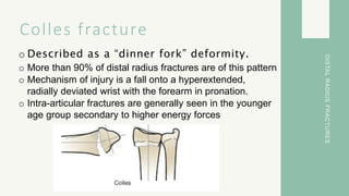
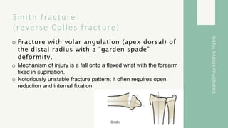
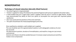
DISTAL RADIUS
FRACTURE
Colles fracture
o Described as a “dinner fork” deformity.
o More than 90% of distal radius fractures are of this pattern
o Mechanism of injury is a fall onto a hyperextended,
radially deviated wrist with the forearm in pronation.
o Intra-articular fractures are generally seen in the younger
age group secondary to higher energy forces
DISTAL
RADIUS
FRACTURES
Smith fracture
(reverse Colles fracture)
o Fracture with volar angulation (apex dorsal) of
the distal radius with a “garden spade”
deformity.
o Mechanism of injury is a fall onto a flexed wrist with the forearm
fixed in supination.
o Notoriously unstable fracture pattern; it often requires open
reduction and internal fixation
DISTAL
RADIUS
FRACTURES
Basic Baby Care
NONOPERATIVE
Indications:
• Nondisplaced or minimally displaced
fractures
• Displaced fractures with a stable fracture
pattern which can be expected to unite
within acceptable radiographic parameters
• Low-demand elderly patients in whom
future functional impairment is less of a
priority than immediate health concerns
and/or operative risks
OPERATIVE
Indications:
• High-energy injury
• Secondary loss of reduction
• Open fractures
• Displaced shear fractures (type II)
• Comminuted and displaced articular fractures with
articular impaction (type III)
• Fracture–dislocations (type IV)
• Combined injuries with metaphyseal–diaphyseal
comminution (type V)
• Fractures complicated by nerve compression,
compartment syndrome, or multiple injuries
• Bilateral distal radius fractures
• An impaired contralateral extremity
TREATMENT
NONOPERATIVE
Technique of closed reduction (dorsally tilted fracture)
• The distal fragment is hyperextended.
• Traction is applied to reduce the distal to the proximal fragment with pressure applied to the distal radius.
• A well-molded long arm (“sugar-tong”) splint is applied, with the wrist in neutral to slight flexion. Studies
have demonstrated the ability of short arm splints to accomplish the same goal with improved patient
satisfaction.
• One must avoid extreme positions of the wrist and hand.
• The splint should leave the metacarpophalangeal joints free.
Once swelling has subsided, a well-molded cast is applied.
The cast should be worn for approximately 6 weeks or until radiographic evidence of union
has occurred.
The ideal forearm position, duration of immobilization, and need for a long arm cast remain
controversial;
Extreme wrist flexion should be avoided because it increases carpal canal pressure (and thus
median nerve compression
Reference
1. Campbell’s Operative Orthopaedics, 4-Volume Set 14th Edition
(2020)
2. Hoppenfeld Surgical Exposures in Orthopaedics: The Anatomic
Approach 5th Edition (2016)
3. Apley’s System of Orthopaedics and Fractures Ninth Edition, 2010

DISTAL RADIUS FRACTURE.pptx

  • 1.
  • 2.
    Colles fracture o Describedas a “dinner fork” deformity. o More than 90% of distal radius fractures are of this pattern o Mechanism of injury is a fall onto a hyperextended, radially deviated wrist with the forearm in pronation. o Intra-articular fractures are generally seen in the younger age group secondary to higher energy forces DISTAL RADIUS FRACTURES
  • 3.
    Smith fracture (reverse Collesfracture) o Fracture with volar angulation (apex dorsal) of the distal radius with a “garden spade” deformity. o Mechanism of injury is a fall onto a flexed wrist with the forearm fixed in supination. o Notoriously unstable fracture pattern; it often requires open reduction and internal fixation DISTAL RADIUS FRACTURES
  • 4.
    Basic Baby Care NONOPERATIVE Indications: •Nondisplaced or minimally displaced fractures • Displaced fractures with a stable fracture pattern which can be expected to unite within acceptable radiographic parameters • Low-demand elderly patients in whom future functional impairment is less of a priority than immediate health concerns and/or operative risks OPERATIVE Indications: • High-energy injury • Secondary loss of reduction • Open fractures • Displaced shear fractures (type II) • Comminuted and displaced articular fractures with articular impaction (type III) • Fracture–dislocations (type IV) • Combined injuries with metaphyseal–diaphyseal comminution (type V) • Fractures complicated by nerve compression, compartment syndrome, or multiple injuries • Bilateral distal radius fractures • An impaired contralateral extremity TREATMENT
  • 5.
    NONOPERATIVE Technique of closedreduction (dorsally tilted fracture) • The distal fragment is hyperextended. • Traction is applied to reduce the distal to the proximal fragment with pressure applied to the distal radius. • A well-molded long arm (“sugar-tong”) splint is applied, with the wrist in neutral to slight flexion. Studies have demonstrated the ability of short arm splints to accomplish the same goal with improved patient satisfaction. • One must avoid extreme positions of the wrist and hand. • The splint should leave the metacarpophalangeal joints free. Once swelling has subsided, a well-molded cast is applied. The cast should be worn for approximately 6 weeks or until radiographic evidence of union has occurred. The ideal forearm position, duration of immobilization, and need for a long arm cast remain controversial; Extreme wrist flexion should be avoided because it increases carpal canal pressure (and thus median nerve compression
  • 6.
    Reference 1. Campbell’s OperativeOrthopaedics, 4-Volume Set 14th Edition (2020) 2. Hoppenfeld Surgical Exposures in Orthopaedics: The Anatomic Approach 5th Edition (2016) 3. Apley’s System of Orthopaedics and Fractures Ninth Edition, 2010