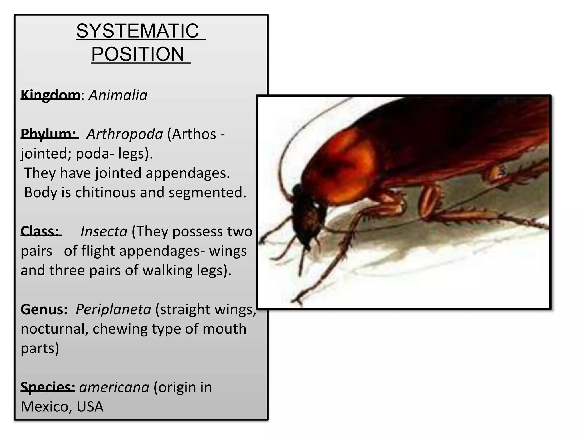
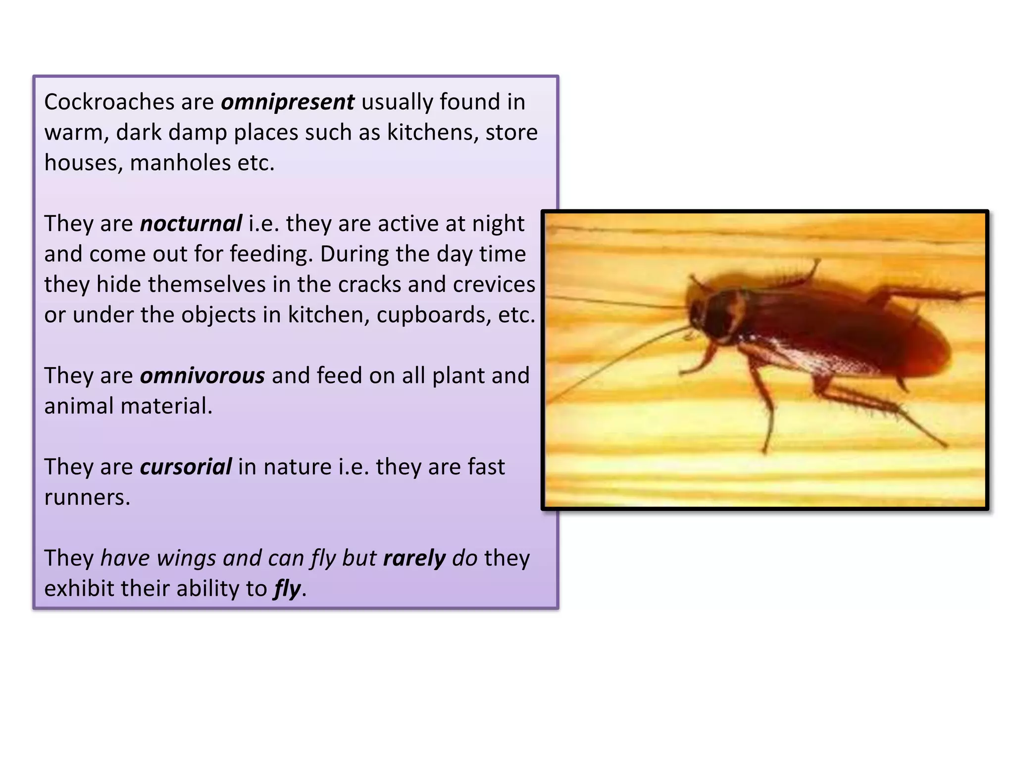
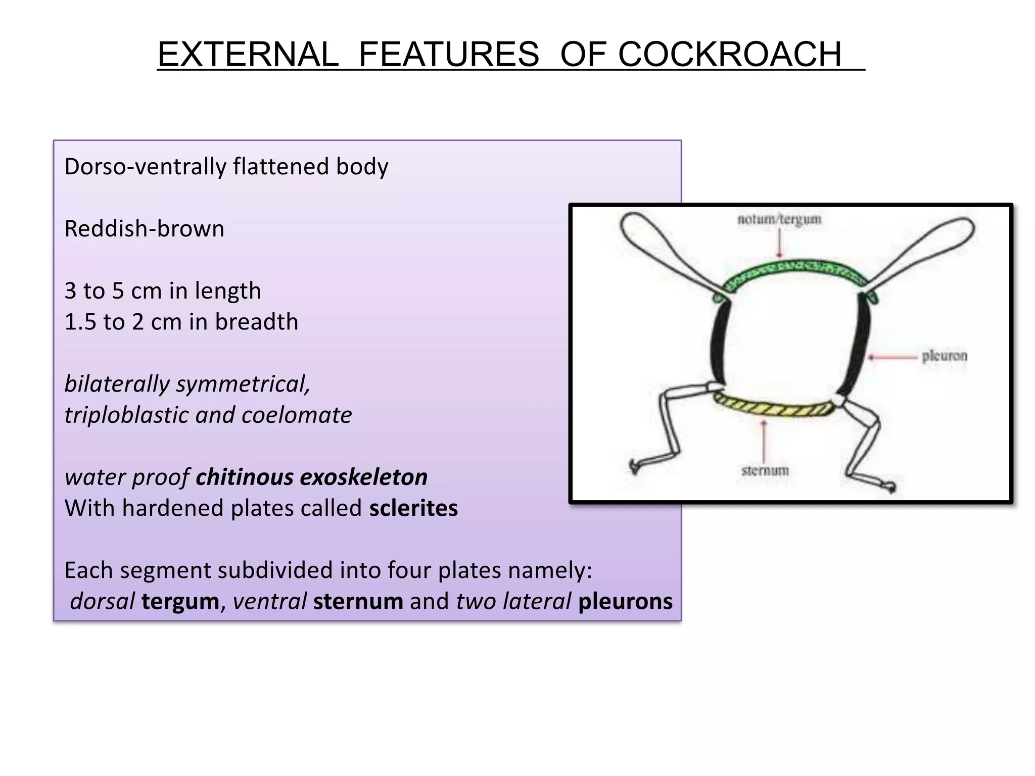
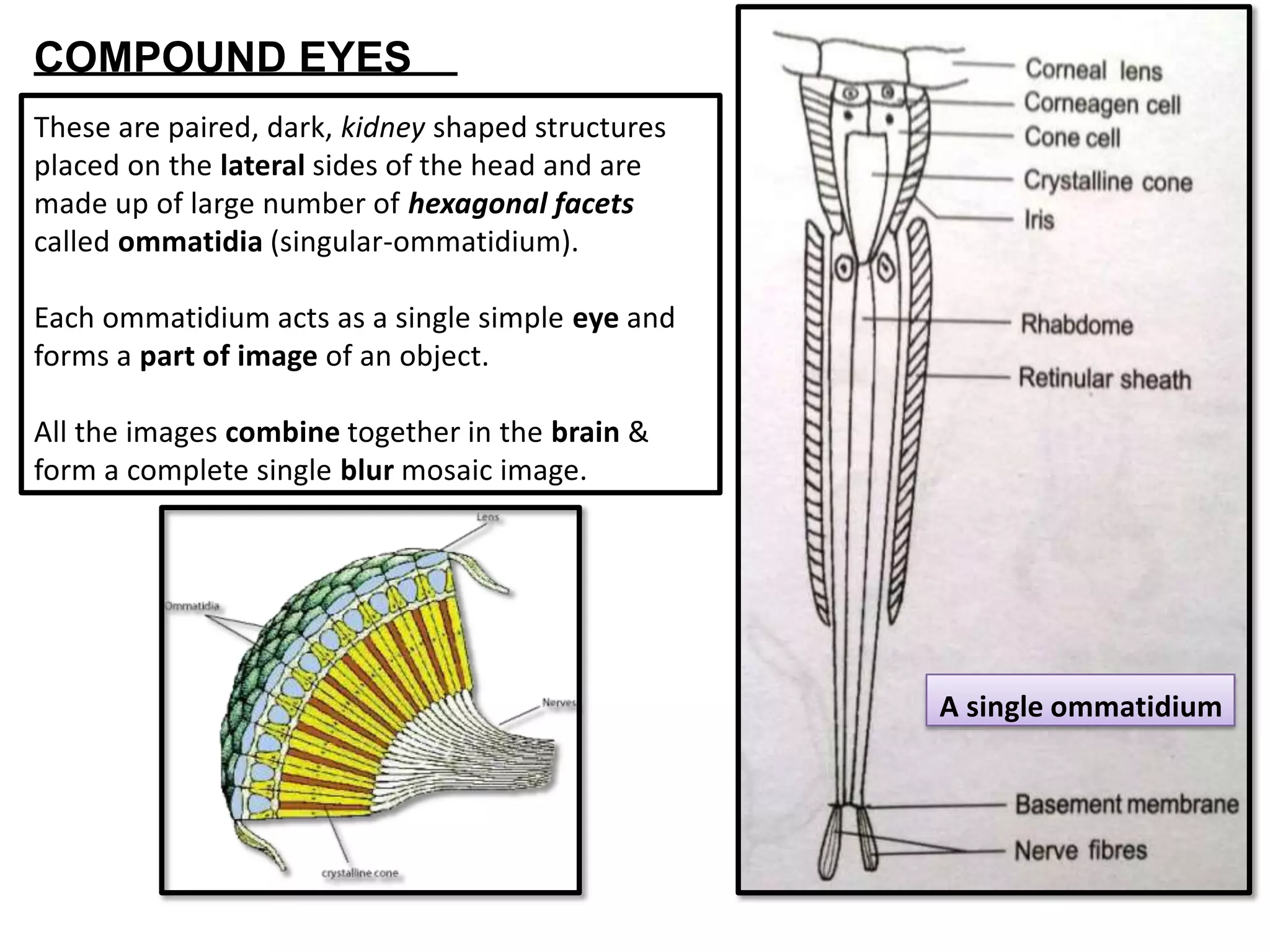
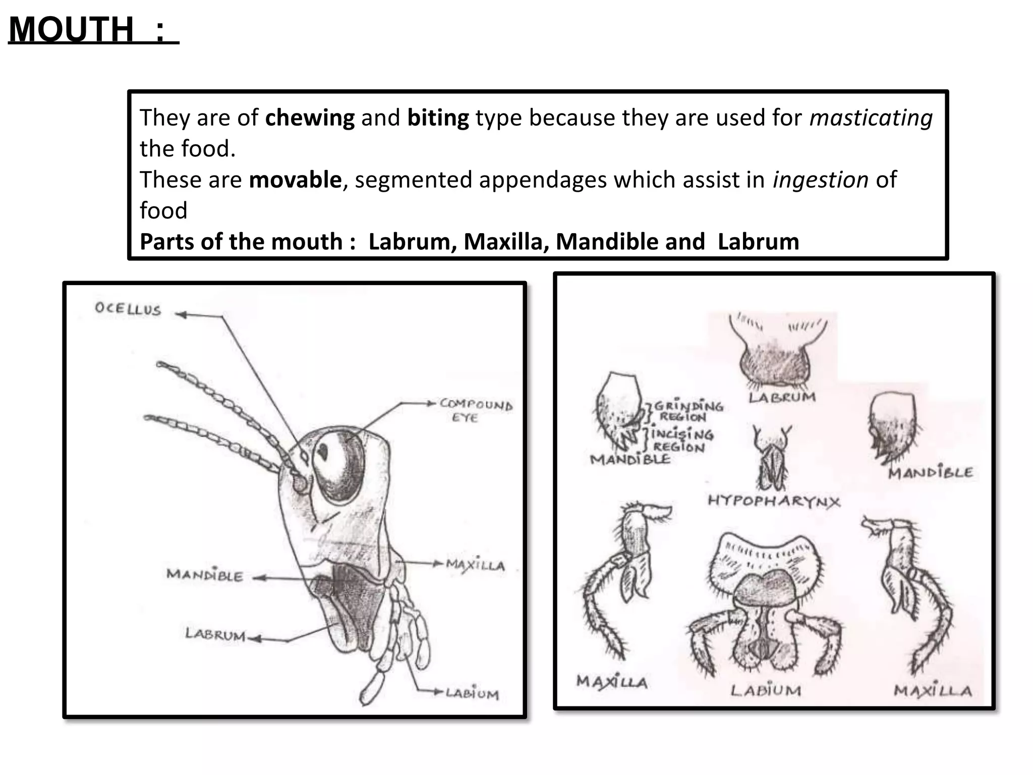
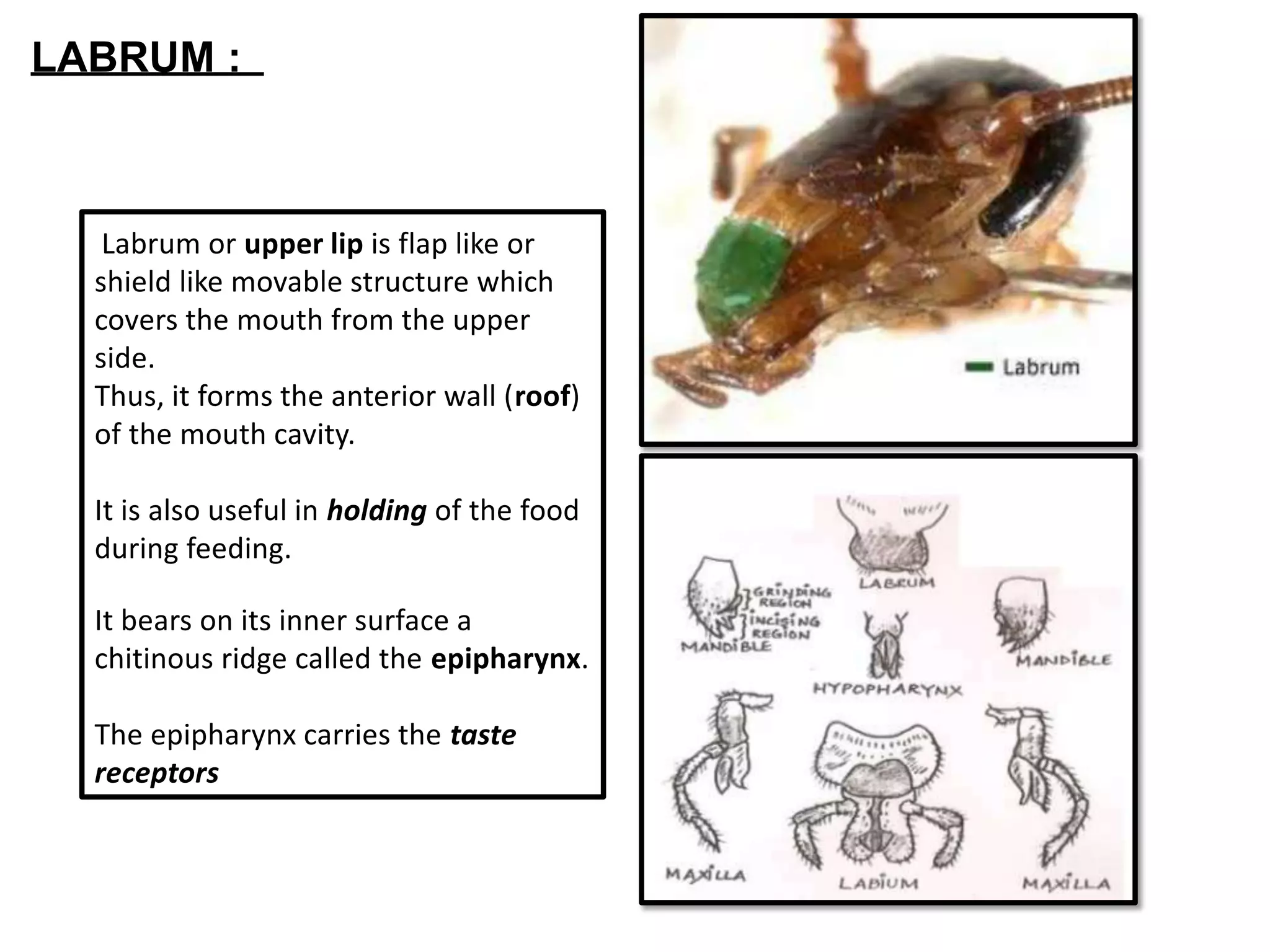
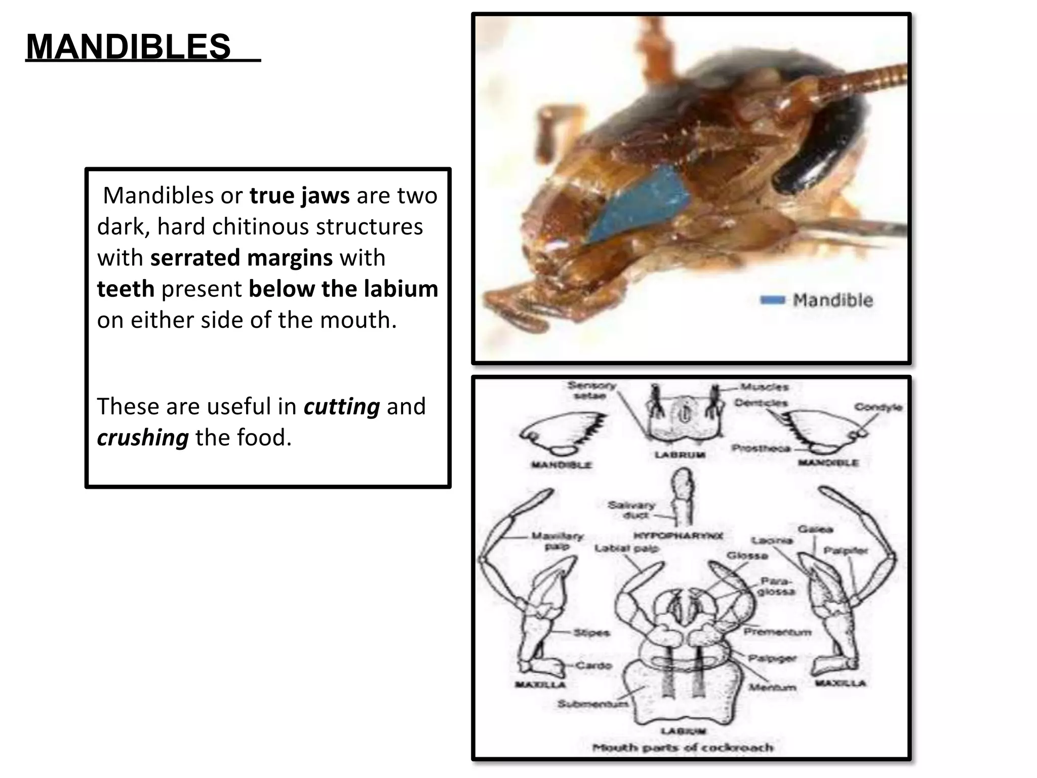
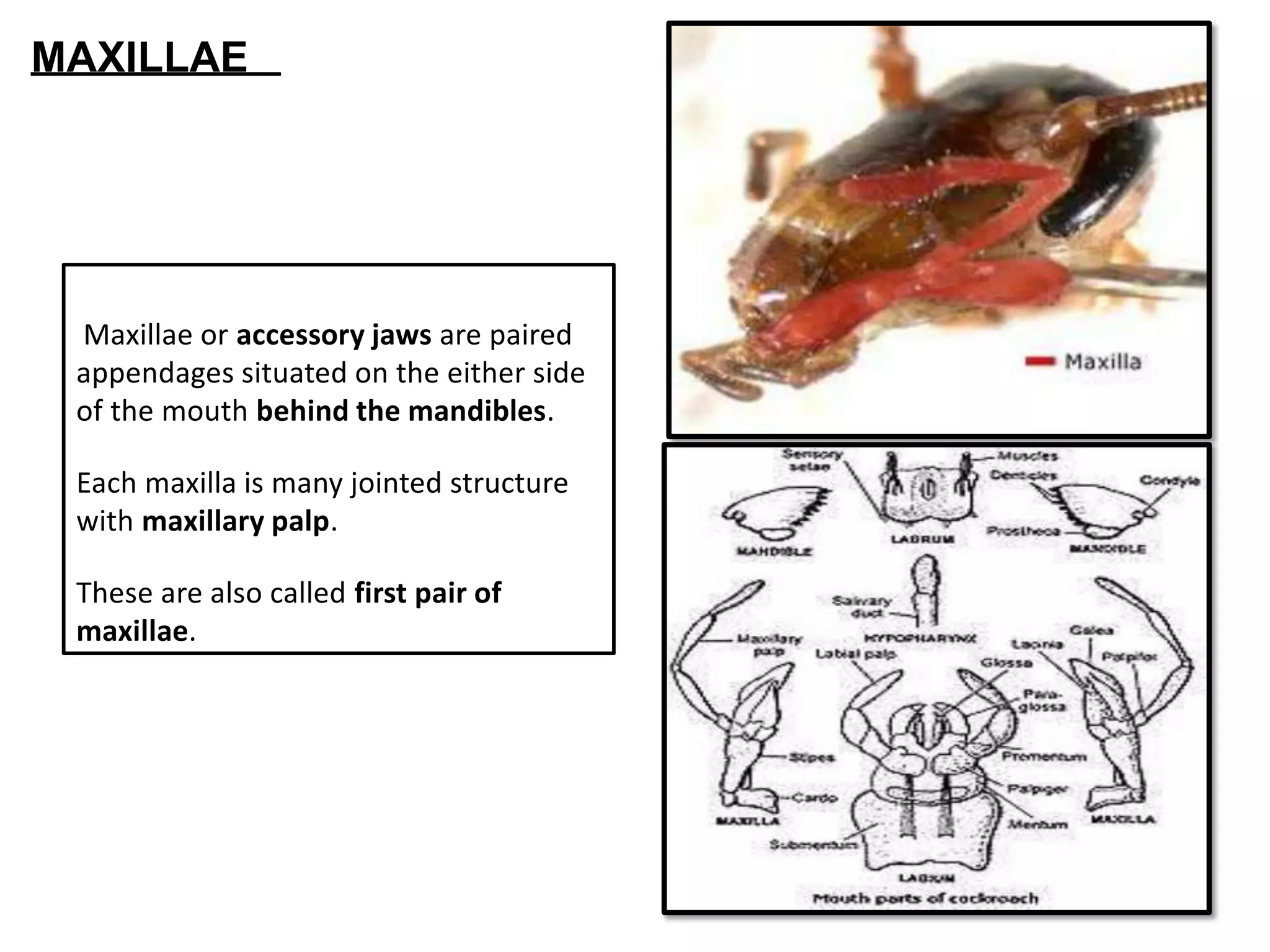
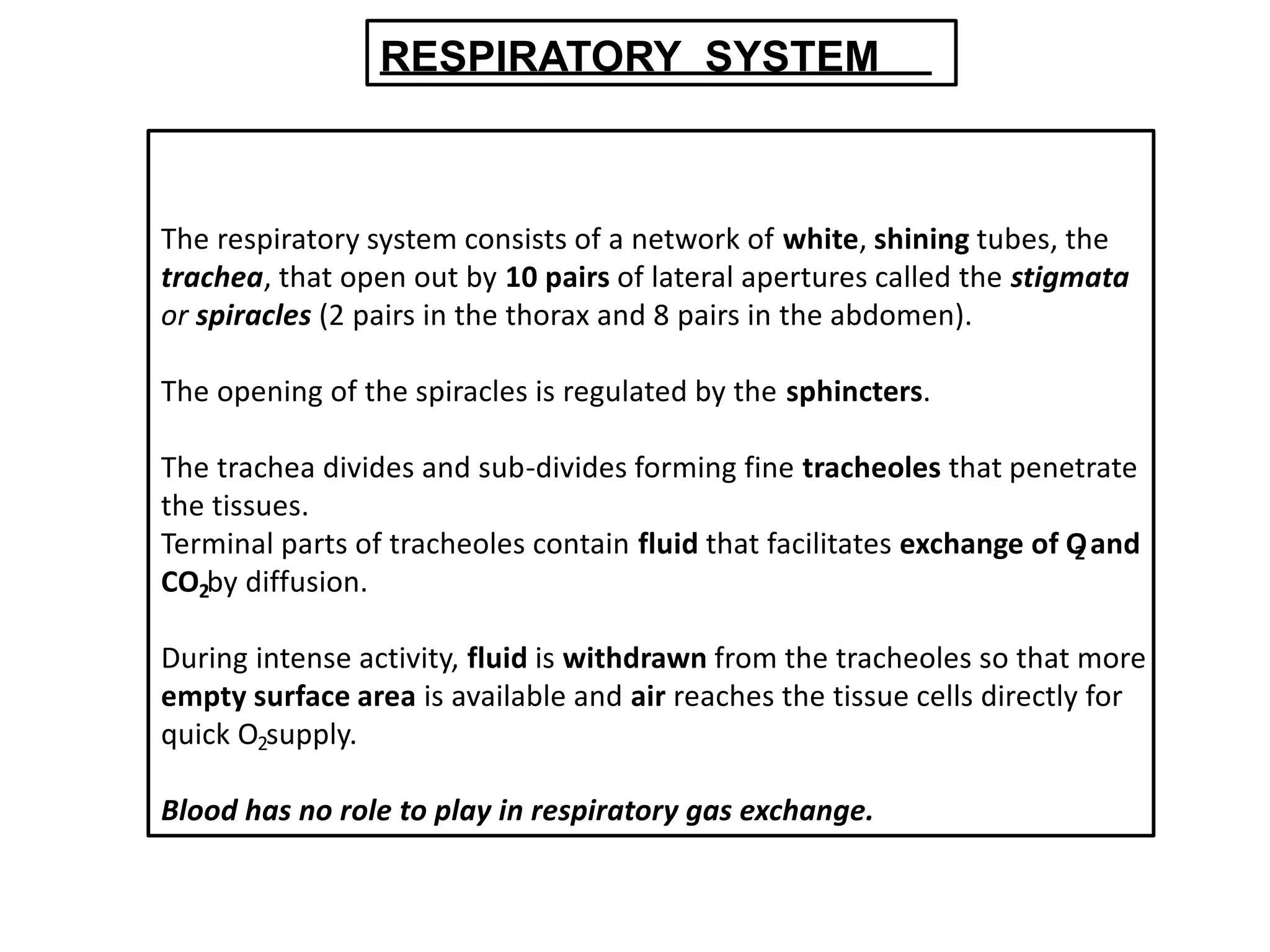
Study of Animal Type
By
Dr. Priti D.Diwan
Assistant Professor
Department of Zoology
J.D.Patil Sangludkar Mahavidyalay
Daryapur
SYSTEMATIC
POSITION
Kingdom: Animalia
Phylum: Arthropoda (Arthos -
jointed; poda- legs).
They have jointed appendages.
Body is chitinous and segmented.
Class: Insecta (They possess two
pairs of flight appendages- wings
and three pairs of walking legs).
Genus: Periplaneta (straight wings,
nocturnal, chewing type of mouth
parts)
Species: americana (origin in
Mexico, USA
Periplaneta americana Blatta orientalis
i. It is lighter in color. i. It is darker in color.
i. It is about 38 mm long. i. It is about 25 mm long.
i. Wings are present in both the sexes
and extend beyond the posterior end
of the body.
i. Wings are rudimentary in the female
and
do not reach the hind end of the body
in
the male.
Cockroaches are omnipresent usually found in
warm, dark damp places such as kitchens, store
houses, manholes etc.
They are nocturnal i.e. they are active at night
and come out for feeding. During the day time
they hide themselves in the cracks and crevices
or under the objects in kitchen, cupboards, etc.
They are omnivorous and feed on all plant and
animal material.
They are cursorial in nature i.e. they are fast
runners.
They have wings and can fly but rarely do they
exhibit their ability to fly.
EXTERNAL FEATURES OF COCKROACH
Dorso-ventrally flattened body
Reddish-brown
3 to 5 cm in length
1.5 to 2 cm in breadth
bilaterally symmetrical,
triploblastic and coelomate
water proof chitinous exoskeleton
With hardened plates called sclerites
Each segment subdivided into four plates namely:
dorsal tergum, ventral sternum and two lateral pleurons
BODY DIVISIONS
HEAD
Ovate, flattened dorso-ventrally ,pear-shaped,
attached at the right angles to the thorax by a
thin, narrow, movable neck or cervicum.
Head is formed of six sclerites. These fuse in
adults and form the head capsule.
Head bears four important parts:
1. COMPOUND EYES
2. ANTENNAE
3. FENESTRAE
4. MOUTH PARTS OR TROPHI-MOUTH PARTS
COMPOUND EYES
These are paired, dark, kidney shaped structures
placed on the lateral sides of the head and are
made up of large number of hexagonal facets
called ommatidia (singular-ommatidium).
Each ommatidium acts as a single simple eye and
forms a part of image of an object.
All the images combine together in the brain &
form a complete single blur mosaic image.
A single ommatidium
ANTENNAE
- They are paired, long slender filamentous,
many jointed structures present between
the compound eyes.
- They are lodged in the membranous pits
called antennal sockets. These are the
tacto-receptors or organs of olfaction
(sense of smell) useful in localizing the food
material in the vicinity.
- Antennae have sensory receptors that help
in monitoring the environment.
- They are also called the oscillar spots
situated at the base of each antenna and
they appear as white spots.
- They are sensitive to light.
FENESTRAE
MOUTH :
They are of chewing and biting type because they are used for masticating
the food.
These are movable, segmented appendages which assist in ingestion of
food
Parts of the mouth : Labrum, Maxilla, Mandible and Labrum
Labrum or upper lip is flap like or
shield like movable structure which
covers the mouth from the upper
side.
Thus, it forms the anterior wall (roof)
of the mouth cavity.
It is also useful in holding of the food
during feeding.
It bears on its inner surface a
chitinous ridge called the epipharynx.
The epipharynx carries the taste
receptors
LABRUM :
Mandibles or true jaws are two
dark, hard chitinous structures
with serrated margins with
teeth present below the labium
on either side of the mouth.
These are useful in cutting and
crushing the food.
MANDIBLES
Maxillae or accessory jaws are paired
appendages situated on the either side
of the mouth behind the mandibles.
Each maxilla is many jointed structure
with maxillary palp.
These are also called first pair of
maxillae.
MAXILLAE
Labium or lower lip is also called the
second maxillae which covers the
mouth from the ventral side and is
firmly attached to the posterior part of
head.
It has labial palp which is three jointed.
These are sensory in function.
Labium is useful in pushing the chewed
food in the preoral cavity and also
prevents the loss of food falling from
mandibles during the chewing.
LABIUM
Hypo-pharynx or tongue is a
long, prominent medially
placed appendage.
It hangs in the preoral cavity
just in front of the labium
between the first maxillae.
It receives saliva from the
salivary duct and directs it on
to the food.
HYPOPHARYNX
THORAX
Thorax is made up of three distinct
segments:
Anterior prothorax,
middle mesothorax and
posterior metathorax.
The exoskeleton of each segment is
made of 4 sclerites, a dorsal tergum, a
ventral sternum and 2 lateral pleurons.
The mesothorax and metathorax bear a
pair of spiracles.
VENTRALLY : a pair of walking legs (all 3
segments)
DORSALLY : a pair of wings (meso and
meta)
Dorsally the thorax bears two pairs of
membranous wings- fore wing and hind wings.
Fore Wings:
Forewings are first pair of dark, opaque,
thick wings which are protective in
function.
Hind wings:
They are thin, broad membranous,
delicate and transparent second pair of wing
attached to tergum of metathorax. These are
useful in flight and hence are also called true
wings.
WINGS
LEGS
Three pairs of thoracic legs are present
on ventral side.
Each leg is five jointed or segmented and
is covered with the sensory bristles and
spines.
Each leg has five podomeres namely:
Coxa, Trochanter, Femur, Tibia, and
Tarsus
Tarsus is the last segment and is made up
of five movable segments or tarsomeres.
Last segment of the tarsus bears a pair of
claws and arolium helpful in clinging.
The abdomen is the largest of the
three regions and contains most of
the viscera.
Its segmentation is conspicuous both
dorsally and ventrally.
The abdomen consists of 11
segments of which the 11th
is
reduced and fused with the 10th
so
that a maximum of ten segments are
distinct and visible.
Dorsally each segment is covered by
a tergite and, ventrally by a sternite.
Laterally, tergites and sternites are
connected by un-sclerotized flexible
pleura.
Cockroaches exhibit sexual dimorphism.
In males, anal styles are present in
segment 9. These are absent in females.
Most abdominal segments lack
appendages but segment 10 bears a pair
of large, segmented cerci, called anal
cerci, which are visible in both dorsal
and ventral views. These are sensitive to
air movements, including sound, and
perhaps to ground vibrations.
Eight pairs of spiracles are present in
the abdomen.
Genital openings are present on
different segments in both the sexes
and are surrounded by the chitinous
plates in both, which are collectively
called gonapophyses.
DIGESTIVE
SYSTEM
Digestive System
Digestive Glands
Alimentary canal
Salivary
Glands
Hepatic
Caecae
Glands of
mesenteron
Foregut Midgut Hindgut
Pre-oral
Cavity
Mouth Pharynx Oesophagus Crop Gizzard
Ileum Colon Rectum
ALIMENTARY CANAL
FORE GUT OR STOMODAEUM:
It consists of pre oral cavity, mouth, pharynx, oesophagus, crop and gizzard or proventriculus.
PRE ORAL CAVITY : It is a space in front of the mouth.
The hypopharynx projects in the pre oral cavity
MOUTH : It is a small narrow opening surrounded by the mouth parts
PHARYNX : Short funnel-like part lying in the head region; it leads to the oesophagus
Both mouth and pharynx possess taste sensillae
OESOPHAGUS : It passes through the neck and opens into a sac like structure called crop
CROP : It is a large, highly distensible, pear-shaped sac that stores food.
It is long extending to fourth or fifth abdominal segment.
From crop food enters into gizzard
GIZZARD :
It is also called proventriculus.
It is provided with a circlet of six chitinous
teeth that help to crush the food.
Behind the chitinous teeth are, backwardly
directed fine chitinous bristles in the grooves
of gizzard.
These are interconnected with each other and
form a filter.
Gizzard has, therefore, an efficient grinding
and straining apparatu
Gizzard marks the end of fore-gut.
MID GUT OR MESENTERON:
It consists of ventriculus (stomach) and eight hepatic caecae.
Hepatic caeca are thin, transparent, blind tubules arranged in a
whorl or rosette fashion in the anterior part of the stomach. They
secrete digestive enzymes.
Ventriculus or stomach is a short and narrow tube of uniform
diameter. It is concerned with digestion of food.
The mid-gut is the major organ of digestion and absorption
HIND GUT OR PROCTODAEUM:
Hind-gut consists of ileum, colon and rectum.
It is slightly broader than the mid-gut.
At its junction with mid-gut, are present a ring of up to 150
yellow thread-like blind Malpighian tubules which are
excretory in nature.
It helps in removal of excretory products from
the haemolymph.
Their product is poured into the ileum.
Ileum is short and narrow
Colon is coiled and wide tube.
Rectum is the last part of the hind-gut. Rectum opens to
the outside by anus which lies below the 10 tergum
th
SALIVARY GLANDS
Cockroach has two salivary glands, one on each
side of crop.
Each gland has two glandular lobes and a
receptacle or reservoir.
Glandular lobes consist of several irregular, white
colored lobules which are provided with
collecting ducts. They secrete saliva.
The two gland ducts of two salivary glands unite
and form a common salivary duct.
Thin walled transparent bag-like structures called
salivary receptacles store saliva.
The common salivary receptacular duct runs
below the oesophagus through the neck and
opens at the base of the tongue or hypo pharynx.
Salivary glands open by the common salivary duct
into the preoral cavity.
Saliva contains amylase, chitinase and cellulase
The respiratory system consists of a network of white, shining tubes, the
trachea, that open out by 10 pairs of lateral apertures called the stigmata
or spiracles (2 pairs in the thorax and 8 pairs in the abdomen).
The opening of the spiracles is regulated by the sphincters.
The trachea divides and sub-divides forming fine tracheoles that penetrate
the tissues.
Terminal parts of tracheoles contain fluid that facilitates exchange of O
2 and
CO by diffusion.
2
During intense activity, fluid is withdrawn from the tracheoles so that more
empty surface area is available and air reaches the tissue cells directly for
quick O supply.
2
Blood has no role to play in respiratory gas exchange.
RESPIRATORY SYSTEM
REPRODUCTIVE SYSTEM
Cockroaches are dioecious, i.e. sexes are separate and distinguishable externally.
That is to say, cockroaches exhibit sexual dimorphism.
Both sexes have well developed reproductive organs
MALE FEMALE
MALE REPRODUCTIVE SYSTEM
1. TESTES : - A pair of testes
- in the 4 /5
th th
abdominal segemnts
- Trilobed, dorsolaterally below the terga
2. VASA DEFERENTIA:- Arises from each testis
- white thread like tube
- pass downwards & enters seminal vesicles
3. SEMINAL VESICLES : - Sac-like structures which store sperms
4. EJACULATORY DUCT: - Formed by the union of 2 seminal vesicles at the base of
utricular gland
- Elongated tube which opens through the male genital pore
5. UTRICULAR GLAND: - present at the junction of vasa deferentia and ejaculatory duct
- formed by several tubules arising from seminal vesicles
- present in the 6th
and 7th
segments
- Utricular gland = long tubules + short tubules + seminal vesicle
6. MALE GENITAL PORE: - between the 9th
and the 10th
abdominal segement, ventrally
7. GONAPOPHYSES / PHALLOMERES: - The male genital pore is surrounded by chitinous
hooks and plates called gonapophyses which help
in copulation
8. SPERMATOPHORES: - The sperms are bundled into spermatophores wherein they are
glued together and covered by a 3-layered protective covering.
- Inner layer ----- by tubules of utricular gland
Middle layer ----- by ejaculatory duct
Outer layer ------ by phallic gland
FEMALE REPRODUCTIVE SYSTEM
1. OVARIES : - pair of Ovaries lying laterally in the 4 – 6
th th
segment
-
-
-
each ovary is made up of 8 ovarioles (broad base and narrow end)
The narrow end of all the 8 ovarioles unite to form a filament.
The ovarioles have linear rows of ova at different stages of development
in the middle region.
- The basal part of the ovariole is tubular and allows passage of the ova; it is
called as the PEDICEL
-
-
The pedicels of all the ovarioles unite to form OVIDUCT.
The right and left oviduct join to form the VAGINA, which opens in the
genital chamber.
2. SPERMATHECAE : - Pair of unequal sized spermathecae in the 6
- The left one stores sperms during copulation
th
segment
The right one is non-functional .
- They open in the genital chamber by a common duct.
3.GONAPOPHYSES: - 3 pairs of chitinous plate like structures present around the
genital aperature.
- Posterior 2 chambers are as ovipositors,which carry the eggs to
the oothecal chamber.
PROCESS OF REPRODUCTION:
- Sperms are transferred through spermatophores.
- Their fertilized eggs are encased in capsules called oothecae.
- Within the oothecae development takes a month to complete and after that the young
ones , the nymph emerges from the oothecae
- The cuticle is cast off periodically called as ECDYSIS or MOULTING.
- These oothecae are dropped or glued to a suitable surface, usually in a crack or crevice
of high relative humidity near a food source.
- On an average, females produce 9-10 oothecae, each containing 14-16 eggs.
- The development of P. Americana is paurometabolous, meaning there is development
through nymphal stage.
- The nymphs look very much like adults.
- The nymph grows by moulting about 13 times to reach adult form.
- The next to last stage has wing pads but only adults have wings.

cockroach

  • 1.
    Study of AnimalType By Dr. Priti D.Diwan Assistant Professor Department of Zoology J.D.Patil Sangludkar Mahavidyalay Daryapur
  • 4.
    SYSTEMATIC POSITION Kingdom: Animalia Phylum: Arthropoda(Arthos - jointed; poda- legs). They have jointed appendages. Body is chitinous and segmented. Class: Insecta (They possess two pairs of flight appendages- wings and three pairs of walking legs). Genus: Periplaneta (straight wings, nocturnal, chewing type of mouth parts) Species: americana (origin in Mexico, USA
  • 5.
    Periplaneta americana Blattaorientalis i. It is lighter in color. i. It is darker in color. i. It is about 38 mm long. i. It is about 25 mm long. i. Wings are present in both the sexes and extend beyond the posterior end of the body. i. Wings are rudimentary in the female and do not reach the hind end of the body in the male.
  • 6.
    Cockroaches are omnipresentusually found in warm, dark damp places such as kitchens, store houses, manholes etc. They are nocturnal i.e. they are active at night and come out for feeding. During the day time they hide themselves in the cracks and crevices or under the objects in kitchen, cupboards, etc. They are omnivorous and feed on all plant and animal material. They are cursorial in nature i.e. they are fast runners. They have wings and can fly but rarely do they exhibit their ability to fly.
  • 7.
    EXTERNAL FEATURES OFCOCKROACH Dorso-ventrally flattened body Reddish-brown 3 to 5 cm in length 1.5 to 2 cm in breadth bilaterally symmetrical, triploblastic and coelomate water proof chitinous exoskeleton With hardened plates called sclerites Each segment subdivided into four plates namely: dorsal tergum, ventral sternum and two lateral pleurons
  • 8.
  • 9.
    HEAD Ovate, flattened dorso-ventrally,pear-shaped, attached at the right angles to the thorax by a thin, narrow, movable neck or cervicum. Head is formed of six sclerites. These fuse in adults and form the head capsule. Head bears four important parts: 1. COMPOUND EYES 2. ANTENNAE 3. FENESTRAE 4. MOUTH PARTS OR TROPHI-MOUTH PARTS
  • 10.
    COMPOUND EYES These arepaired, dark, kidney shaped structures placed on the lateral sides of the head and are made up of large number of hexagonal facets called ommatidia (singular-ommatidium). Each ommatidium acts as a single simple eye and forms a part of image of an object. All the images combine together in the brain & form a complete single blur mosaic image. A single ommatidium
  • 11.
    ANTENNAE - They arepaired, long slender filamentous, many jointed structures present between the compound eyes. - They are lodged in the membranous pits called antennal sockets. These are the tacto-receptors or organs of olfaction (sense of smell) useful in localizing the food material in the vicinity. - Antennae have sensory receptors that help in monitoring the environment. - They are also called the oscillar spots situated at the base of each antenna and they appear as white spots. - They are sensitive to light. FENESTRAE
  • 12.
    MOUTH : They areof chewing and biting type because they are used for masticating the food. These are movable, segmented appendages which assist in ingestion of food Parts of the mouth : Labrum, Maxilla, Mandible and Labrum
  • 13.
    Labrum or upperlip is flap like or shield like movable structure which covers the mouth from the upper side. Thus, it forms the anterior wall (roof) of the mouth cavity. It is also useful in holding of the food during feeding. It bears on its inner surface a chitinous ridge called the epipharynx. The epipharynx carries the taste receptors LABRUM :
  • 14.
    Mandibles or truejaws are two dark, hard chitinous structures with serrated margins with teeth present below the labium on either side of the mouth. These are useful in cutting and crushing the food. MANDIBLES
  • 16.
    Maxillae or accessoryjaws are paired appendages situated on the either side of the mouth behind the mandibles. Each maxilla is many jointed structure with maxillary palp. These are also called first pair of maxillae. MAXILLAE
  • 17.
    Labium or lowerlip is also called the second maxillae which covers the mouth from the ventral side and is firmly attached to the posterior part of head. It has labial palp which is three jointed. These are sensory in function. Labium is useful in pushing the chewed food in the preoral cavity and also prevents the loss of food falling from mandibles during the chewing. LABIUM
  • 18.
    Hypo-pharynx or tongueis a long, prominent medially placed appendage. It hangs in the preoral cavity just in front of the labium between the first maxillae. It receives saliva from the salivary duct and directs it on to the food. HYPOPHARYNX
  • 19.
    THORAX Thorax is madeup of three distinct segments: Anterior prothorax, middle mesothorax and posterior metathorax. The exoskeleton of each segment is made of 4 sclerites, a dorsal tergum, a ventral sternum and 2 lateral pleurons. The mesothorax and metathorax bear a pair of spiracles. VENTRALLY : a pair of walking legs (all 3 segments) DORSALLY : a pair of wings (meso and meta)
  • 20.
    Dorsally the thoraxbears two pairs of membranous wings- fore wing and hind wings. Fore Wings: Forewings are first pair of dark, opaque, thick wings which are protective in function. Hind wings: They are thin, broad membranous, delicate and transparent second pair of wing attached to tergum of metathorax. These are useful in flight and hence are also called true wings. WINGS
  • 21.
    LEGS Three pairs ofthoracic legs are present on ventral side. Each leg is five jointed or segmented and is covered with the sensory bristles and spines. Each leg has five podomeres namely: Coxa, Trochanter, Femur, Tibia, and Tarsus Tarsus is the last segment and is made up of five movable segments or tarsomeres. Last segment of the tarsus bears a pair of claws and arolium helpful in clinging.
  • 23.
    The abdomen isthe largest of the three regions and contains most of the viscera. Its segmentation is conspicuous both dorsally and ventrally. The abdomen consists of 11 segments of which the 11th is reduced and fused with the 10th so that a maximum of ten segments are distinct and visible. Dorsally each segment is covered by a tergite and, ventrally by a sternite. Laterally, tergites and sternites are connected by un-sclerotized flexible pleura.
  • 24.
    Cockroaches exhibit sexualdimorphism. In males, anal styles are present in segment 9. These are absent in females. Most abdominal segments lack appendages but segment 10 bears a pair of large, segmented cerci, called anal cerci, which are visible in both dorsal and ventral views. These are sensitive to air movements, including sound, and perhaps to ground vibrations. Eight pairs of spiracles are present in the abdomen. Genital openings are present on different segments in both the sexes and are surrounded by the chitinous plates in both, which are collectively called gonapophyses.
  • 25.
  • 26.
    Digestive System Digestive Glands Alimentarycanal Salivary Glands Hepatic Caecae Glands of mesenteron Foregut Midgut Hindgut Pre-oral Cavity Mouth Pharynx Oesophagus Crop Gizzard Ileum Colon Rectum
  • 27.
  • 28.
    FORE GUT ORSTOMODAEUM: It consists of pre oral cavity, mouth, pharynx, oesophagus, crop and gizzard or proventriculus. PRE ORAL CAVITY : It is a space in front of the mouth. The hypopharynx projects in the pre oral cavity MOUTH : It is a small narrow opening surrounded by the mouth parts PHARYNX : Short funnel-like part lying in the head region; it leads to the oesophagus Both mouth and pharynx possess taste sensillae OESOPHAGUS : It passes through the neck and opens into a sac like structure called crop CROP : It is a large, highly distensible, pear-shaped sac that stores food. It is long extending to fourth or fifth abdominal segment. From crop food enters into gizzard
  • 29.
    GIZZARD : It isalso called proventriculus. It is provided with a circlet of six chitinous teeth that help to crush the food. Behind the chitinous teeth are, backwardly directed fine chitinous bristles in the grooves of gizzard. These are interconnected with each other and form a filter. Gizzard has, therefore, an efficient grinding and straining apparatu Gizzard marks the end of fore-gut.
  • 30.
    MID GUT ORMESENTERON: It consists of ventriculus (stomach) and eight hepatic caecae. Hepatic caeca are thin, transparent, blind tubules arranged in a whorl or rosette fashion in the anterior part of the stomach. They secrete digestive enzymes. Ventriculus or stomach is a short and narrow tube of uniform diameter. It is concerned with digestion of food. The mid-gut is the major organ of digestion and absorption
  • 31.
    HIND GUT ORPROCTODAEUM: Hind-gut consists of ileum, colon and rectum. It is slightly broader than the mid-gut. At its junction with mid-gut, are present a ring of up to 150 yellow thread-like blind Malpighian tubules which are excretory in nature. It helps in removal of excretory products from the haemolymph. Their product is poured into the ileum. Ileum is short and narrow Colon is coiled and wide tube. Rectum is the last part of the hind-gut. Rectum opens to the outside by anus which lies below the 10 tergum th
  • 32.
    SALIVARY GLANDS Cockroach hastwo salivary glands, one on each side of crop. Each gland has two glandular lobes and a receptacle or reservoir. Glandular lobes consist of several irregular, white colored lobules which are provided with collecting ducts. They secrete saliva. The two gland ducts of two salivary glands unite and form a common salivary duct. Thin walled transparent bag-like structures called salivary receptacles store saliva. The common salivary receptacular duct runs below the oesophagus through the neck and opens at the base of the tongue or hypo pharynx. Salivary glands open by the common salivary duct into the preoral cavity. Saliva contains amylase, chitinase and cellulase
  • 33.
    The respiratory systemconsists of a network of white, shining tubes, the trachea, that open out by 10 pairs of lateral apertures called the stigmata or spiracles (2 pairs in the thorax and 8 pairs in the abdomen). The opening of the spiracles is regulated by the sphincters. The trachea divides and sub-divides forming fine tracheoles that penetrate the tissues. Terminal parts of tracheoles contain fluid that facilitates exchange of O 2 and CO by diffusion. 2 During intense activity, fluid is withdrawn from the tracheoles so that more empty surface area is available and air reaches the tissue cells directly for quick O supply. 2 Blood has no role to play in respiratory gas exchange. RESPIRATORY SYSTEM
  • 35.
    REPRODUCTIVE SYSTEM Cockroaches aredioecious, i.e. sexes are separate and distinguishable externally. That is to say, cockroaches exhibit sexual dimorphism. Both sexes have well developed reproductive organs MALE FEMALE
  • 36.
  • 37.
    1. TESTES :- A pair of testes - in the 4 /5 th th abdominal segemnts - Trilobed, dorsolaterally below the terga 2. VASA DEFERENTIA:- Arises from each testis - white thread like tube - pass downwards & enters seminal vesicles 3. SEMINAL VESICLES : - Sac-like structures which store sperms 4. EJACULATORY DUCT: - Formed by the union of 2 seminal vesicles at the base of utricular gland - Elongated tube which opens through the male genital pore 5. UTRICULAR GLAND: - present at the junction of vasa deferentia and ejaculatory duct - formed by several tubules arising from seminal vesicles - present in the 6th and 7th segments - Utricular gland = long tubules + short tubules + seminal vesicle 6. MALE GENITAL PORE: - between the 9th and the 10th abdominal segement, ventrally
  • 38.
    7. GONAPOPHYSES /PHALLOMERES: - The male genital pore is surrounded by chitinous hooks and plates called gonapophyses which help in copulation 8. SPERMATOPHORES: - The sperms are bundled into spermatophores wherein they are glued together and covered by a 3-layered protective covering. - Inner layer ----- by tubules of utricular gland Middle layer ----- by ejaculatory duct Outer layer ------ by phallic gland
  • 39.
  • 40.
    1. OVARIES :- pair of Ovaries lying laterally in the 4 – 6 th th segment - - - each ovary is made up of 8 ovarioles (broad base and narrow end) The narrow end of all the 8 ovarioles unite to form a filament. The ovarioles have linear rows of ova at different stages of development in the middle region. - The basal part of the ovariole is tubular and allows passage of the ova; it is called as the PEDICEL - - The pedicels of all the ovarioles unite to form OVIDUCT. The right and left oviduct join to form the VAGINA, which opens in the genital chamber. 2. SPERMATHECAE : - Pair of unequal sized spermathecae in the 6 - The left one stores sperms during copulation th segment The right one is non-functional . - They open in the genital chamber by a common duct. 3.GONAPOPHYSES: - 3 pairs of chitinous plate like structures present around the genital aperature. - Posterior 2 chambers are as ovipositors,which carry the eggs to the oothecal chamber.
  • 41.
    PROCESS OF REPRODUCTION: -Sperms are transferred through spermatophores. - Their fertilized eggs are encased in capsules called oothecae. - Within the oothecae development takes a month to complete and after that the young ones , the nymph emerges from the oothecae - The cuticle is cast off periodically called as ECDYSIS or MOULTING. - These oothecae are dropped or glued to a suitable surface, usually in a crack or crevice of high relative humidity near a food source. - On an average, females produce 9-10 oothecae, each containing 14-16 eggs. - The development of P. Americana is paurometabolous, meaning there is development through nymphal stage. - The nymphs look very much like adults. - The nymph grows by moulting about 13 times to reach adult form. - The next to last stage has wing pads but only adults have wings.