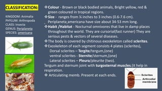
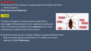
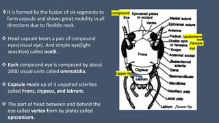
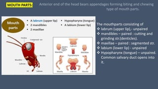
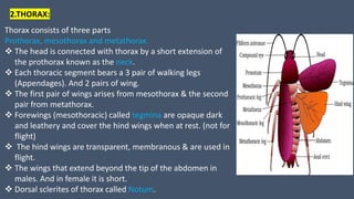
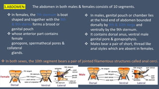
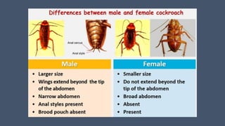
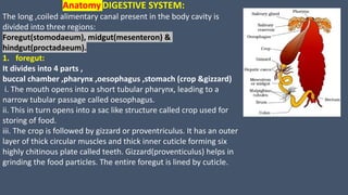
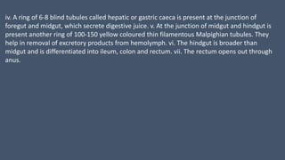
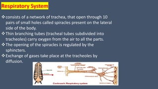
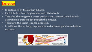
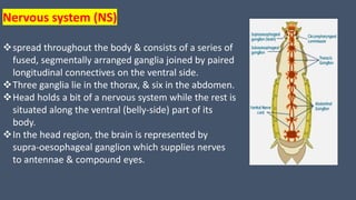
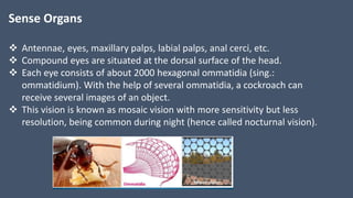
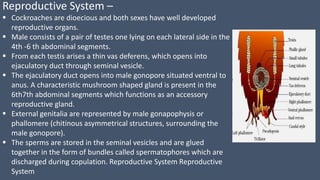
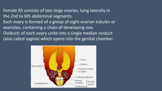
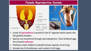
COCKROACH
(Periplaneta americana)
BY MINAXI PATIL
CLASSIFICATION:
KINGDOM: Animalia
PHYLUM: Arthropoda
CLASS: Insecta
GENUS: Periplaneta
SPECIES: americana
Colour - Brown or black bodied animals, Bright yellow, red &
green coloured in tropical regions.
Size - ranges from ¼ inches to 3 inches (0.6-7.6 cm).
Periplaneta americana have size about 34-53 mm long.
Habit /Habitat - Nocturnal omnivores that live in damp places
throughout the world. They are cursorial(fast runner) They are
serious pests & vectors of several diseases.
The body is covered by chitinous exoskeleton called sclerites.
Exoskeleton of each segment consists 4 plates (sclerites),
Dorsal sclerites – Tergite/tergum,(one)
ventral sclerites - Sternite/sternum,(one)
Lateral sclerites – Pleura/pleurite (two).
Tergum and sternum joint with turgosternal muscles.(it help in
respiration.
 Articulating memb. Present at each ends.
Morphology :
 The body of the cockroach is segmented and divisible into three
distinct regions –
Head, thorax and abdomen.
1. HEAD:
 Head is triangular in shape and lies anteriorly at
right angles (Perpendicular) to the longitudinal body axis.
A pair of long thread (filiform) like antennae arises from
membranous sockets lying in front of eyes.
 Antennae have sensory receptor, thigmo receptor,(vibration) that
help in monitoring the environment. It is made up of many
segments called Podomeres.
compound
/Simple
eye
/podomere
/lower lip
Upper lip
It is formed by the fusion of six segments to
form capsule and shows great mobility in all
directions due to flexible neck.
 Head capsule bears a pair of compound
eyes(visual eye). And simple eye(light
sensitive) called ocelli.
 Each compound eye is composed by about
2000 visual units called ommatidia.
 Capsule made up of 3 unpaired sclerites
called Frons, clypeus, and labrum.
 The part of head between and behind the
eye called vertex form by plates called
epicranium.
MOUTH PARTS:
The mouthparts consisting of
 labrum (upper lip),- unpaired
 mandibles – paired : cutting and
grinding str.(denticles).
 maxillae – paired : segmented str.
 labium (lower lip) - unpaired
 Hypopharynx (tongue) – unpaired.
Common salivary duct opens into
it.
Anterior end of the head bears appendages forming biting and chewing
type of mouth parts.
2.THORAX:
Thorax consists of three parts
Prothorax, mesothorax and metathorax.
 The head is connected with thorax by a short extension of
the prothorax known as the neck.
 Each thoracic segment bears a 3 pair of walking legs
(Appendages). And 2 pairs of wing.
 The first pair of wings arises from mesothorax & the second
pair from metathorax.
 Forewings (mesothoracic) called tegmina are opaque dark
and leathery and cover the hind wings when at rest. (not for
flight)
 The hind wings are transparent, membranous & are used in
flight.
 The wings that extend beyond the tip of the abdomen in
males. And in female it is short.
 Dorsal sclerites of thorax called Notum.
3.ABDOMEN: The abdomen in both males & females consists of 10 segments.
 In females, the 7th sternum is boat
shaped and together with the 8th
& 9th sterna forms a brood or
genital pouch
 whose anterior part contains
female
gonopore, spermathecal pores &
collateral
glands.
 In males, genital pouch or chamber lies
at the hind end of abdomen bounded
dorsally by 9th & 10th terga and
ventrally by the 9th sternum.
 It contains dorsal anus, ventral male
genital pore & gonapophysis.
 Males bear a pair of short, thread like
anal styles which are absent in females.
 In both sexes, the 10th segment bears a pair of jointed filamentous structures called anal cerci.
AnatomyDIGESTIVE SYSTEM:
The long ,coiled alimentary canal present in the body cavity is
divided into three regions:
Foregut(stomodaeum), midgut(mesenteron) &
hindgut(proctadaeum).
1. foregut:
It divides into 4 parts ,
buccal chamber ,pharynx ,oesophagus ,stomach (crop &gizzard)
i. The mouth opens into a short tubular pharynx, leading to a
narrow tubular passage called oesophagus.
ii. This in turn opens into a sac like structure called crop used for
storing of food.
iii. The crop is followed by gizzard or proventriculus. It has an outer
layer of thick circular muscles and thick inner cuticle forming six
highly chitinous plate called teeth. Gizzard(proventiculus) helps in
grinding the food particles. The entire foregut is lined by cuticle.
iv. A ring of 6-8 blind tubules called hepatic or gastric caeca is present at the junction of
foregut and midgut, which secrete digestive juice. v. At the junction of midgut and hindgut is
present another ring of 100-150 yellow coloured thin filamentous Malpighian tubules. They
help in removal of excretory products from hemolymph. vi. The hindgut is broader than
midgut and is differentiated into ileum, colon and rectum. vii. The rectum opens out through
anus.
 An open type.
 Blood vessels are poorly developed and open into space
(haemocoel).
 Visceral organs located in the haemocoel are bathed in blood
(haemolymph).
 The haemolymph is composed of colorless plasma and haemocytes.
 Heart of cockroach consists of elongated muscular tube lying along
mid dorsal line of thorax and abdomen.
 It is differentiated into funnel shaped chambers with ostia on either
side.
 Blood from sinuses enter heart through ostia & is pumped anteriorly
to sinuses again.
Blood Vascular System
Respiratory System
consists of a network of trachea, that open through 10
pairs of small holes called spiracles present on the lateral
side of the body.
Thin branching tubes (tracheal tubes subdivided into
tracheoles) carry oxygen from the air to all the parts.
The opening of the spiracles is regulated by the
sphincters.
Exchange of gases take place at the tracheoles by
diffusion.
Excretion
• Is performed by Malpighian tubules.
• Each tubule is lined by glandular and ciliated cells.
• They absorb nitrogenous waste products and convert them into uric
acid which is excreted out through the hindgut.
• Therefore, this insect is called uricotelic.
• In addition, the fat body, nephrocytes and urecose glands also help in
excretion.
Nervous system (NS)
spread throughout the body & consists of a series of
fused, segmentally arranged ganglia joined by paired
longitudinal connectives on the ventral side.
Three ganglia lie in the thorax, & six in the abdomen.
Head holds a bit of a nervous system while the rest is
situated along the ventral (belly-side) part of its
body.
In the head region, the brain is represented by
supra-oesophageal ganglion which supplies nerves
to antennae & compound eyes.
Sense Organs
 Antennae, eyes, maxillary palps, labial palps, anal cerci, etc.
 Compound eyes are situated at the dorsal surface of the head.
 Each eye consists of about 2000 hexagonal ommatidia (sing.:
ommatidium). With the help of several ommatidia, a cockroach can
receive several images of an object.
 This vision is known as mosaic vision with more sensitivity but less
resolution, being common during night (hence called nocturnal vision).
Reproductive System –
 Cockroaches are dioecious and both sexes have well developed
reproductive organs.
 Male consists of a pair of testes one lying on each lateral side in the
4th -6 th abdominal segments.
 From each testis arises a thin vas deferens, which opens into
ejaculatory duct through seminal vesicle.
 The ejaculatory duct opens into male gonopore situated ventral to
anus. A characteristic mushroom shaped gland is present in the
6th7th abdominal segments which functions as an accessory
reproductive gland.
 External genitalia are represented by male gonapophysis or
phallomere (chitinous asymmetrical structures, surrounding the
male gonopore).
 The sperms are stored in the seminal vesicles and are glued
together in the form of bundles called spermatophores which are
discharged during copulation. Reproductive System Reproductive
System
Female RS consists of two large ovaries, lying laterally in
the 2nd to 6th abdominal segments.
Each ovary is formed of a group of eight ovarian tubules or
ovarioles, containing a chain of developing ova.
Oviducts of each ovary unite into a single median oviduct
(also called vagina) which opens into the genital chamber.
COCKROACH ( structural organization in animal) full pdf

COCKROACH ( structural organization in animal) full pdf

  • 1.
  • 2.
    CLASSIFICATION: KINGDOM: Animalia PHYLUM: Arthropoda CLASS:Insecta GENUS: Periplaneta SPECIES: americana Colour - Brown or black bodied animals, Bright yellow, red & green coloured in tropical regions. Size - ranges from ¼ inches to 3 inches (0.6-7.6 cm). Periplaneta americana have size about 34-53 mm long. Habit /Habitat - Nocturnal omnivores that live in damp places throughout the world. They are cursorial(fast runner) They are serious pests & vectors of several diseases. The body is covered by chitinous exoskeleton called sclerites. Exoskeleton of each segment consists 4 plates (sclerites), Dorsal sclerites – Tergite/tergum,(one) ventral sclerites - Sternite/sternum,(one) Lateral sclerites – Pleura/pleurite (two). Tergum and sternum joint with turgosternal muscles.(it help in respiration.  Articulating memb. Present at each ends.
  • 3.
    Morphology :  Thebody of the cockroach is segmented and divisible into three distinct regions – Head, thorax and abdomen. 1. HEAD:  Head is triangular in shape and lies anteriorly at right angles (Perpendicular) to the longitudinal body axis. A pair of long thread (filiform) like antennae arises from membranous sockets lying in front of eyes.  Antennae have sensory receptor, thigmo receptor,(vibration) that help in monitoring the environment. It is made up of many segments called Podomeres.
  • 4.
    compound /Simple eye /podomere /lower lip Upper lip Itis formed by the fusion of six segments to form capsule and shows great mobility in all directions due to flexible neck.  Head capsule bears a pair of compound eyes(visual eye). And simple eye(light sensitive) called ocelli.  Each compound eye is composed by about 2000 visual units called ommatidia.  Capsule made up of 3 unpaired sclerites called Frons, clypeus, and labrum.  The part of head between and behind the eye called vertex form by plates called epicranium.
  • 5.
    MOUTH PARTS: The mouthpartsconsisting of  labrum (upper lip),- unpaired  mandibles – paired : cutting and grinding str.(denticles).  maxillae – paired : segmented str.  labium (lower lip) - unpaired  Hypopharynx (tongue) – unpaired. Common salivary duct opens into it. Anterior end of the head bears appendages forming biting and chewing type of mouth parts.
  • 6.
    2.THORAX: Thorax consists ofthree parts Prothorax, mesothorax and metathorax.  The head is connected with thorax by a short extension of the prothorax known as the neck.  Each thoracic segment bears a 3 pair of walking legs (Appendages). And 2 pairs of wing.  The first pair of wings arises from mesothorax & the second pair from metathorax.  Forewings (mesothoracic) called tegmina are opaque dark and leathery and cover the hind wings when at rest. (not for flight)  The hind wings are transparent, membranous & are used in flight.  The wings that extend beyond the tip of the abdomen in males. And in female it is short.  Dorsal sclerites of thorax called Notum.
  • 7.
    3.ABDOMEN: The abdomenin both males & females consists of 10 segments.  In females, the 7th sternum is boat shaped and together with the 8th & 9th sterna forms a brood or genital pouch  whose anterior part contains female gonopore, spermathecal pores & collateral glands.  In males, genital pouch or chamber lies at the hind end of abdomen bounded dorsally by 9th & 10th terga and ventrally by the 9th sternum.  It contains dorsal anus, ventral male genital pore & gonapophysis.  Males bear a pair of short, thread like anal styles which are absent in females.  In both sexes, the 10th segment bears a pair of jointed filamentous structures called anal cerci.
  • 9.
    AnatomyDIGESTIVE SYSTEM: The long,coiled alimentary canal present in the body cavity is divided into three regions: Foregut(stomodaeum), midgut(mesenteron) & hindgut(proctadaeum). 1. foregut: It divides into 4 parts , buccal chamber ,pharynx ,oesophagus ,stomach (crop &gizzard) i. The mouth opens into a short tubular pharynx, leading to a narrow tubular passage called oesophagus. ii. This in turn opens into a sac like structure called crop used for storing of food. iii. The crop is followed by gizzard or proventriculus. It has an outer layer of thick circular muscles and thick inner cuticle forming six highly chitinous plate called teeth. Gizzard(proventiculus) helps in grinding the food particles. The entire foregut is lined by cuticle.
  • 10.
    iv. A ringof 6-8 blind tubules called hepatic or gastric caeca is present at the junction of foregut and midgut, which secrete digestive juice. v. At the junction of midgut and hindgut is present another ring of 100-150 yellow coloured thin filamentous Malpighian tubules. They help in removal of excretory products from hemolymph. vi. The hindgut is broader than midgut and is differentiated into ileum, colon and rectum. vii. The rectum opens out through anus.
  • 11.
     An opentype.  Blood vessels are poorly developed and open into space (haemocoel).  Visceral organs located in the haemocoel are bathed in blood (haemolymph).  The haemolymph is composed of colorless plasma and haemocytes.  Heart of cockroach consists of elongated muscular tube lying along mid dorsal line of thorax and abdomen.  It is differentiated into funnel shaped chambers with ostia on either side.  Blood from sinuses enter heart through ostia & is pumped anteriorly to sinuses again. Blood Vascular System
  • 12.
    Respiratory System consists ofa network of trachea, that open through 10 pairs of small holes called spiracles present on the lateral side of the body. Thin branching tubes (tracheal tubes subdivided into tracheoles) carry oxygen from the air to all the parts. The opening of the spiracles is regulated by the sphincters. Exchange of gases take place at the tracheoles by diffusion.
  • 13.
    Excretion • Is performedby Malpighian tubules. • Each tubule is lined by glandular and ciliated cells. • They absorb nitrogenous waste products and convert them into uric acid which is excreted out through the hindgut. • Therefore, this insect is called uricotelic. • In addition, the fat body, nephrocytes and urecose glands also help in excretion.
  • 14.
    Nervous system (NS) spreadthroughout the body & consists of a series of fused, segmentally arranged ganglia joined by paired longitudinal connectives on the ventral side. Three ganglia lie in the thorax, & six in the abdomen. Head holds a bit of a nervous system while the rest is situated along the ventral (belly-side) part of its body. In the head region, the brain is represented by supra-oesophageal ganglion which supplies nerves to antennae & compound eyes.
  • 15.
    Sense Organs  Antennae,eyes, maxillary palps, labial palps, anal cerci, etc.  Compound eyes are situated at the dorsal surface of the head.  Each eye consists of about 2000 hexagonal ommatidia (sing.: ommatidium). With the help of several ommatidia, a cockroach can receive several images of an object.  This vision is known as mosaic vision with more sensitivity but less resolution, being common during night (hence called nocturnal vision).
  • 16.
    Reproductive System – Cockroaches are dioecious and both sexes have well developed reproductive organs.  Male consists of a pair of testes one lying on each lateral side in the 4th -6 th abdominal segments.  From each testis arises a thin vas deferens, which opens into ejaculatory duct through seminal vesicle.  The ejaculatory duct opens into male gonopore situated ventral to anus. A characteristic mushroom shaped gland is present in the 6th7th abdominal segments which functions as an accessory reproductive gland.  External genitalia are represented by male gonapophysis or phallomere (chitinous asymmetrical structures, surrounding the male gonopore).  The sperms are stored in the seminal vesicles and are glued together in the form of bundles called spermatophores which are discharged during copulation. Reproductive System Reproductive System
  • 17.
    Female RS consistsof two large ovaries, lying laterally in the 2nd to 6th abdominal segments. Each ovary is formed of a group of eight ovarian tubules or ovarioles, containing a chain of developing ova. Oviducts of each ovary unite into a single median oviduct (also called vagina) which opens into the genital chamber.