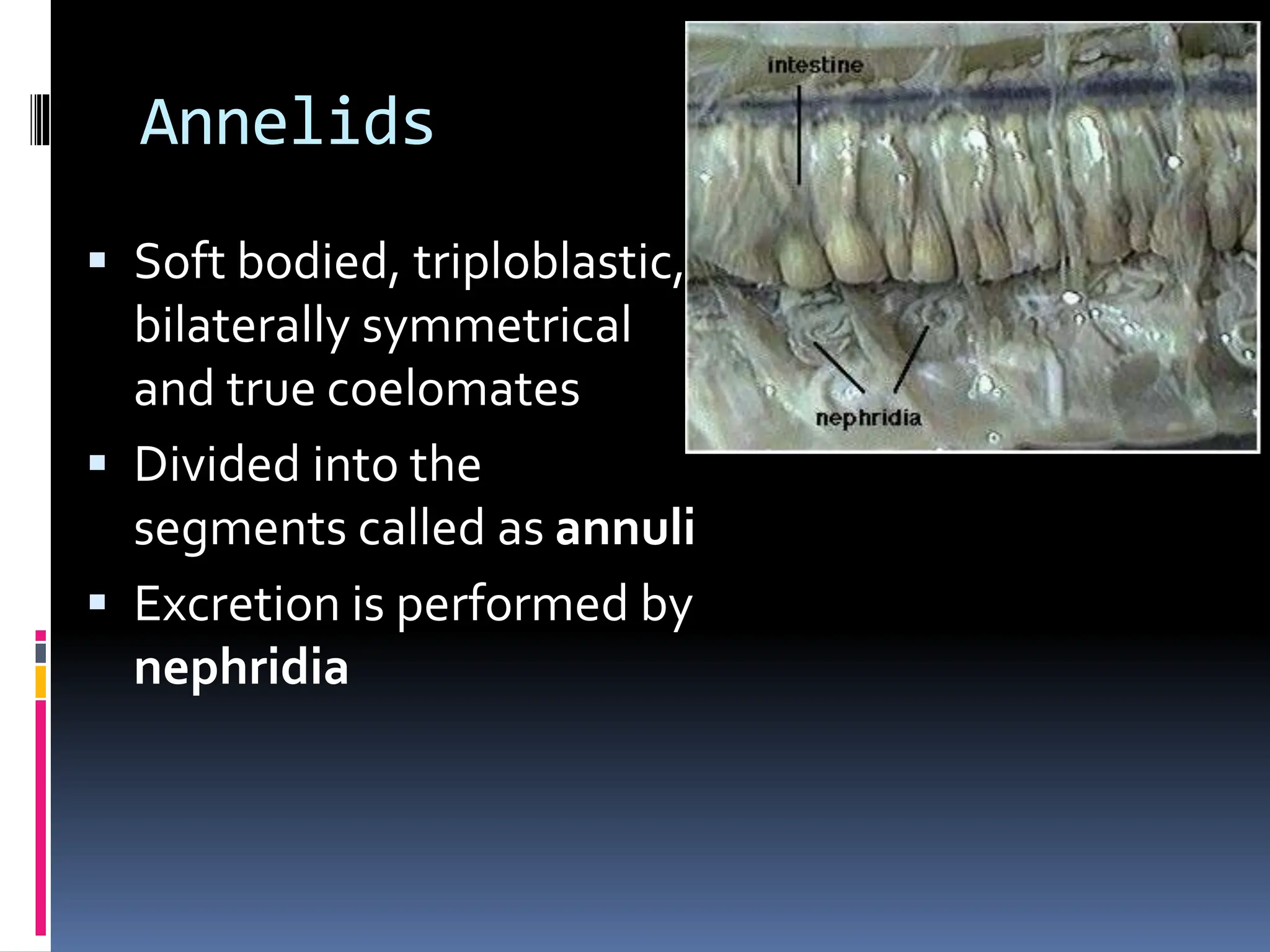
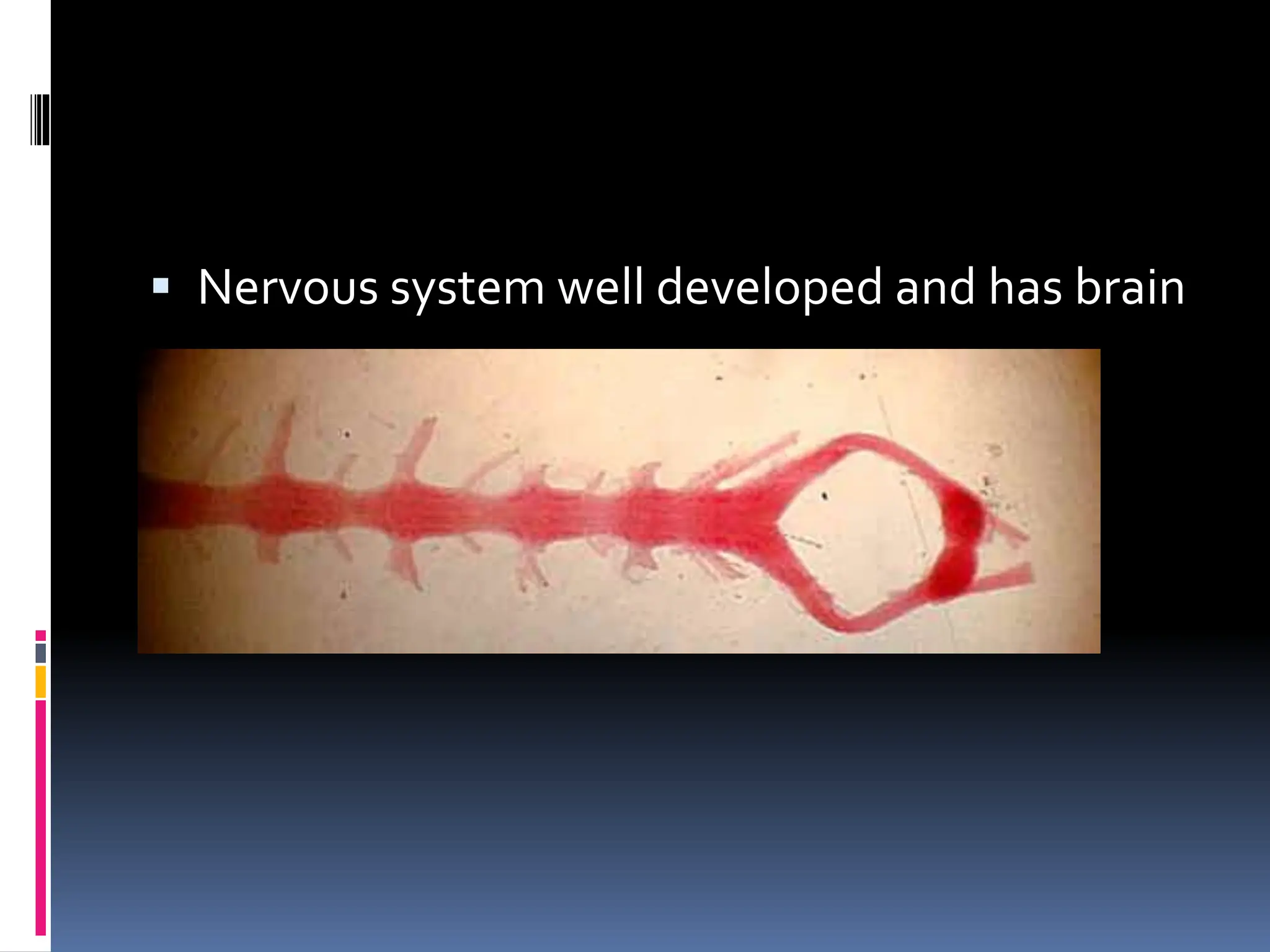
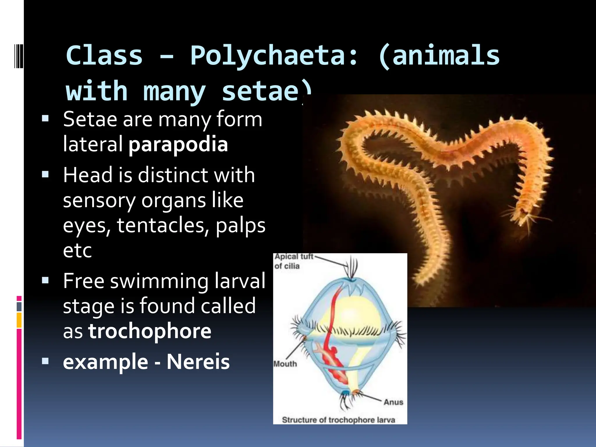
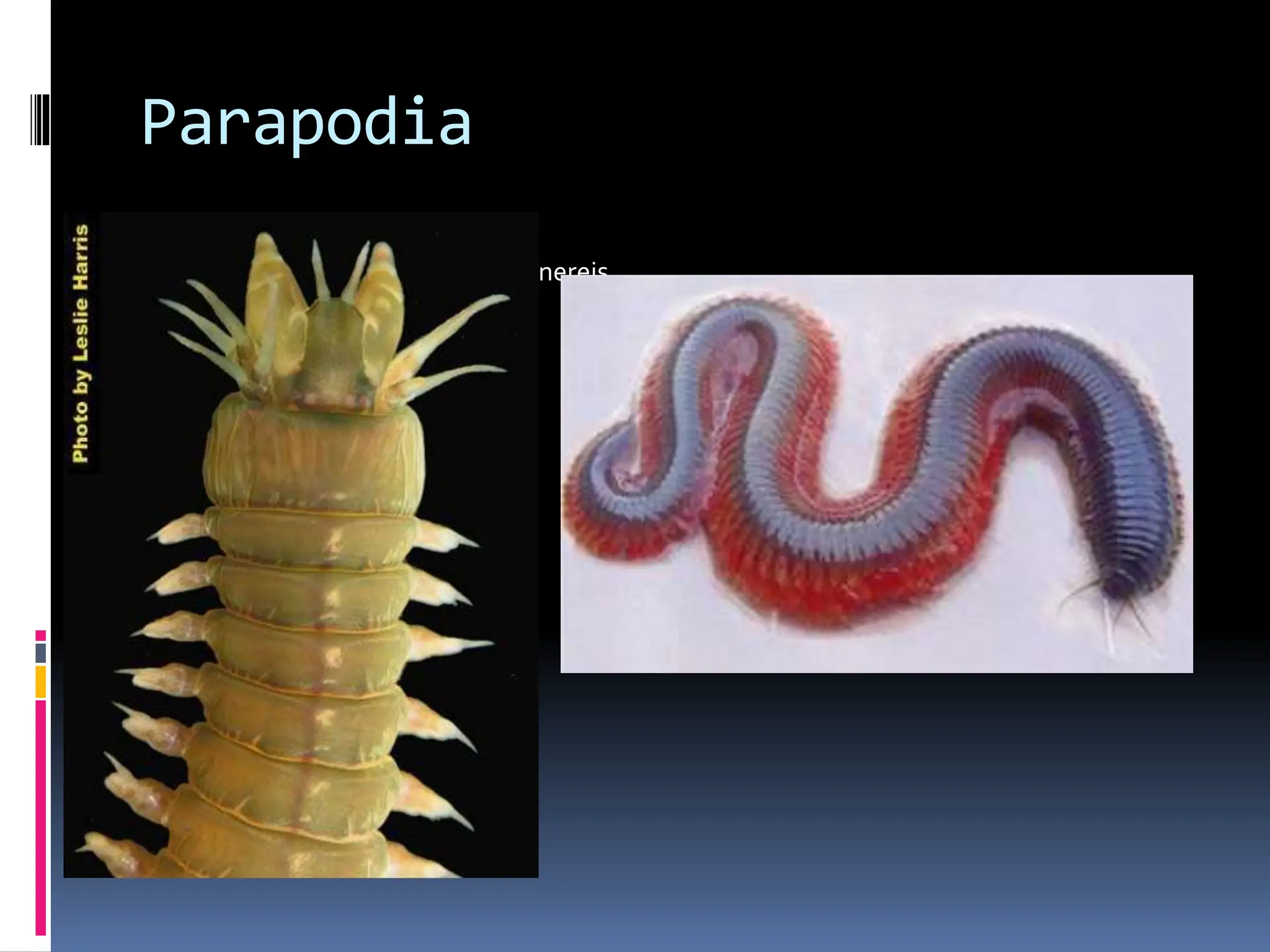
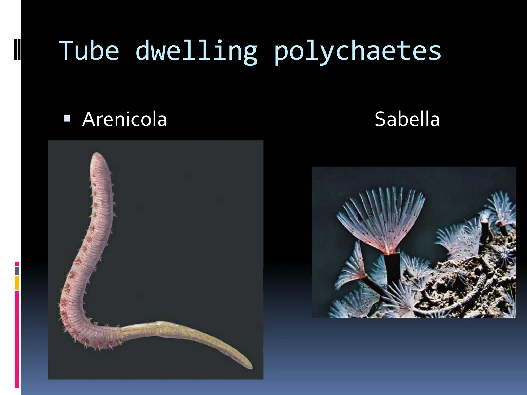
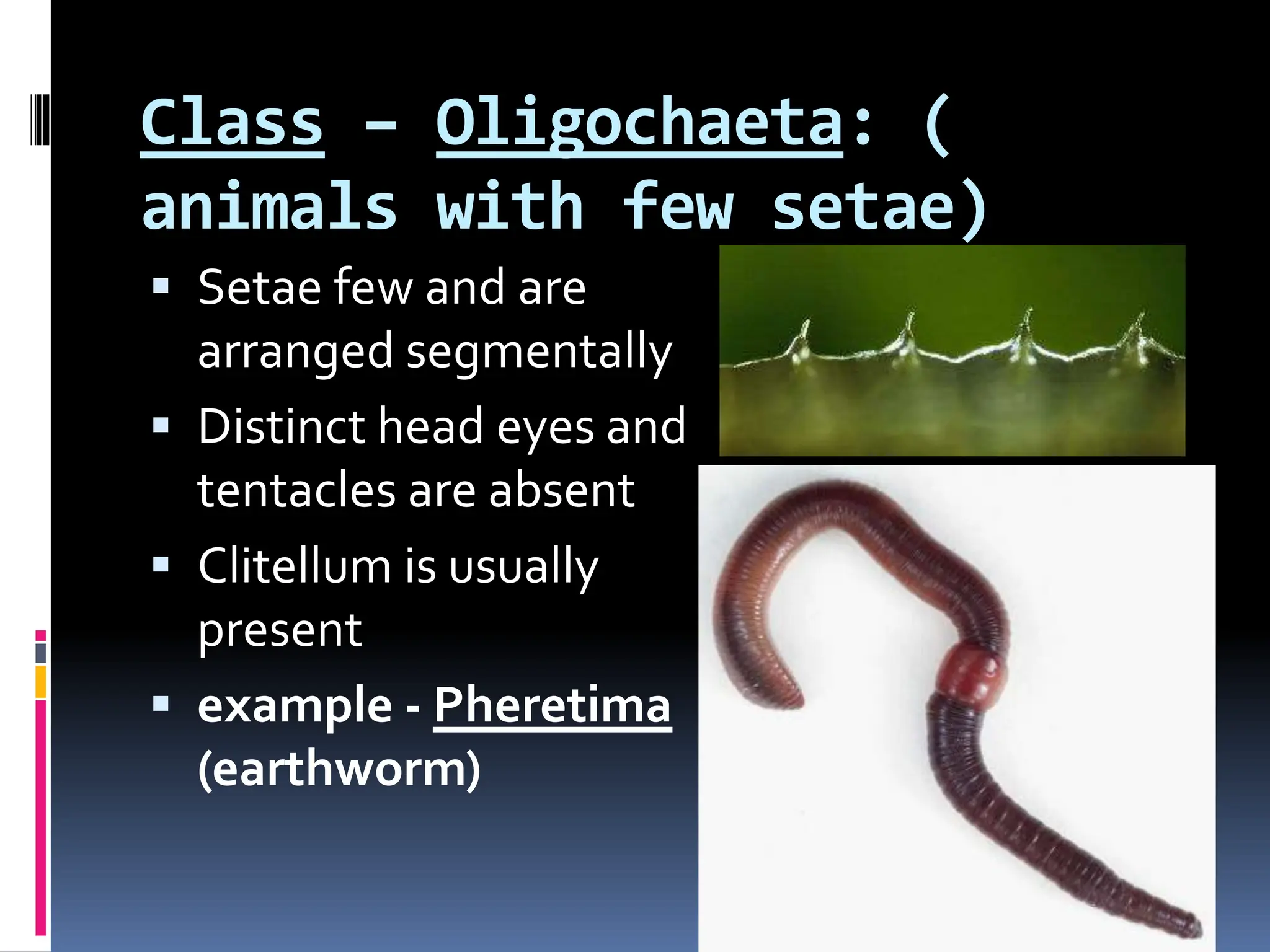
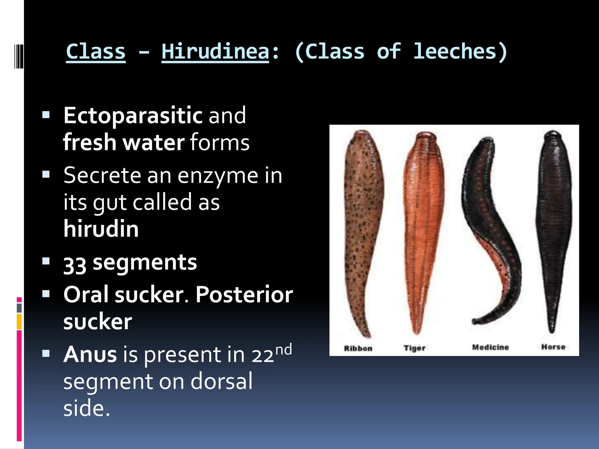
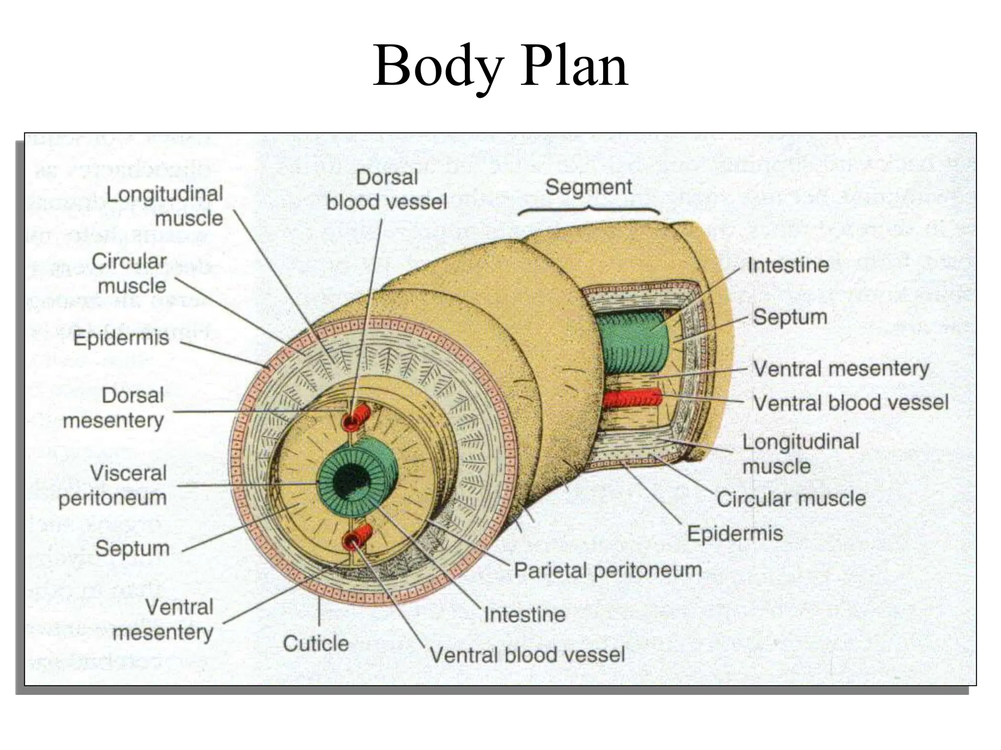
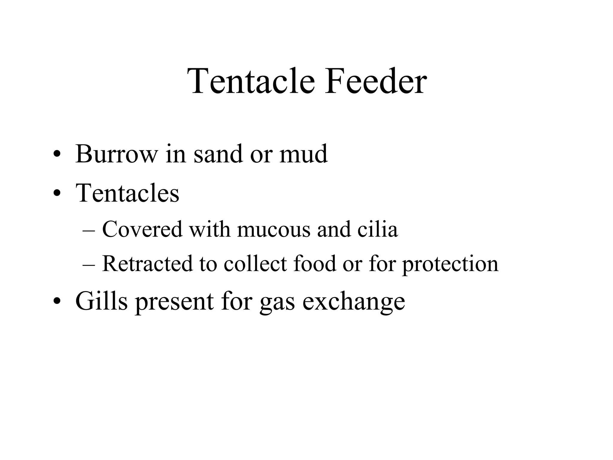
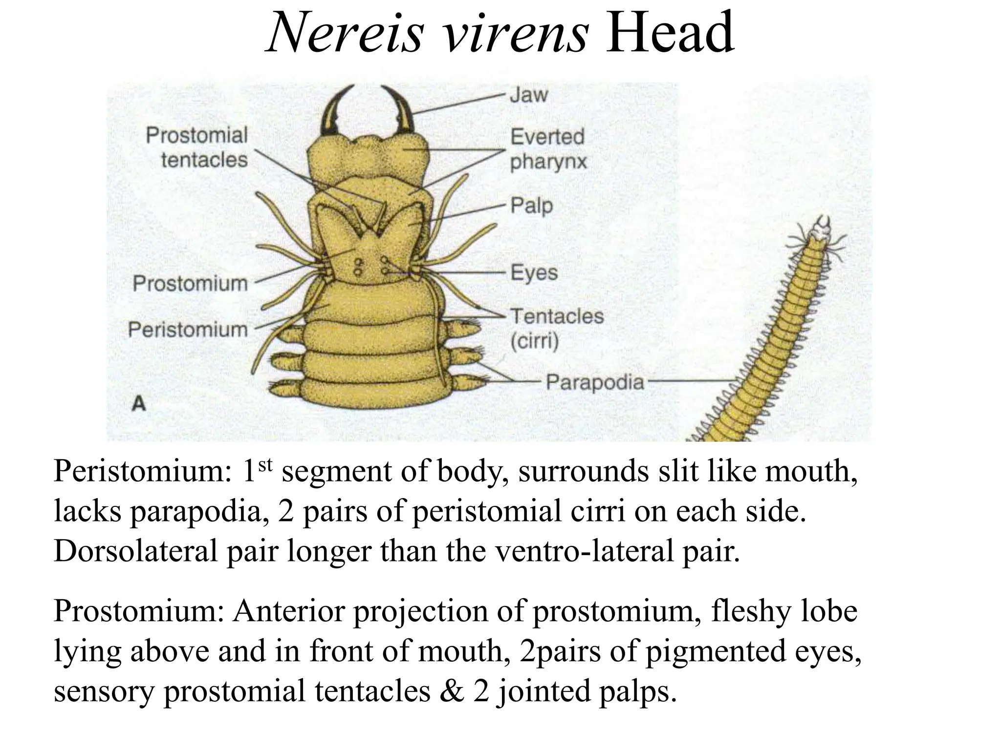
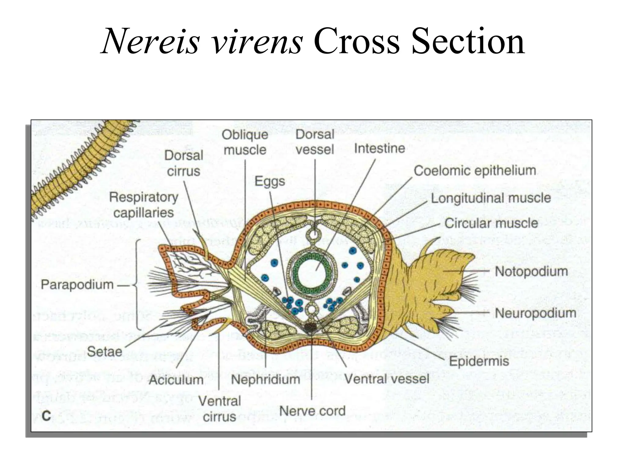
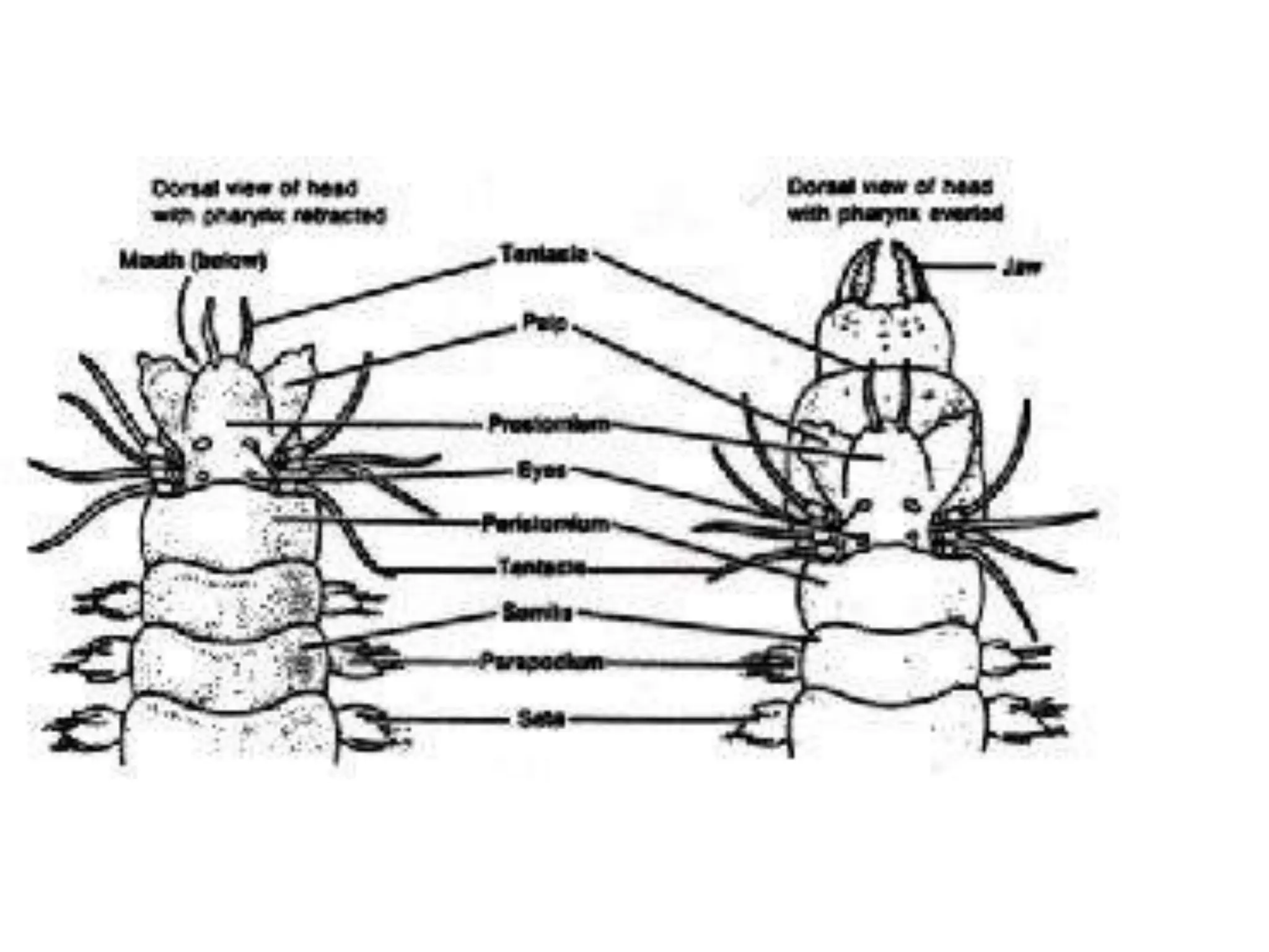
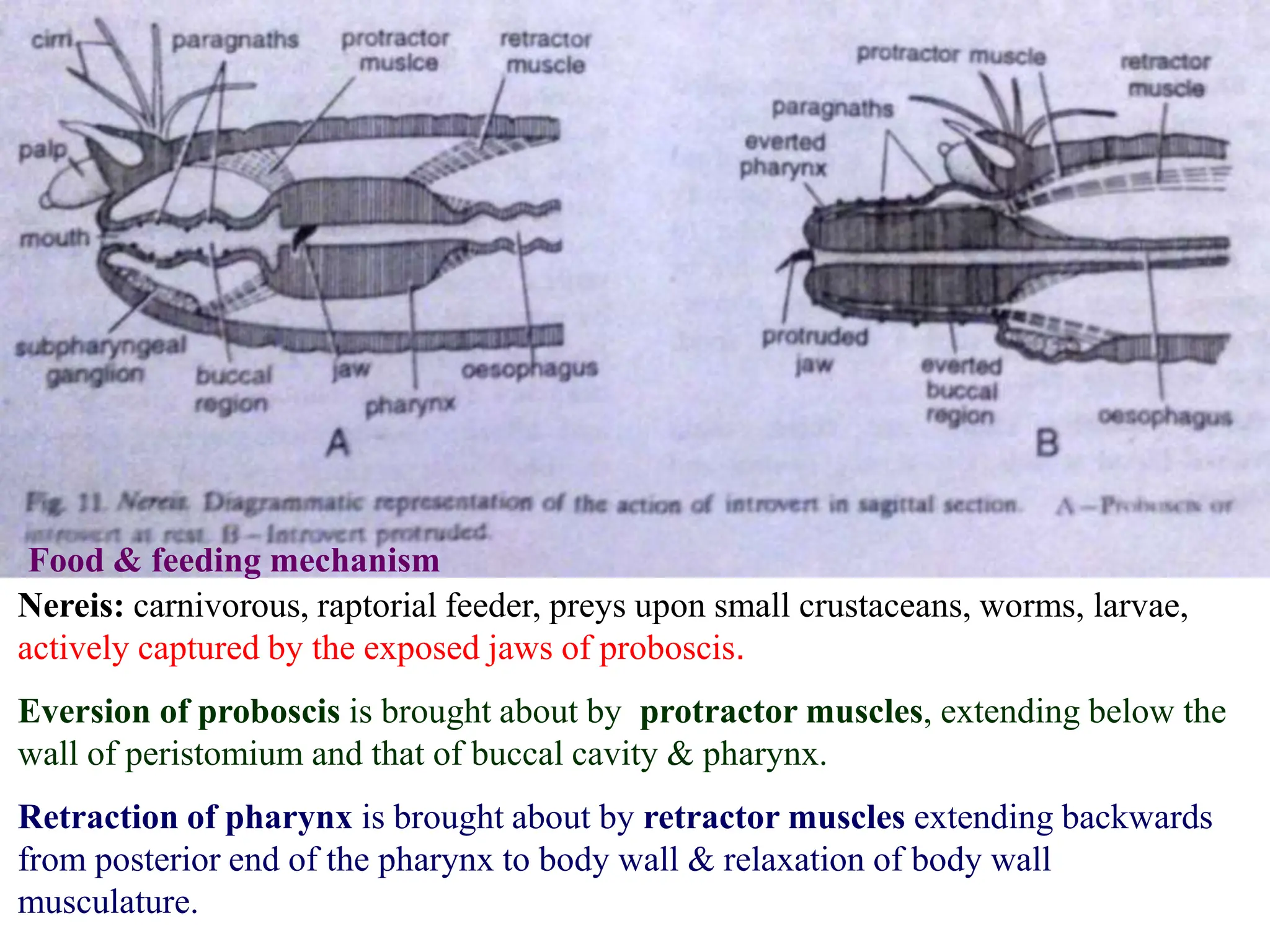
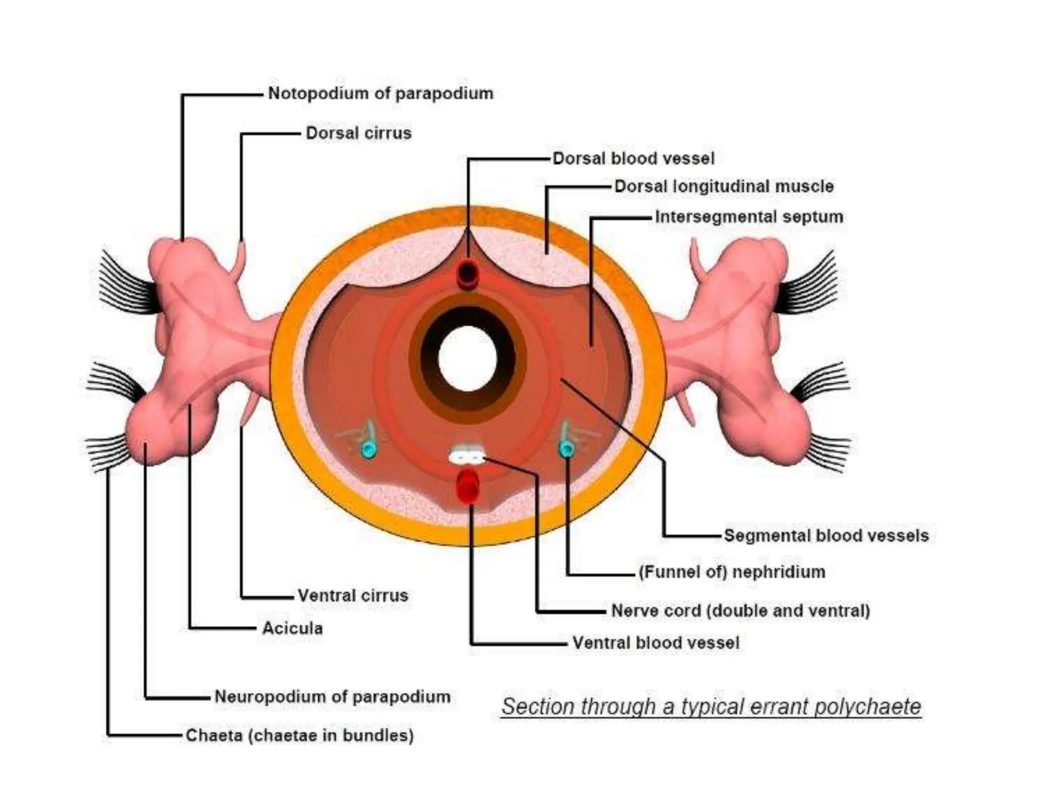
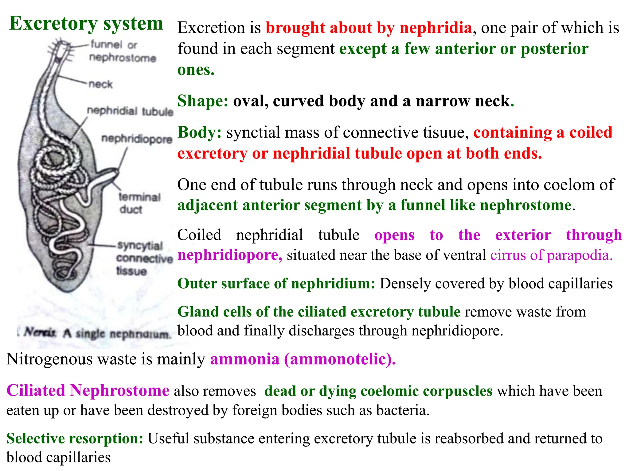
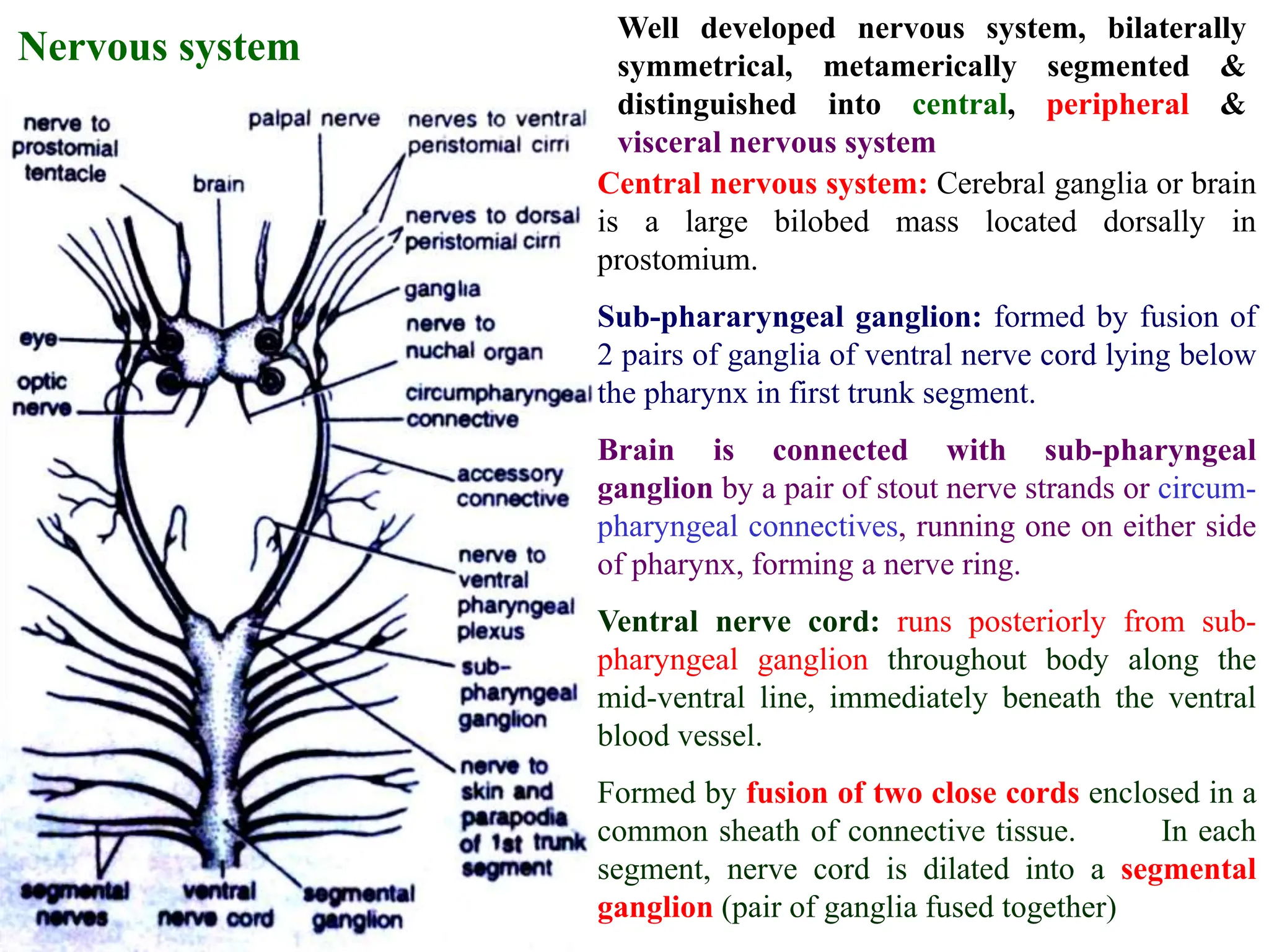
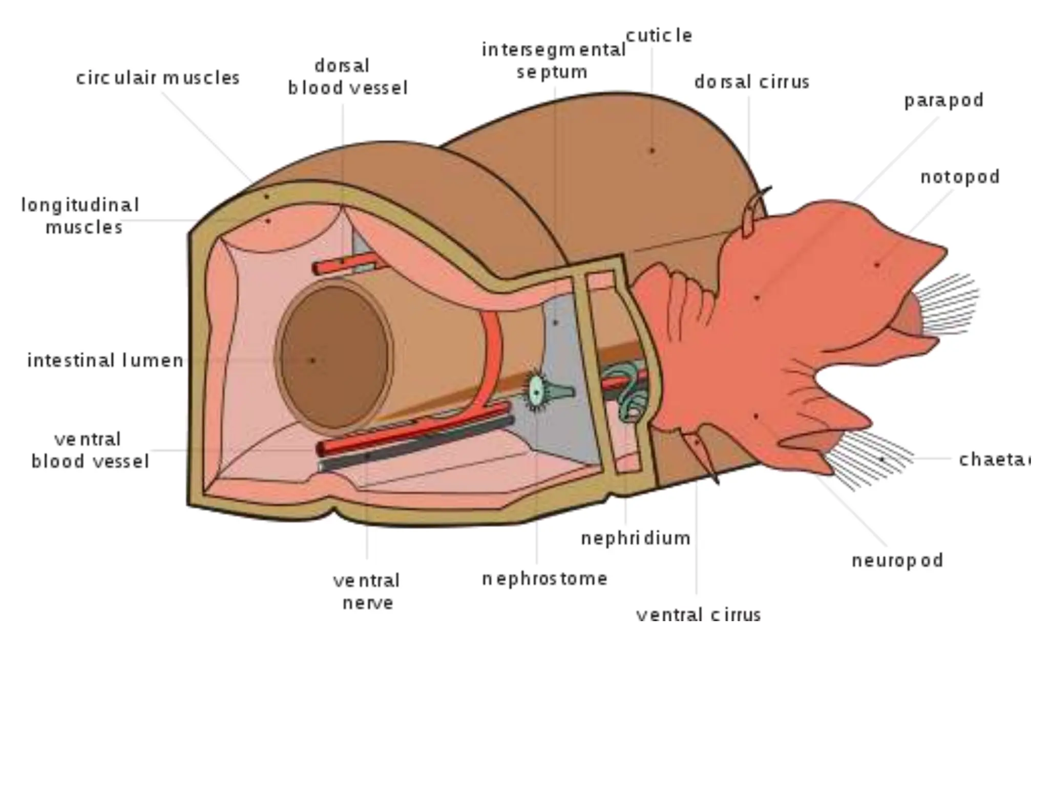
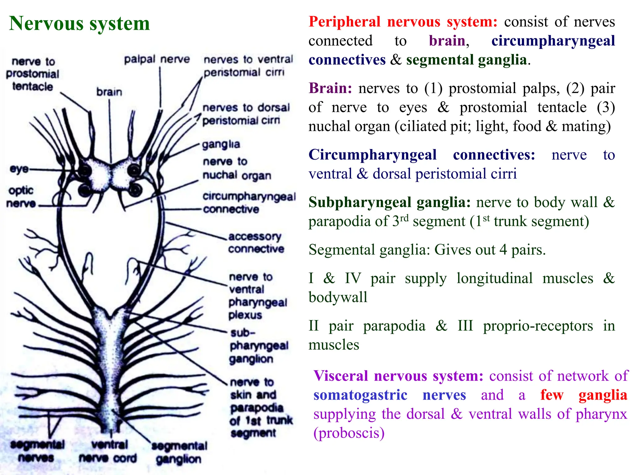
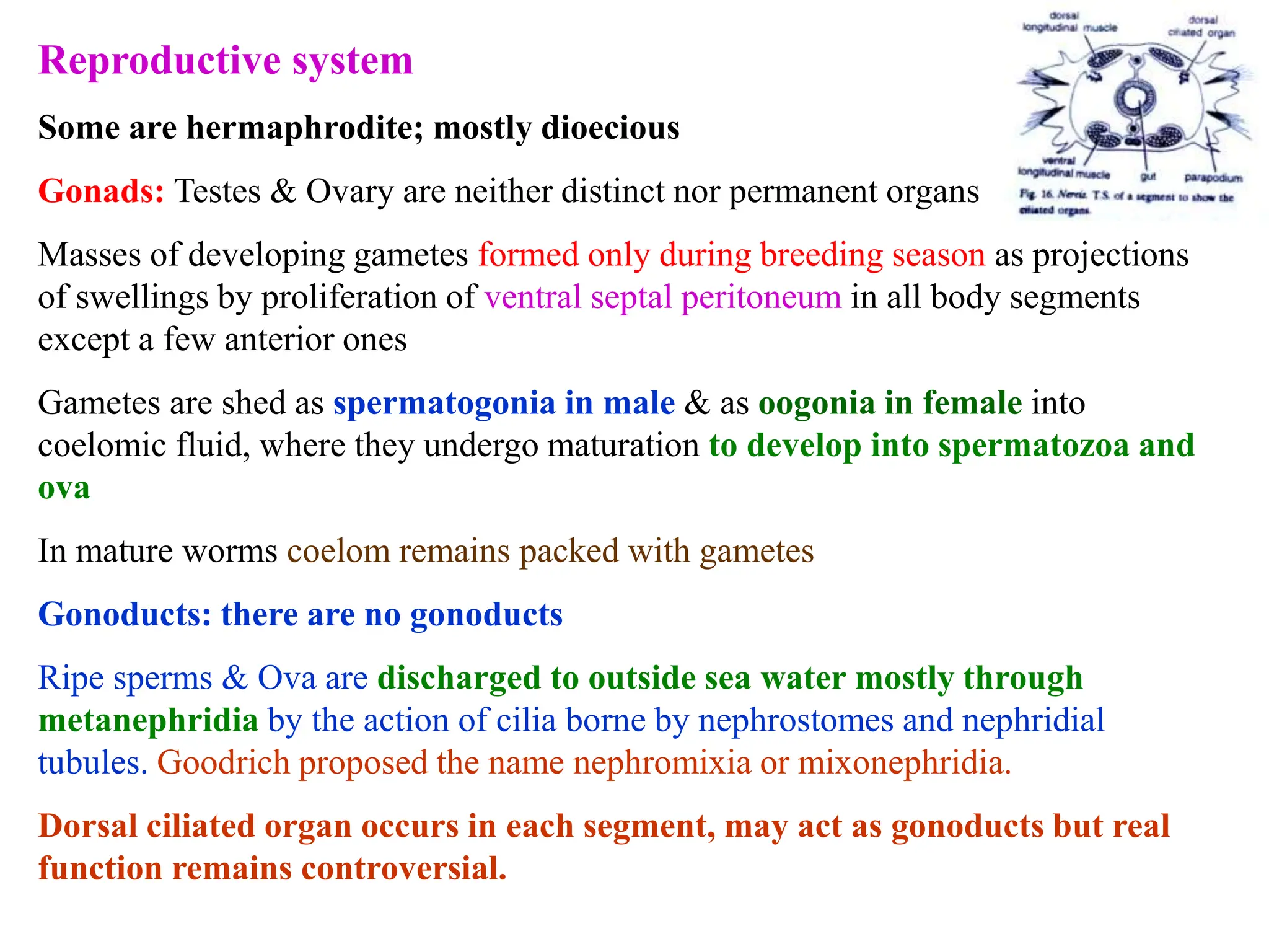
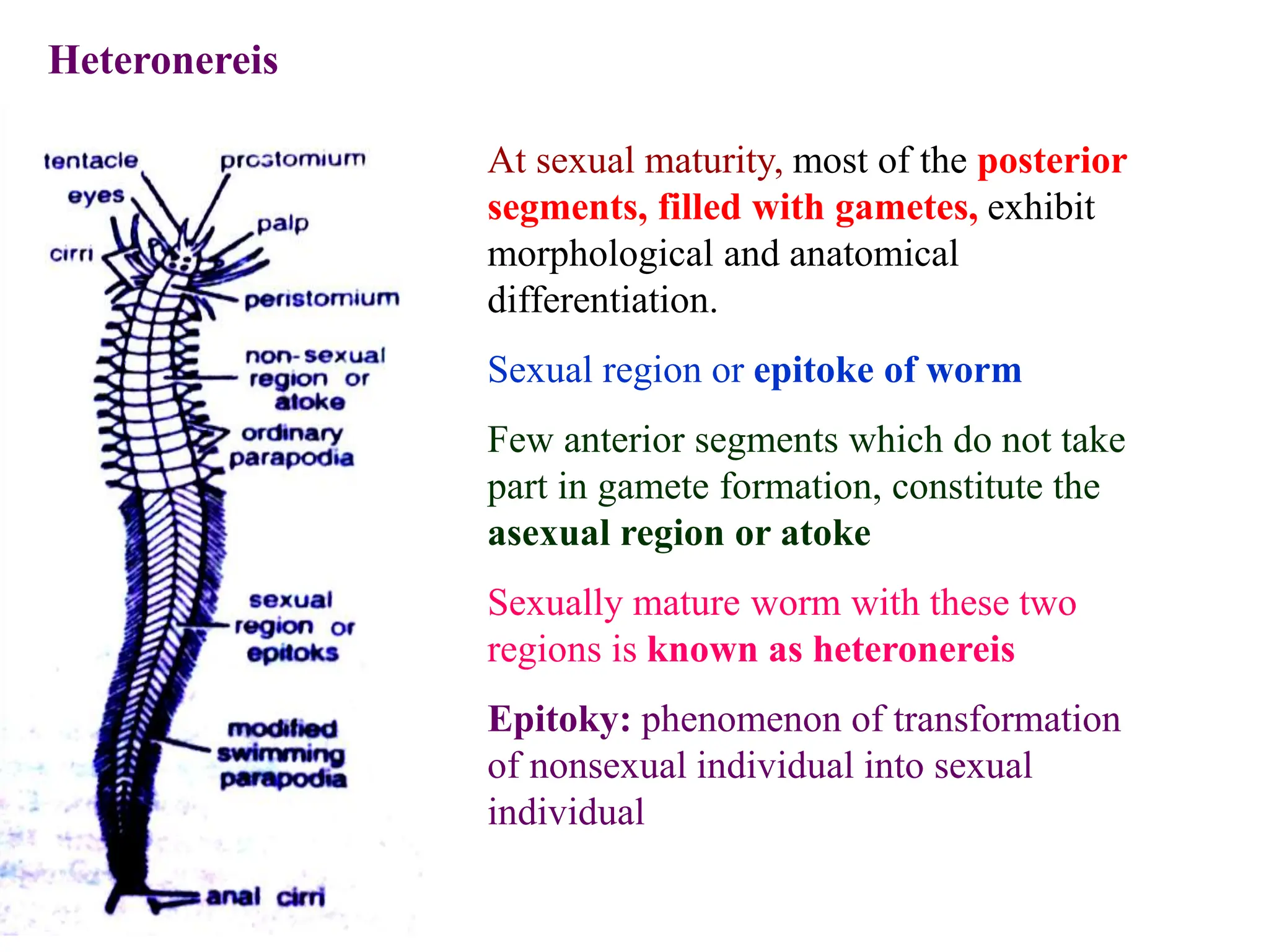
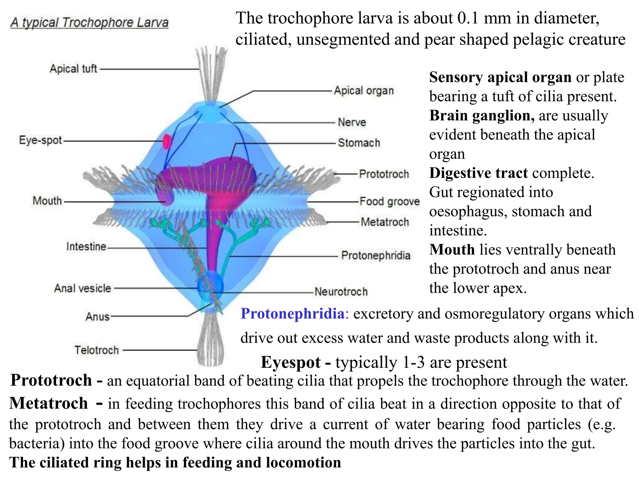
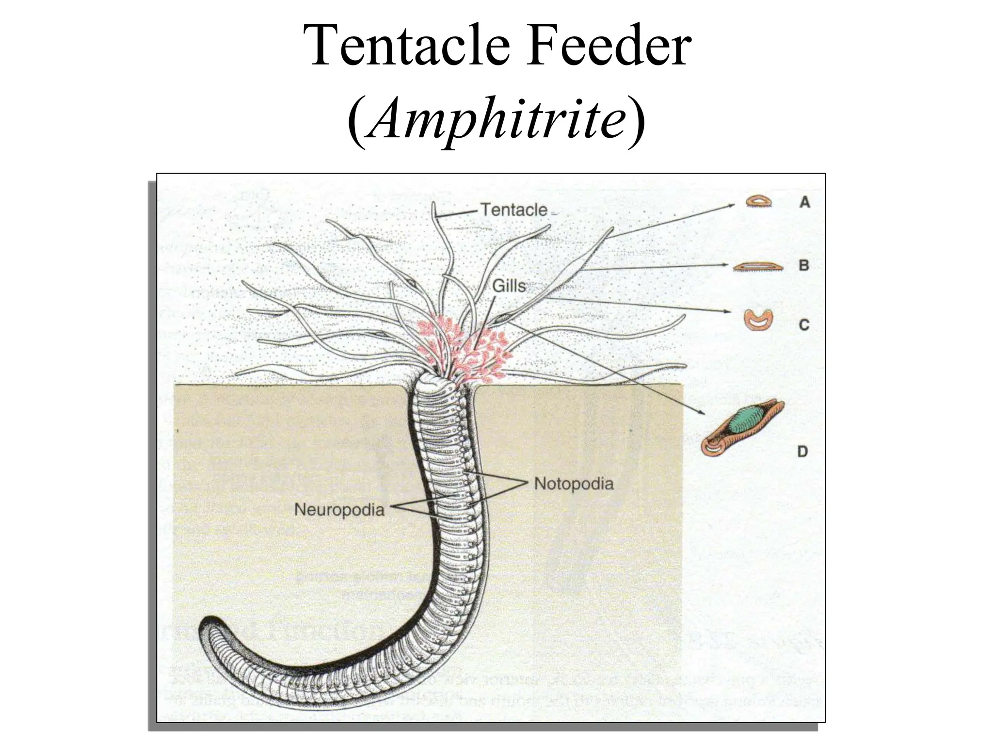
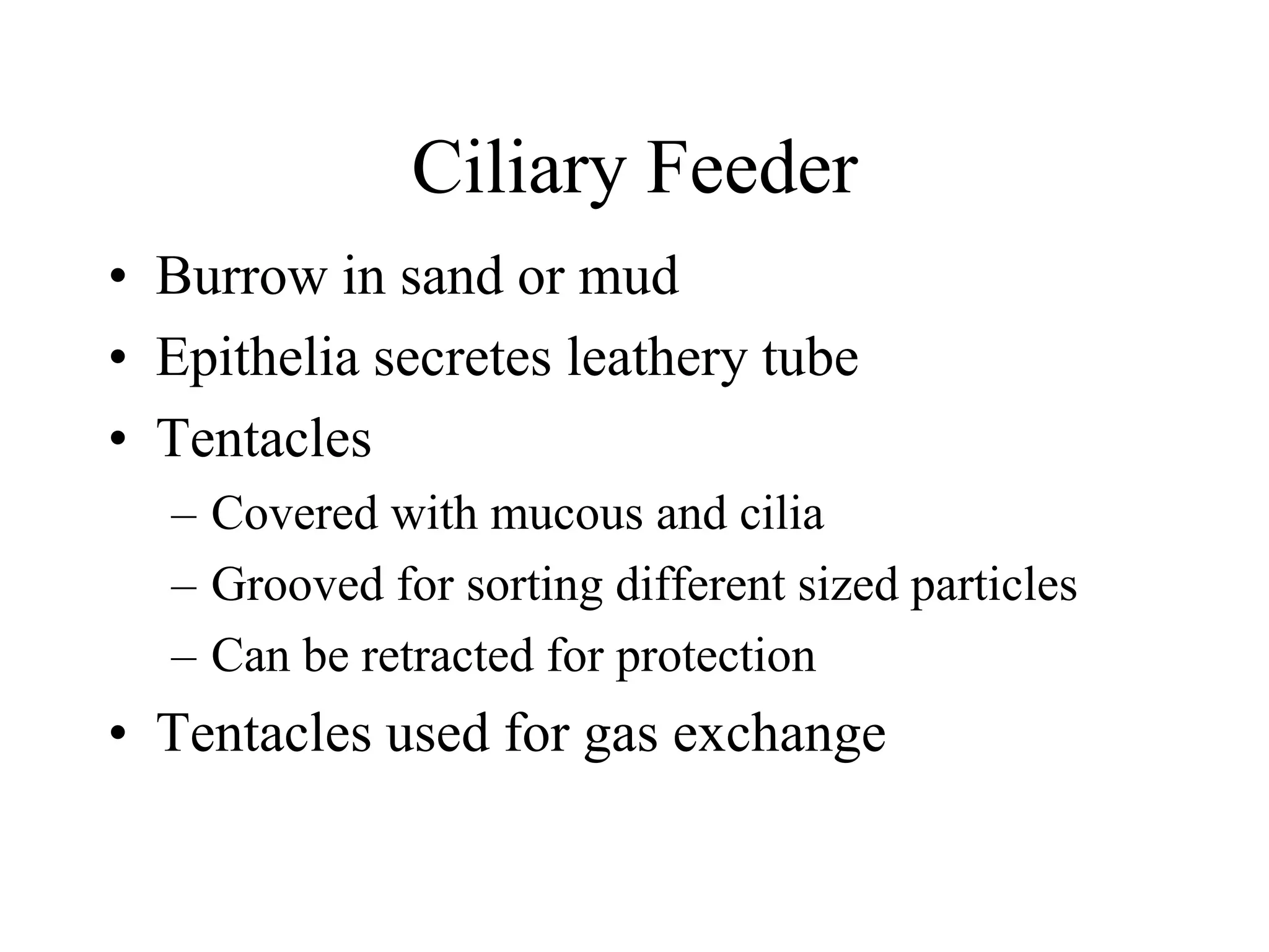
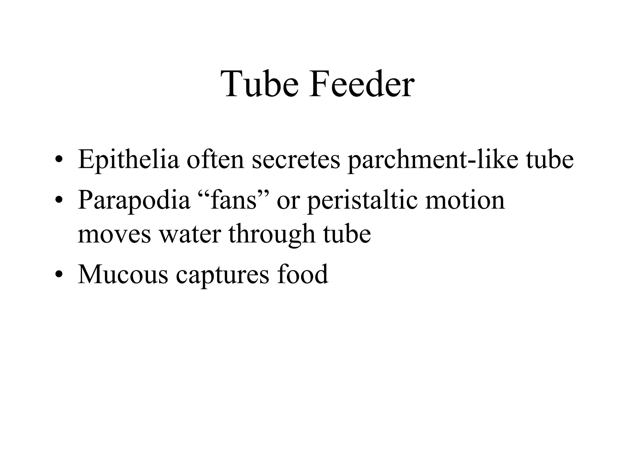
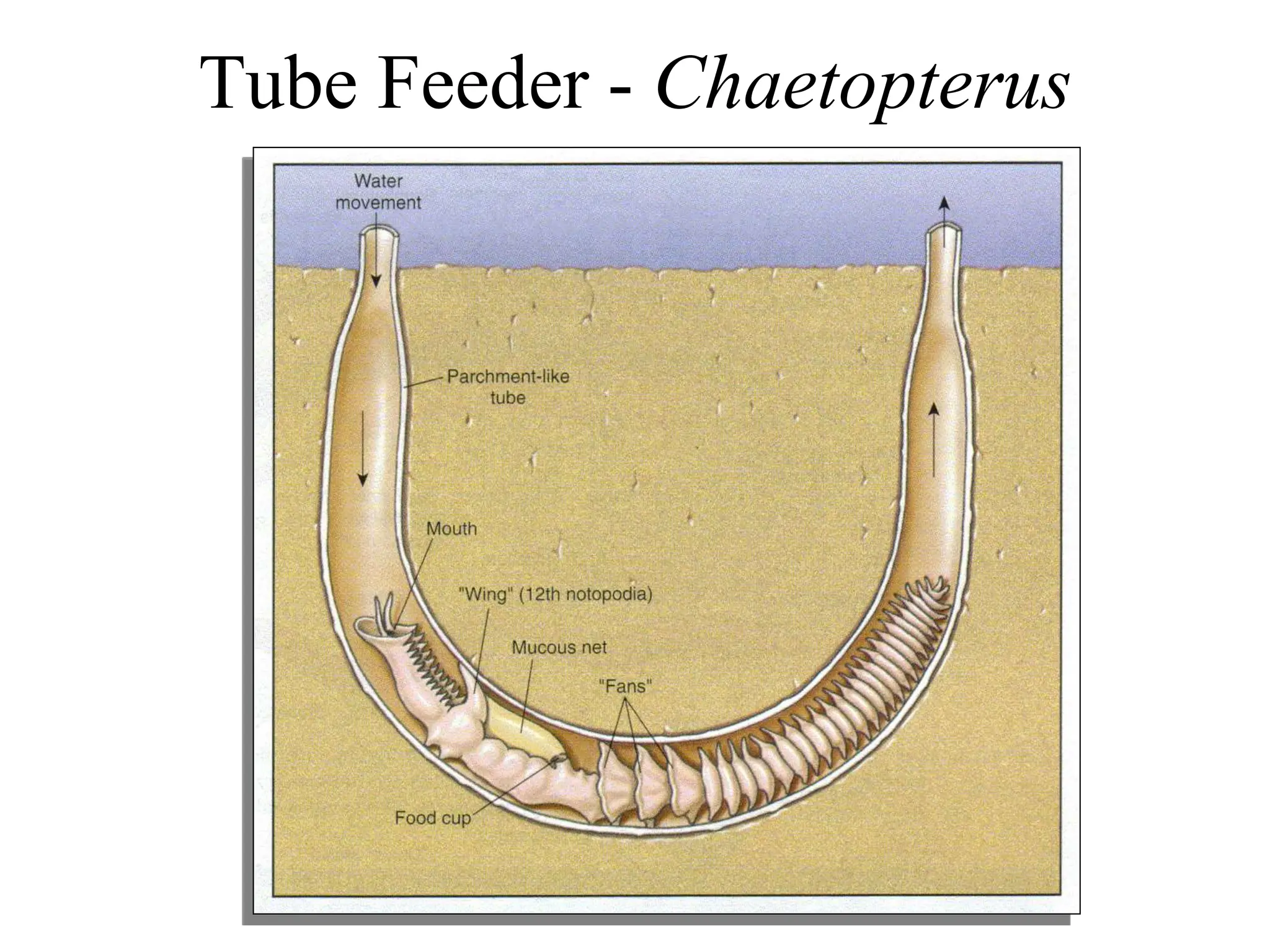
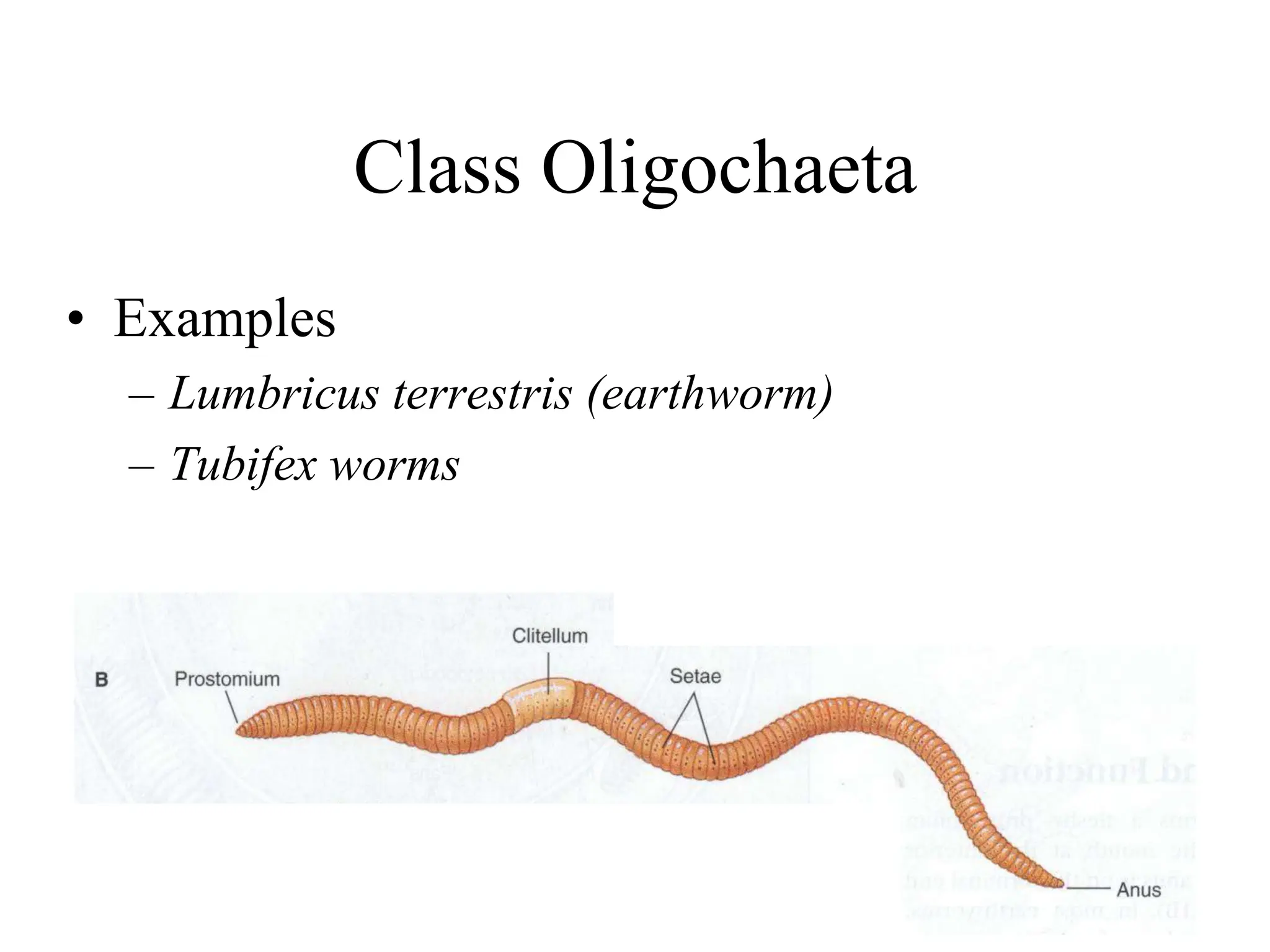
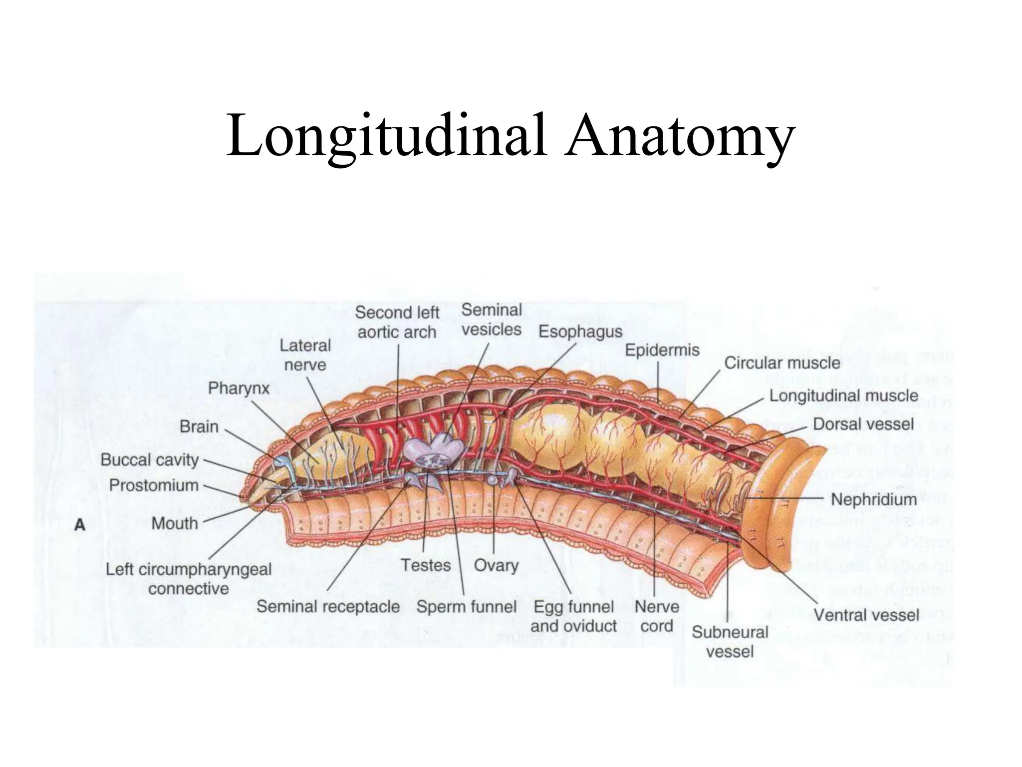
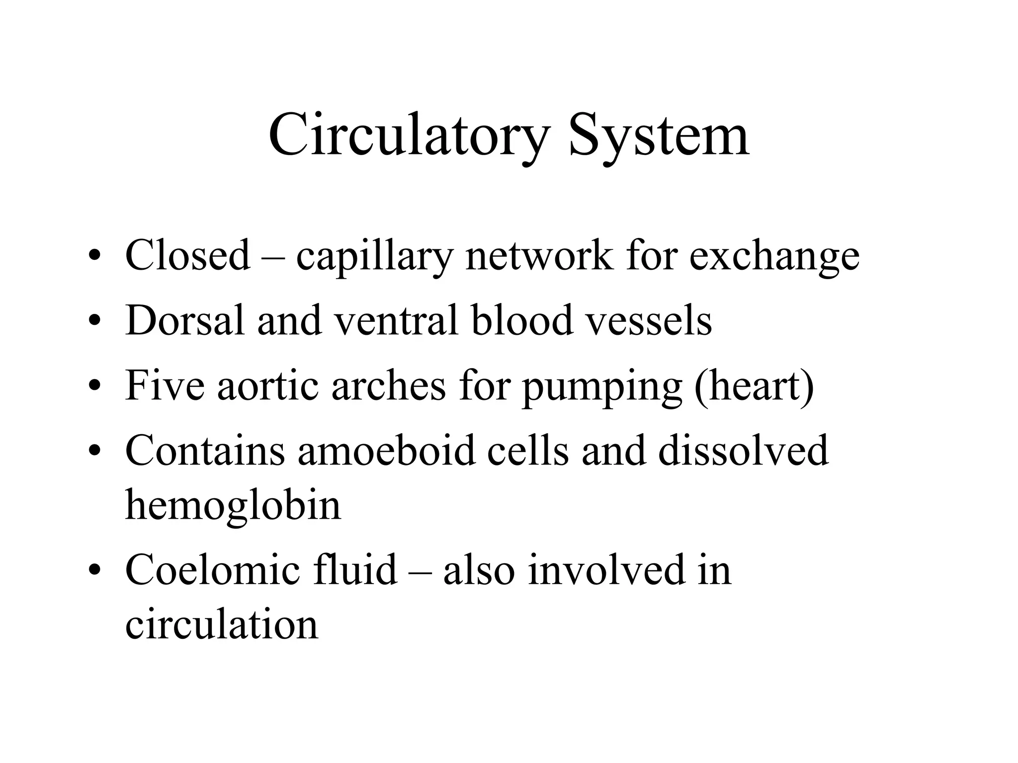
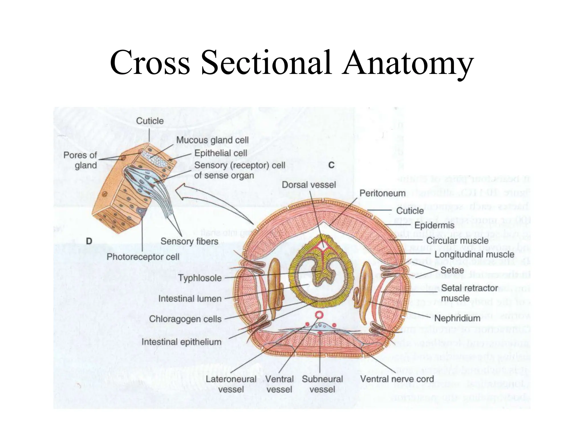
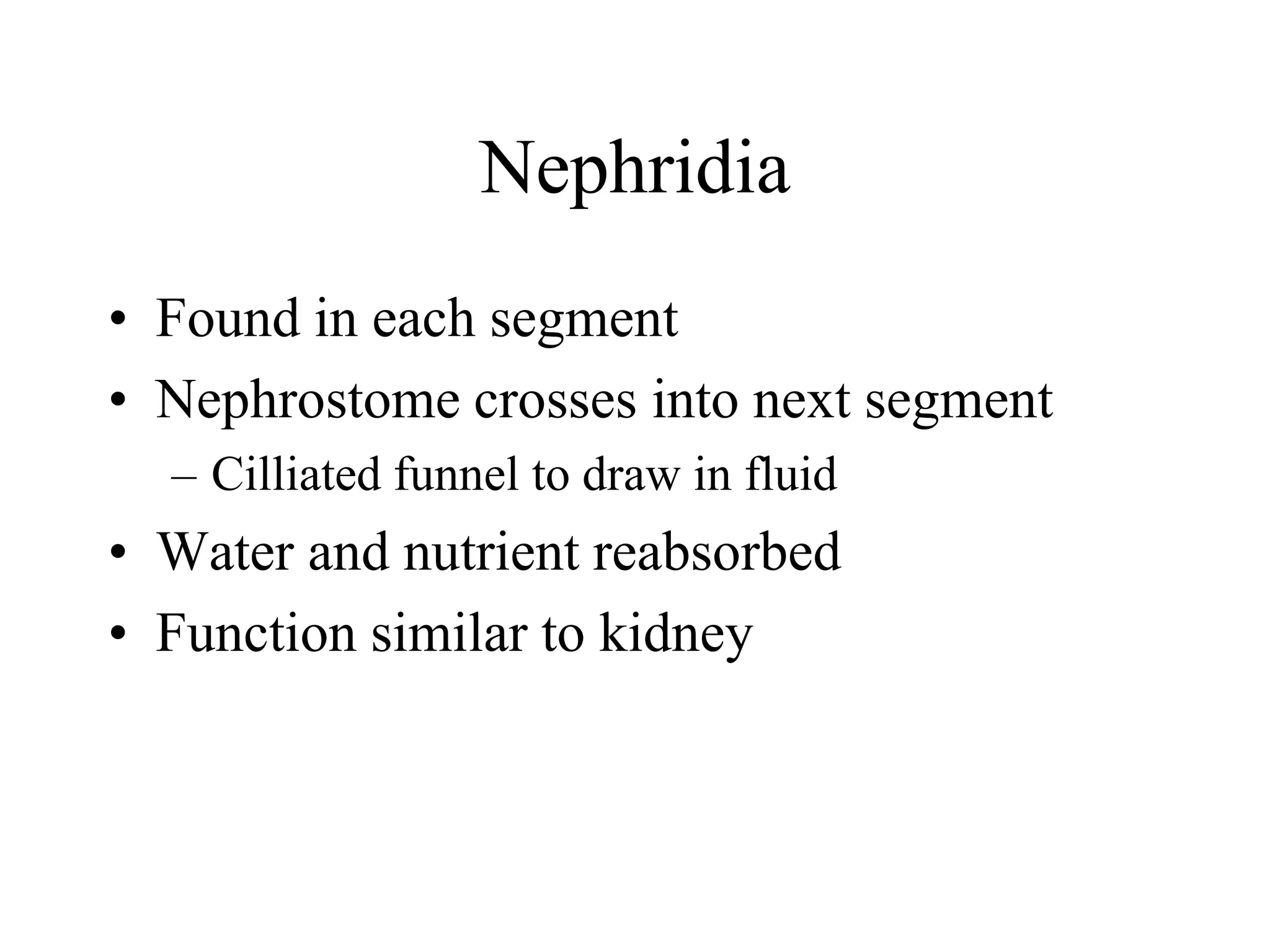
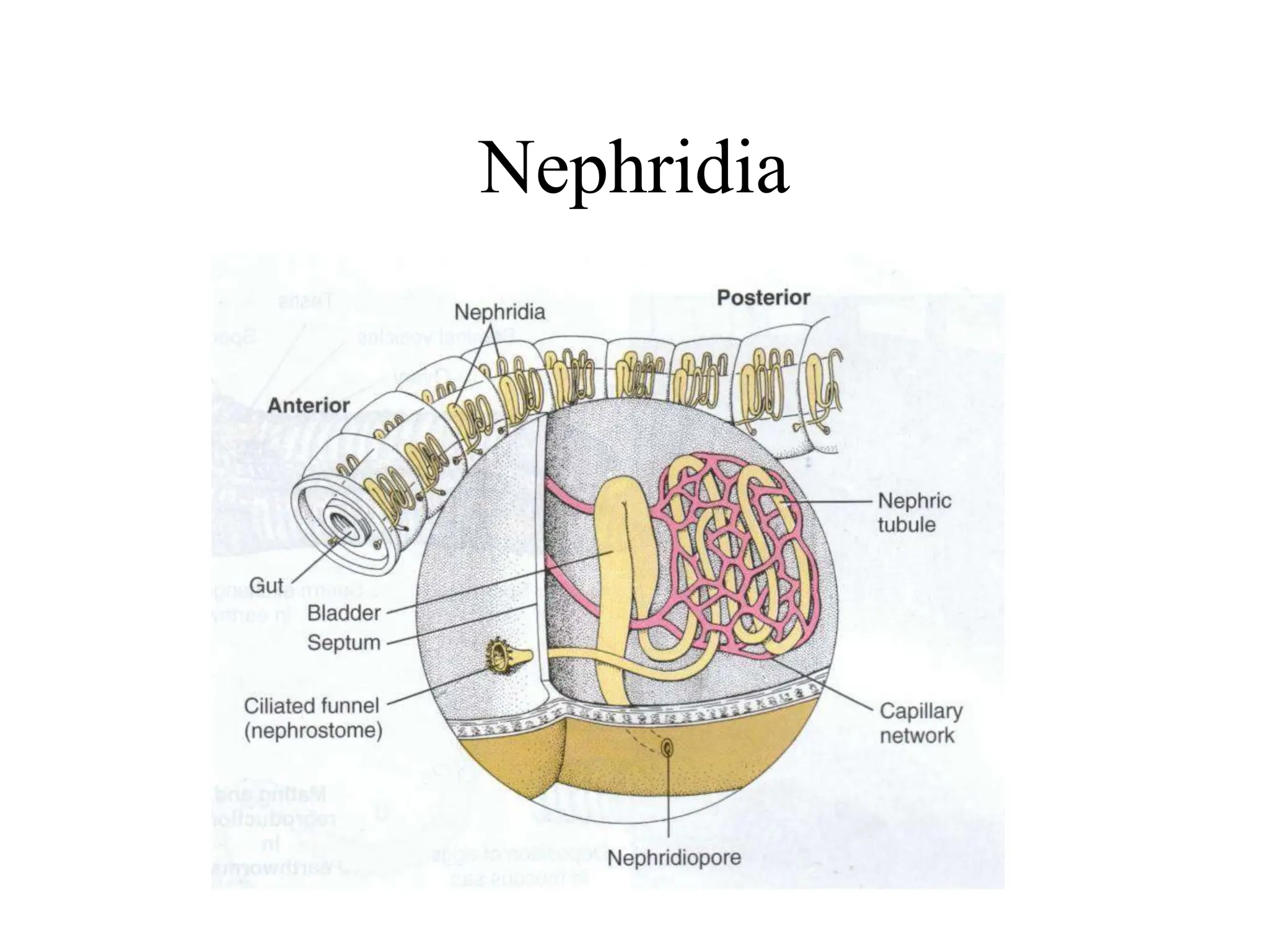
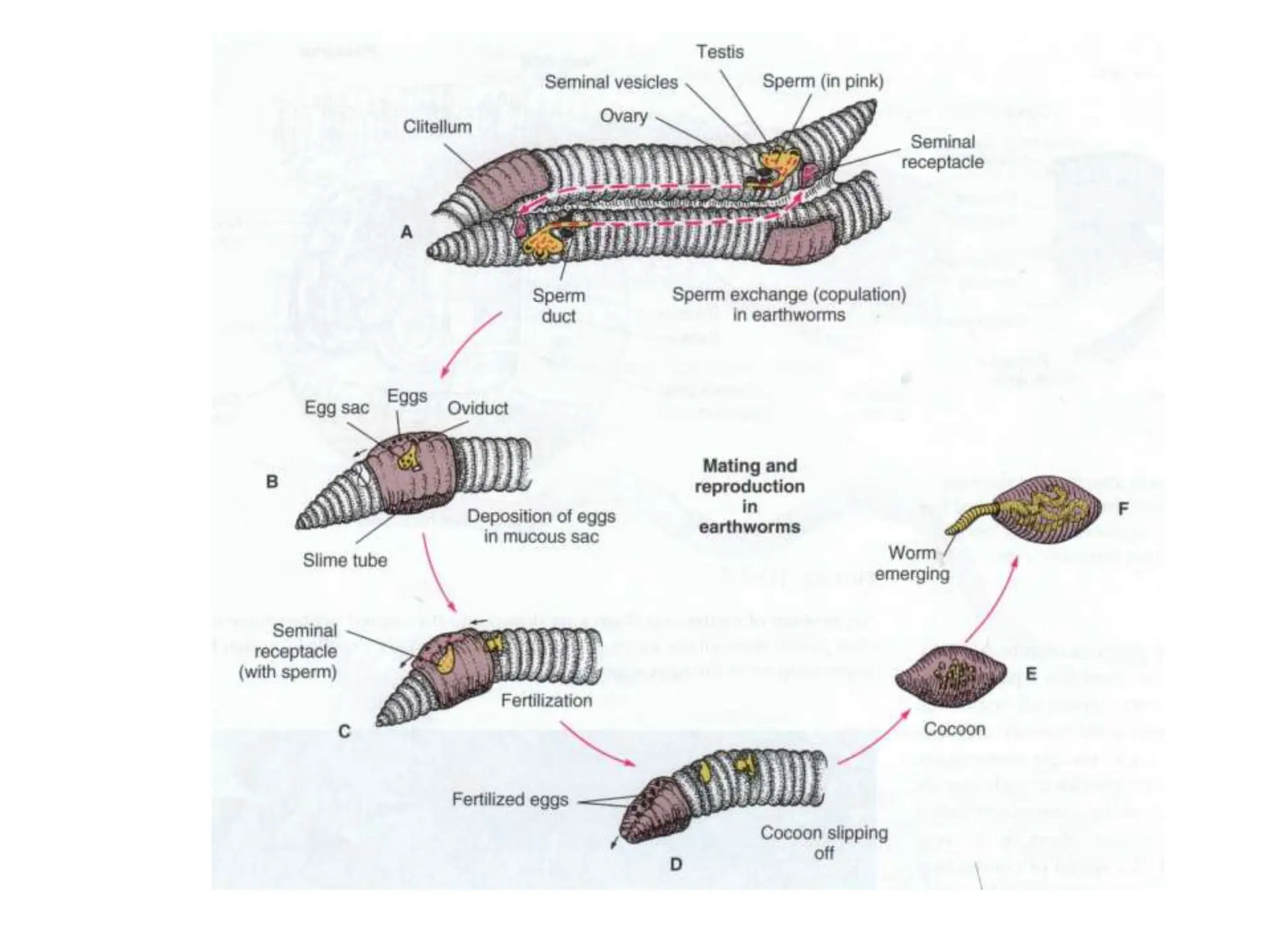
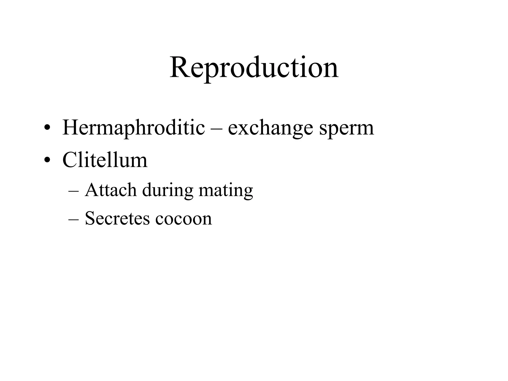
PHYLUM ANNELIDA
(Metamerically segmented and coelomate animals)
Annelids
 Soft bodied, triploblastic,
bilaterally symmetrical
and true coelomates
 Divided into the
segments called as annuli
 Excretion is performed by
nephridia
 Nervous system well developed and has brain
Class – Polychaeta: (animals
with many setae)
 Setae are many form
lateral parapodia
 Head is distinct with
sensory organs like
eyes, tentacles, palps
etc
 Free swimming larval
stage is found called
as trochophore
 example - Nereis
Parapodia
 Heteronereis
Tube dwelling polychaetes
 Arenicola Sabella
Class – Oligochaeta: (
animals with few setae)
 Setae few and are
arranged segmentally
 Distinct head eyes and
tentacles are absent
 Clitellum is usually
present
 example - Pheretima
(earthworm)
Class – Hirudinea: (Class of leeches)
 Ectoparasitic and
fresh water forms
 Secrete an enzyme in
its gut called as
hirudin
 33 segments
 Oral sucker. Posterior
sucker
 Anus is present in 22nd
segment on dorsal
side.
Leech bite
Nereis (Clamworm or sand worm)
Systematic position
Phylum: Annelida
Class: Polychaeta
Subclass: Errantia
Habits and Habitat
Nereis: marine polychaetes, usually found on sandy shore
between tidemarks.
Lives in burrows which it forms with the help of jaws.
Burrow is U shaped (upto 60cm) coated with mucous secreted
from body, which binds together fine particles of sand.
During breeding season worm lreaves its burrow permanently
and swims about actively in water (Heteronereis).
Length varies from few to about 40 cm.
N. pelagica is brownish, N. cultrifera is greenish, N. virens is steel
blue
Body divisions:
Metamerically segmented and number is fairly constant for a
species: about 80 in N. cultrifera & N. dumerilli and about 200
in N. virens.
3 distinct region body regions: Head, trunk and abdomen
Class Polychaeta
• Complex head structures
– Eyes
– Tentacles
– Jaws
– Filtering devices
• Parapodia – leg-like appendages at each
segment
• Setae – chitinous fibers
Body Plan – Key Features
• Segmented (metamers)
• Septa separate coelem at each segment
• Longitudinal and circular muscles
• Peristaltic movement
• Closed circulatory system
• Cerebral ganglia with two nerve cords
• Advanced nephridia
Free Swimming Predator
Nereis virens
• Parapodia used to swim and burrow
• Chitinous jaws to capture prey
• Eye spots and small tentacle
• Gas exchange at parapodia
Parapodia: Flattened, fleshy,
vertical flap like outgrowth
of body wall on lateral sides
of trunk segments.
Biramous: Dorsal
notopodium & Ventral
neuropodium
Ramus divided ito 2 leaf like
lobes or ligulae (dorsal
superior & ventral inferior
ligula
Each part bears tentacular
process known as cirrus;
dorsal cirrus larger than
ventral
Aciculum: Parapodia supported
internally by a long, stout and black
chitinous rod
Setae/Chaetae: Each part also bears a bundle of long, fine, stiff
chitinous bristles
Body Plan
Tentacle Feeder
• Burrow in sand or mud
• Tentacles
– Covered with mucous and cilia
– Retracted to collect food or for protection
• Gills present for gas exchange
Nereis virens Head
Peristomium: 1st segment of body, surrounds slit like mouth,
lacks parapodia, 2 pairs of peristomial cirri on each side.
Dorsolateral pair longer than the ventro-lateral pair.
Prostomium: Anterior projection of prostomium, fleshy lobe
lying above and in front of mouth, 2pairs of pigmented eyes,
sensory prostomial tentacles & 2 jointed palps.
Nereis virens Cross Section
Alimentary canal: Straight tube ext. from ant. to post
suspened in the body cavity by dorsal mesentery & inter-
segmental septa. Opening at ant. End mouth, post end anus.
Foregut: comprises buccal cavity and pharynx
Midgut: Oesophagus and stomach intestine
Hindgut: Rectum occupies the last body segment
(pygidium), opens to the exterior through a terminal anus
Mouth: transverse elongated slit opening ventrally below
prostomium, bordered by peristomium leading to buccal cavity
Buccal cavity: wide chamber succeeded by the highly muscular
pharynx.
Denticles: Cuticle lining their lumen is thickened at places to form
denticles or teeth or paragnaths
Chitinous jaw: embedded laterally in the wall of pharynx is a
stout, mobile, incurved, pointed, notched at apex with inner
margins serrated
Oesophagus: Narrow tube extending 5 segments behind pharynx
Oesophageal gland: long, unbranched, sacculated glandular
pouches. Secretes proteolytic enzymes.
Stomach intestine: segmentally constricted, straight, thin walled tube. Epithelial lining contains scattered
gland cells, secrete digestive enzymes.
Digestive System
Buccal cavity and pharynx are wrapped in a common muscular
coat and together they extend upto 4th or 5th trunk segment.
They can be fully everted to form a proboscis or introvert.
This operation exposes the tip of the jaw for capturing prey.
Food & feeding mechanism
Nereis: carnivorous, raptorial feeder, preys upon small crustaceans, worms, larvae,
actively captured by the exposed jaws of proboscis.
Eversion of proboscis is brought about by protractor muscles, extending below the
wall of peristomium and that of buccal cavity & pharynx.
Retraction of pharynx is brought about by retractor muscles extending backwards
from posterior end of the pharynx to body wall & relaxation of body wall
musculature.
Filter feeding:
Nereis diversicolor secretes mucous at one end of its burrow.
By dorso-ventral undulations of its body sets up a constant
current of water entering through the burrow through that end
and leaving through the other.
Mucous cone behave like a strainer, holding the food particles.
Food laden mucous cone is finally ingested by the worm.
Digestion, absorption and egestion
Engulfed food passes backwards through midgut by peristaltic action of gut
wall
Oesophageal glands and gland cells of mid-gut epithelium secrete juices
containing digestive enzymes like:
Amylases: catalyzes the hydrolysis of starch into sugar
Lipases: catalyzes the breakdown or hydrolysis of fats (lipids)
Proteases: catalyzes the hydrolysis of the peptide bonds
Digestion and absorption takes place mainly in the stomach intestine.
Indigested food substances pass into rectum and are finally egested through
anus as faecal matter.
Respiration:
Gills or any other special organs are absent
Respiration is carried out by whole body surface but more
specifically by the thin, flattened lobes of parapodia, which
possess extensive capillary networks, lying very close to the
surface.
Blood circulates throughout the body through
a system of closed tubes or blood vessels.
Blood: Fluid filled plasma containing
nucleated, colourless amoeboid cells or
corpuscles and dissolved haemoglobin, which
serves as the respiratory pigment.
Brings about transportation of gases, food,
excretory materials.
Blood vessels: Three main longitudinal blood
vessels: dorsal, ventral and a peri-neural
Dorsal vessel: runs above the alimentary canal, through dorsal mesentery, from hind
end forward to 5th segment.
Strong peristaltic waves pass from its posterior to anterior end, driving blood anteriorly.
Behind oesophagus, it acts as a collecting vessel receiving blood from body wall,
parapodia, nephridia and alimentary canal.
Ventral vessel: Mid ventrally below the alimentary canal, entire length of body.
Blood flows from anterior to posterior end.
Peri-neural vessel: It runs ventrally surrounding the nerve cord, collects blood from the
ventral body wall and conveys to the ventral vessel.
Excretory system Excretion is brought about by nephridia, one pair of which is
found in each segment except a few anterior or posterior
ones.
Shape: oval, curved body and a narrow neck.
Body: synctial mass of connective tisuue, containing a coiled
excretory or nephridial tubule open at both ends.
One end of tubule runs through neck and opens into coelom of
adjacent anterior segment by a funnel like nephrostome.
Coiled nephridial tubule opens to the exterior through
nephridiopore, situated near the base of ventral cirrus of parapodia.
Outer surface of nephridium: Densely covered by blood capillaries
Gland cells of the ciliated excretory tubule remove waste from
blood and finally discharges through nephridiopore.
Nitrogenous waste is mainly ammonia (ammonotelic).
Ciliated Nephrostome also removes dead or dying coelomic corpuscles which have been
eaten up or have been destroyed by foreign bodies such as bacteria.
Selective resorption: Useful substance entering excretory tubule is reabsorbed and returned to
blood capillaries
Nervous system
Well developed nervous system, bilaterally
symmetrical, metamerically segmented &
distinguished into central, peripheral &
visceral nervous system
Central nervous system: Cerebral ganglia or brain
is a large bilobed mass located dorsally in
prostomium.
Sub-phararyngeal ganglion: formed by fusion of
2 pairs of ganglia of ventral nerve cord lying below
the pharynx in first trunk segment.
Brain is connected with sub-pharyngeal
ganglion by a pair of stout nerve strands or circum-
pharyngeal connectives, running one on either side
of pharynx, forming a nerve ring.
Ventral nerve cord: runs posteriorly from sub-
pharyngeal ganglion throughout body along the
mid-ventral line, immediately beneath the ventral
blood vessel.
Formed by fusion of two close cords enclosed in a
common sheath of connective tissue. In each
segment, nerve cord is dilated into a segmental
ganglion (pair of ganglia fused together)
Nervous system Peripheral nervous system: consist of nerves
connected to brain, circumpharyngeal
connectives & segmental ganglia.
Brain: nerves to (1) prostomial palps, (2) pair
of nerve to eyes & prostomial tentacle (3)
nuchal organ (ciliated pit; light, food & mating)
Circumpharyngeal connectives: nerve to
ventral & dorsal peristomial cirri
Subpharyngeal ganglia: nerve to body wall &
parapodia of 3rd segment (1st trunk segment)
Segmental ganglia: Gives out 4 pairs.
I & IV pair supply longitudinal muscles &
bodywall
II pair parapodia & III proprio-receptors in
muscles
Visceral nervous system: consist of network of
somatogastric nerves and a few ganglia
supplying the dorsal & ventral walls of pharynx
(proboscis)
Reproductive system
Some are hermaphrodite; mostly dioecious
Gonads: Testes & Ovary are neither distinct nor permanent organs
Masses of developing gametes formed only during breeding season as projections
of swellings by proliferation of ventral septal peritoneum in all body segments
except a few anterior ones
Gametes are shed as spermatogonia in male & as oogonia in female into
coelomic fluid, where they undergo maturation to develop into spermatozoa and
ova
In mature worms coelom remains packed with gametes
Gonoducts: there are no gonoducts
Ripe sperms & Ova are discharged to outside sea water mostly through
metanephridia by the action of cilia borne by nephrostomes and nephridial
tubules. Goodrich proposed the name nephromixia or mixonephridia.
Dorsal ciliated organ occurs in each segment, may act as gonoducts but real
function remains controversial.
Heteronereis
At sexual maturity, most of the posterior
segments, filled with gametes, exhibit
morphological and anatomical
differentiation.
Sexual region or epitoke of worm
Few anterior segments which do not take
part in gamete formation, constitute the
asexual region or atoke
Sexually mature worm with these two
regions is known as heteronereis
Epitoky: phenomenon of transformation
of nonsexual individual into sexual
individual
Swarming: Sexually mature individuals swim to the surface of sea water in
order to shed sperms and ova
Female produces a substance; fertilium, which attracts the males and
stimulates shedding of sperms, which in turn stimulates the shedding of eggs.
Fertilization is mostly external in Nereis virens and takes place in sea water.
In Platynereis megalops, male wraps around female & inserts his anus into her
mouth and injects sperms.
Sperms pass directly into coelom of female where fertilization of egg occurs.
Fertilized eggs are shed from the posterior end of the females body.
Nereis diversicolor does not attain epitokous form and fertilization takes
place in burrow of female or on the surface of mud.
Characteristic features of heteronereis
• Instead of creeping about on sea buttom, heteroneris swims actively in
surface water
• Body divisible into atoke and epitoke
• Parapodia: posterior region becomes larger, develop flattened leaf
like outgrowth for more rapid respiration. Normal setae are replaced by
oar-shaped setae.
• Eyes are greatly enlarged and become conspicuous
• Prostomial palps become reduced but peristomial cirri becomes
longer
• Intenstine becomes compressed and functionless due to much
developed gonads
• Pygidium develops special sensory papillae
The trochophore larva is about 0.1 mm in diameter,
ciliated, unsegmented and pear shaped pelagic creature
Prototroch - an equatorial band of beating cilia that propels the trochophore through the water.
Metatroch - in feeding trochophores this band of cilia beat in a direction opposite to that of
the prototroch and between them they drive a current of water bearing food particles (e.g.
bacteria) into the food groove where cilia around the mouth drives the particles into the gut.
The ciliated ring helps in feeding and locomotion
Eyespot - typically 1-3 are present
Sensory apical organ or plate
bearing a tuft of cilia present.
Brain ganglion, are usually
evident beneath the apical
organ
Digestive tract complete.
Gut regionated into
oesophagus, stomach and
intestine.
Mouth lies ventrally beneath
the prototroch and anus near
the lower apex.
Protonephridia: excretory and osmoregulatory organs which
drive out excess water and waste products along with it.
Metamorphosis of trochophore larva
Trochophore is a pelagic creature drifting about in sea
Metamorphosis results in termination of planktonic existence.
This results in loss of many larval structures like protonephridia,
muscle band and ciliated girdles.
Cells of apical plate form the prostomium and brain
Larva gradually develops in length with the formation and
development of trunk segments, setal sacs and setae.
Mouth region fuses with the first trunk segment to form the
peristomium without setae.
Larva eventually sinks to the bottom to complete post larval
development and assume the habit of adults
Significance of trochophore
Helps in dispersal of the species.
Phylogenetic significance: It appears in the development of
several phyla such as Annelida, Mollusca, Bryozoa, etc.
suggesting that all these groups of animals all descended from a
common hypothetical ancestor trocozoon (Trocophore theory)
Tentacle Feeder
(Amphitrite)
Ciliary Feeder
• Burrow in sand or mud
• Epithelia secretes leathery tube
• Tentacles
– Covered with mucous and cilia
– Grooved for sorting different sized particles
– Can be retracted for protection
• Tentacles used for gas exchange
Tube Feeder
• Epithelia often secretes parchment-like tube
• Parapodia “fans” or peristaltic motion
moves water through tube
• Mucous captures food
Tube Feeder - Chaetopterus
Class Oligochaeta
• Examples
– Lumbricus terrestris (earthworm)
– Tubifex worms
Longitudinal Anatomy
Circulatory System
• Closed – capillary network for exchange
• Dorsal and ventral blood vessels
• Five aortic arches for pumping (heart)
• Contains amoeboid cells and dissolved
hemoglobin
• Coelomic fluid – also involved in
circulation
Cross Sectional Anatomy
Nephridia
• Found in each segment
• Nephrostome crosses into next segment
– Cilliated funnel to draw in fluid
• Water and nutrient reabsorbed
• Function similar to kidney
Nephridia
Reproduction
• Hermaphroditic – exchange sperm
• Clitellum
– Attach during mating
– Secretes cocoon

Nereis Type Study for BSc 1st semester.ppt

  • 1.
  • 2.
    Annelids  Soft bodied,triploblastic, bilaterally symmetrical and true coelomates  Divided into the segments called as annuli  Excretion is performed by nephridia
  • 3.
     Nervous systemwell developed and has brain
  • 4.
    Class – Polychaeta:(animals with many setae)  Setae are many form lateral parapodia  Head is distinct with sensory organs like eyes, tentacles, palps etc  Free swimming larval stage is found called as trochophore  example - Nereis
  • 5.
  • 6.
  • 7.
    Class – Oligochaeta:( animals with few setae)  Setae few and are arranged segmentally  Distinct head eyes and tentacles are absent  Clitellum is usually present  example - Pheretima (earthworm)
  • 8.
    Class – Hirudinea:(Class of leeches)  Ectoparasitic and fresh water forms  Secrete an enzyme in its gut called as hirudin  33 segments  Oral sucker. Posterior sucker  Anus is present in 22nd segment on dorsal side.
  • 9.
  • 10.
    Nereis (Clamworm orsand worm) Systematic position Phylum: Annelida Class: Polychaeta Subclass: Errantia
  • 11.
    Habits and Habitat Nereis:marine polychaetes, usually found on sandy shore between tidemarks. Lives in burrows which it forms with the help of jaws. Burrow is U shaped (upto 60cm) coated with mucous secreted from body, which binds together fine particles of sand. During breeding season worm lreaves its burrow permanently and swims about actively in water (Heteronereis). Length varies from few to about 40 cm. N. pelagica is brownish, N. cultrifera is greenish, N. virens is steel blue
  • 12.
    Body divisions: Metamerically segmentedand number is fairly constant for a species: about 80 in N. cultrifera & N. dumerilli and about 200 in N. virens. 3 distinct region body regions: Head, trunk and abdomen
  • 13.
    Class Polychaeta • Complexhead structures – Eyes – Tentacles – Jaws – Filtering devices • Parapodia – leg-like appendages at each segment • Setae – chitinous fibers
  • 14.
    Body Plan –Key Features • Segmented (metamers) • Septa separate coelem at each segment • Longitudinal and circular muscles • Peristaltic movement • Closed circulatory system • Cerebral ganglia with two nerve cords • Advanced nephridia
  • 15.
    Free Swimming Predator Nereisvirens • Parapodia used to swim and burrow • Chitinous jaws to capture prey • Eye spots and small tentacle • Gas exchange at parapodia
  • 16.
    Parapodia: Flattened, fleshy, verticalflap like outgrowth of body wall on lateral sides of trunk segments. Biramous: Dorsal notopodium & Ventral neuropodium Ramus divided ito 2 leaf like lobes or ligulae (dorsal superior & ventral inferior ligula Each part bears tentacular process known as cirrus; dorsal cirrus larger than ventral Aciculum: Parapodia supported internally by a long, stout and black chitinous rod Setae/Chaetae: Each part also bears a bundle of long, fine, stiff chitinous bristles
  • 17.
  • 18.
    Tentacle Feeder • Burrowin sand or mud • Tentacles – Covered with mucous and cilia – Retracted to collect food or for protection • Gills present for gas exchange
  • 19.
    Nereis virens Head Peristomium:1st segment of body, surrounds slit like mouth, lacks parapodia, 2 pairs of peristomial cirri on each side. Dorsolateral pair longer than the ventro-lateral pair. Prostomium: Anterior projection of prostomium, fleshy lobe lying above and in front of mouth, 2pairs of pigmented eyes, sensory prostomial tentacles & 2 jointed palps.
  • 20.
  • 22.
    Alimentary canal: Straighttube ext. from ant. to post suspened in the body cavity by dorsal mesentery & inter- segmental septa. Opening at ant. End mouth, post end anus. Foregut: comprises buccal cavity and pharynx Midgut: Oesophagus and stomach intestine Hindgut: Rectum occupies the last body segment (pygidium), opens to the exterior through a terminal anus Mouth: transverse elongated slit opening ventrally below prostomium, bordered by peristomium leading to buccal cavity Buccal cavity: wide chamber succeeded by the highly muscular pharynx. Denticles: Cuticle lining their lumen is thickened at places to form denticles or teeth or paragnaths Chitinous jaw: embedded laterally in the wall of pharynx is a stout, mobile, incurved, pointed, notched at apex with inner margins serrated Oesophagus: Narrow tube extending 5 segments behind pharynx Oesophageal gland: long, unbranched, sacculated glandular pouches. Secretes proteolytic enzymes. Stomach intestine: segmentally constricted, straight, thin walled tube. Epithelial lining contains scattered gland cells, secrete digestive enzymes. Digestive System
  • 23.
    Buccal cavity andpharynx are wrapped in a common muscular coat and together they extend upto 4th or 5th trunk segment. They can be fully everted to form a proboscis or introvert. This operation exposes the tip of the jaw for capturing prey.
  • 27.
    Food & feedingmechanism Nereis: carnivorous, raptorial feeder, preys upon small crustaceans, worms, larvae, actively captured by the exposed jaws of proboscis. Eversion of proboscis is brought about by protractor muscles, extending below the wall of peristomium and that of buccal cavity & pharynx. Retraction of pharynx is brought about by retractor muscles extending backwards from posterior end of the pharynx to body wall & relaxation of body wall musculature.
  • 28.
    Filter feeding: Nereis diversicolorsecretes mucous at one end of its burrow. By dorso-ventral undulations of its body sets up a constant current of water entering through the burrow through that end and leaving through the other. Mucous cone behave like a strainer, holding the food particles. Food laden mucous cone is finally ingested by the worm.
  • 29.
    Digestion, absorption andegestion Engulfed food passes backwards through midgut by peristaltic action of gut wall Oesophageal glands and gland cells of mid-gut epithelium secrete juices containing digestive enzymes like: Amylases: catalyzes the hydrolysis of starch into sugar Lipases: catalyzes the breakdown or hydrolysis of fats (lipids) Proteases: catalyzes the hydrolysis of the peptide bonds Digestion and absorption takes place mainly in the stomach intestine. Indigested food substances pass into rectum and are finally egested through anus as faecal matter.
  • 30.
    Respiration: Gills or anyother special organs are absent Respiration is carried out by whole body surface but more specifically by the thin, flattened lobes of parapodia, which possess extensive capillary networks, lying very close to the surface.
  • 33.
    Blood circulates throughoutthe body through a system of closed tubes or blood vessels. Blood: Fluid filled plasma containing nucleated, colourless amoeboid cells or corpuscles and dissolved haemoglobin, which serves as the respiratory pigment. Brings about transportation of gases, food, excretory materials. Blood vessels: Three main longitudinal blood vessels: dorsal, ventral and a peri-neural Dorsal vessel: runs above the alimentary canal, through dorsal mesentery, from hind end forward to 5th segment. Strong peristaltic waves pass from its posterior to anterior end, driving blood anteriorly. Behind oesophagus, it acts as a collecting vessel receiving blood from body wall, parapodia, nephridia and alimentary canal. Ventral vessel: Mid ventrally below the alimentary canal, entire length of body. Blood flows from anterior to posterior end. Peri-neural vessel: It runs ventrally surrounding the nerve cord, collects blood from the ventral body wall and conveys to the ventral vessel.
  • 37.
    Excretory system Excretionis brought about by nephridia, one pair of which is found in each segment except a few anterior or posterior ones. Shape: oval, curved body and a narrow neck. Body: synctial mass of connective tisuue, containing a coiled excretory or nephridial tubule open at both ends. One end of tubule runs through neck and opens into coelom of adjacent anterior segment by a funnel like nephrostome. Coiled nephridial tubule opens to the exterior through nephridiopore, situated near the base of ventral cirrus of parapodia. Outer surface of nephridium: Densely covered by blood capillaries Gland cells of the ciliated excretory tubule remove waste from blood and finally discharges through nephridiopore. Nitrogenous waste is mainly ammonia (ammonotelic). Ciliated Nephrostome also removes dead or dying coelomic corpuscles which have been eaten up or have been destroyed by foreign bodies such as bacteria. Selective resorption: Useful substance entering excretory tubule is reabsorbed and returned to blood capillaries
  • 38.
    Nervous system Well developednervous system, bilaterally symmetrical, metamerically segmented & distinguished into central, peripheral & visceral nervous system Central nervous system: Cerebral ganglia or brain is a large bilobed mass located dorsally in prostomium. Sub-phararyngeal ganglion: formed by fusion of 2 pairs of ganglia of ventral nerve cord lying below the pharynx in first trunk segment. Brain is connected with sub-pharyngeal ganglion by a pair of stout nerve strands or circum- pharyngeal connectives, running one on either side of pharynx, forming a nerve ring. Ventral nerve cord: runs posteriorly from sub- pharyngeal ganglion throughout body along the mid-ventral line, immediately beneath the ventral blood vessel. Formed by fusion of two close cords enclosed in a common sheath of connective tissue. In each segment, nerve cord is dilated into a segmental ganglion (pair of ganglia fused together)
  • 40.
    Nervous system Peripheralnervous system: consist of nerves connected to brain, circumpharyngeal connectives & segmental ganglia. Brain: nerves to (1) prostomial palps, (2) pair of nerve to eyes & prostomial tentacle (3) nuchal organ (ciliated pit; light, food & mating) Circumpharyngeal connectives: nerve to ventral & dorsal peristomial cirri Subpharyngeal ganglia: nerve to body wall & parapodia of 3rd segment (1st trunk segment) Segmental ganglia: Gives out 4 pairs. I & IV pair supply longitudinal muscles & bodywall II pair parapodia & III proprio-receptors in muscles Visceral nervous system: consist of network of somatogastric nerves and a few ganglia supplying the dorsal & ventral walls of pharynx (proboscis)
  • 41.
    Reproductive system Some arehermaphrodite; mostly dioecious Gonads: Testes & Ovary are neither distinct nor permanent organs Masses of developing gametes formed only during breeding season as projections of swellings by proliferation of ventral septal peritoneum in all body segments except a few anterior ones Gametes are shed as spermatogonia in male & as oogonia in female into coelomic fluid, where they undergo maturation to develop into spermatozoa and ova In mature worms coelom remains packed with gametes Gonoducts: there are no gonoducts Ripe sperms & Ova are discharged to outside sea water mostly through metanephridia by the action of cilia borne by nephrostomes and nephridial tubules. Goodrich proposed the name nephromixia or mixonephridia. Dorsal ciliated organ occurs in each segment, may act as gonoducts but real function remains controversial.
  • 42.
    Heteronereis At sexual maturity,most of the posterior segments, filled with gametes, exhibit morphological and anatomical differentiation. Sexual region or epitoke of worm Few anterior segments which do not take part in gamete formation, constitute the asexual region or atoke Sexually mature worm with these two regions is known as heteronereis Epitoky: phenomenon of transformation of nonsexual individual into sexual individual
  • 43.
    Swarming: Sexually matureindividuals swim to the surface of sea water in order to shed sperms and ova Female produces a substance; fertilium, which attracts the males and stimulates shedding of sperms, which in turn stimulates the shedding of eggs. Fertilization is mostly external in Nereis virens and takes place in sea water. In Platynereis megalops, male wraps around female & inserts his anus into her mouth and injects sperms. Sperms pass directly into coelom of female where fertilization of egg occurs. Fertilized eggs are shed from the posterior end of the females body. Nereis diversicolor does not attain epitokous form and fertilization takes place in burrow of female or on the surface of mud.
  • 44.
    Characteristic features ofheteronereis • Instead of creeping about on sea buttom, heteroneris swims actively in surface water • Body divisible into atoke and epitoke • Parapodia: posterior region becomes larger, develop flattened leaf like outgrowth for more rapid respiration. Normal setae are replaced by oar-shaped setae. • Eyes are greatly enlarged and become conspicuous • Prostomial palps become reduced but peristomial cirri becomes longer • Intenstine becomes compressed and functionless due to much developed gonads • Pygidium develops special sensory papillae
  • 46.
    The trochophore larvais about 0.1 mm in diameter, ciliated, unsegmented and pear shaped pelagic creature Prototroch - an equatorial band of beating cilia that propels the trochophore through the water. Metatroch - in feeding trochophores this band of cilia beat in a direction opposite to that of the prototroch and between them they drive a current of water bearing food particles (e.g. bacteria) into the food groove where cilia around the mouth drives the particles into the gut. The ciliated ring helps in feeding and locomotion Eyespot - typically 1-3 are present Sensory apical organ or plate bearing a tuft of cilia present. Brain ganglion, are usually evident beneath the apical organ Digestive tract complete. Gut regionated into oesophagus, stomach and intestine. Mouth lies ventrally beneath the prototroch and anus near the lower apex. Protonephridia: excretory and osmoregulatory organs which drive out excess water and waste products along with it.
  • 47.
    Metamorphosis of trochophorelarva Trochophore is a pelagic creature drifting about in sea Metamorphosis results in termination of planktonic existence. This results in loss of many larval structures like protonephridia, muscle band and ciliated girdles. Cells of apical plate form the prostomium and brain Larva gradually develops in length with the formation and development of trunk segments, setal sacs and setae. Mouth region fuses with the first trunk segment to form the peristomium without setae. Larva eventually sinks to the bottom to complete post larval development and assume the habit of adults
  • 48.
    Significance of trochophore Helpsin dispersal of the species. Phylogenetic significance: It appears in the development of several phyla such as Annelida, Mollusca, Bryozoa, etc. suggesting that all these groups of animals all descended from a common hypothetical ancestor trocozoon (Trocophore theory)
  • 49.
  • 52.
    Ciliary Feeder • Burrowin sand or mud • Epithelia secretes leathery tube • Tentacles – Covered with mucous and cilia – Grooved for sorting different sized particles – Can be retracted for protection • Tentacles used for gas exchange
  • 53.
    Tube Feeder • Epitheliaoften secretes parchment-like tube • Parapodia “fans” or peristaltic motion moves water through tube • Mucous captures food
  • 54.
    Tube Feeder -Chaetopterus
  • 55.
    Class Oligochaeta • Examples –Lumbricus terrestris (earthworm) – Tubifex worms
  • 56.
  • 57.
    Circulatory System • Closed– capillary network for exchange • Dorsal and ventral blood vessels • Five aortic arches for pumping (heart) • Contains amoeboid cells and dissolved hemoglobin • Coelomic fluid – also involved in circulation
  • 58.
  • 59.
    Nephridia • Found ineach segment • Nephrostome crosses into next segment – Cilliated funnel to draw in fluid • Water and nutrient reabsorbed • Function similar to kidney
  • 60.
  • 62.
    Reproduction • Hermaphroditic –exchange sperm • Clitellum – Attach during mating – Secretes cocoon