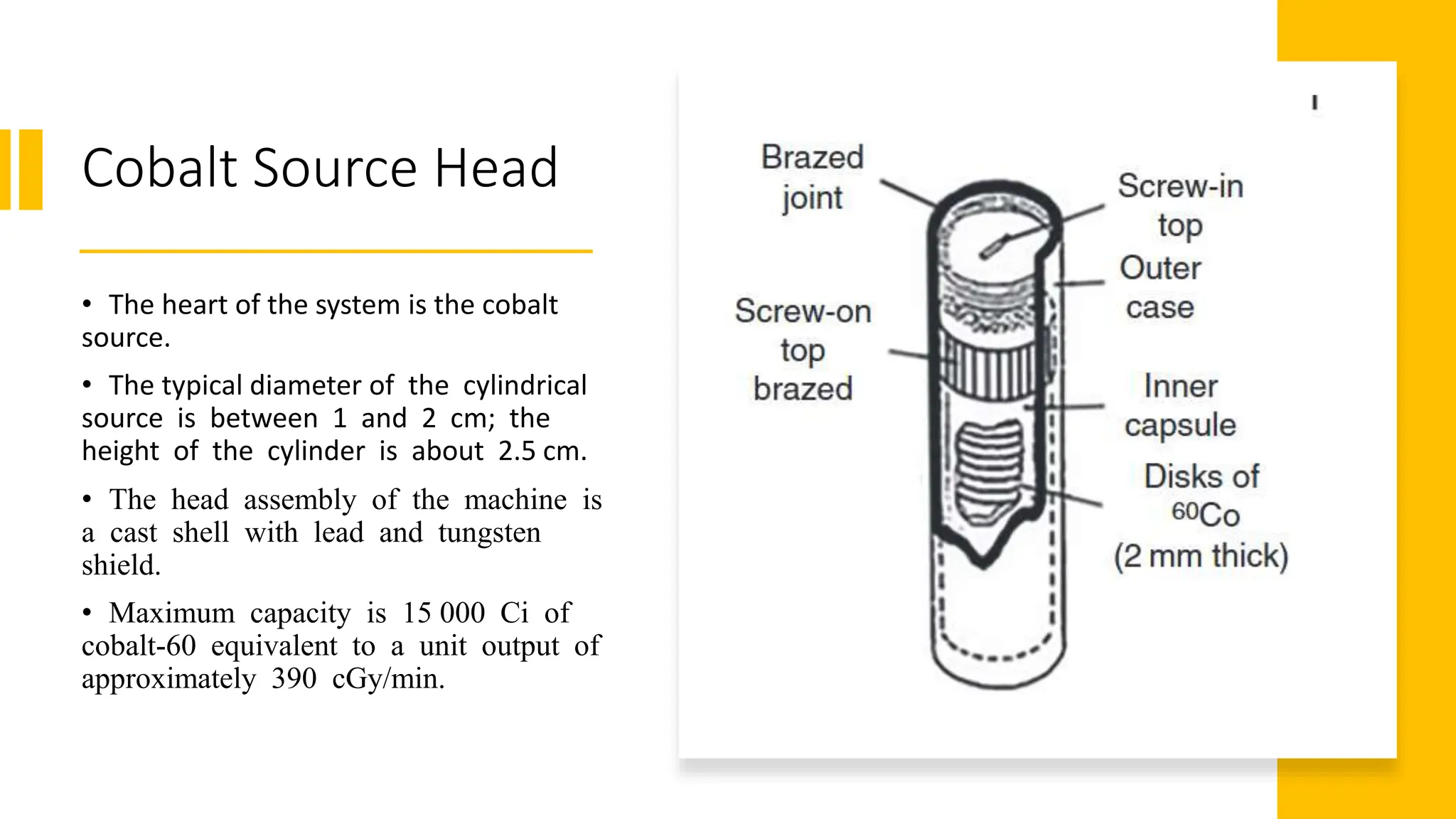
Cobalt 60
Nuclear & Radiating Systems | MDE 227
Ibram Ayad Saad| 20190002
Under supervision of Dr. Nour
Content
• Overview
• Constructional Details
• Cobalt-60 Machine Physical Factors
• Uses
Overview
• cobalt-60 machine is mature technologies for external beam radiation.
• Cobalt-60, is a synthetic radioactive isotope.
• It has a half-life of 5.2714 years.
• It is produced by neutron irradiation in a nuclear reactor.
• 60Co decays by beta decay to the stable isotope nickel- 60.
• The activated nickel nucleus emits two gamma rays with energies of 1.17 and
1.33 MeV.
External Beam Radiation Therapy
• External beam radiation therapy comes from a machine that aims radiation
at your cancer. The machine is large and may be noisy. It does not touch you,
but rotates around you, sending radiation to your body from many
directions.
• External beam radiation therapy is a local treatment, meaning that the
radiation is aimed only at a specific part of your body. For example, if you
have lung cancer, you will get radiation to your chest only and not the rest of
your body.
Working principle
• An external sealed source of cobalt 60is used in teletherapy unit as a source
of intense gamma radiation for the treatment of a variety of cancers.
• Cobalt 60 is used as tracer that is injected into the body, then cobalt 60
source emits gamma rays, that's scanned by machine
Constructional
Details
A cobalt machine consists of the following
major subsystems:
• Cobalt source head comprising a radioactive
source and its housing.
• Head mounts for source movement
mechanism (gantry and stand).
• Beam collimator.
• Treatment table or patient support assembly.
• Control console and safety interlocks.
Cobalt Source Head
• The heart of the system is the cobalt
source.
• The typical diameter of the cylindrical
source is between 1 and 2 cm; the
height of the cylinder is about 2.5 cm.
• The head assembly of the machine is
a cast shell with lead and tungsten
shield.
• Maximum capacity is 15 000 Ci of
cobalt‐60 equivalent to a unit output of
approximately 390 cGy/min.
Cobalt Source Head
• The lead‐filled container is 25 cm thick in all directions from the source. Its
design should ensure that the leakage radiation coming through its thickness would not
cause an overexposure to anyone staying at its surface for prolonged periods of
time.
• The source is placed near the center of a large, lead‐filled steel container. Several
methods have been devised for moving the source from the ‘off ’ to the ‘on’ position.
• In one method, the source is mounted in a heavy metal like tungsten wheel that
is rotated through 180° to carry it from the ‘off ’ position to the ‘on’ position. The
transfer between ‘on’ and ‘off ’ positions takes place in less than two seconds for
pinpoint exposure accuracy. The motor drive acts against a heavy duty torque spring that
instantly returns the source to the ‘off ’ position for normal exposure termination and in
the event of an electrical power failure.
Cobalt Source Head
• In another method, the source is mounted in a sliding plug or drawer, which
carries the source from the ‘off ’ to the ‘on’ position. This method has
become the most commonly used one in commercial machines. All
machines must be arranged so that they are fail ‘safe’, that is, the source must
be held in the ‘on’ position by the continuous application of a force so that if
the power fails, it must return quickly to the ‘off ’ position. In one machine, the
safety facility is in the form of total closure of the diaphragms electrically (0
cm × 0 cm field size) during source stuck position using a safety interlock.
Gantry
Gantry
• There are two types of arrangements of mounting radiation treatment units. In the
earlier designs, the head of the unit was held in a yoke which can be moved
up and down, or back and forth, and can be rotated about the axis. In this case, the
head mount is suspended from a set of rails attached to the ceiling. In the
later models, the unit was mounted on a column with vertical motion and a pivot for
head rotation. The distance from the source to the skin of the patient source to skin
distance (SSD) is usually fixed at 80 cm, and the focus of the set‐up is the surface of
the patient. The alternative mount is the so‐called isocentric or fixed source–axis
distance (SAD) mount. The isocentre is defined as the point where the collimator and
gantry rotational axes intersect. This point lies within a sphere of 1 mm radius
as the gantry rotates through 360°.
Gantry
• The head is encased in a streamlined plastic cover and is mounted on a gantry that can rotate
about a horizontal axis. The gantry supports the source head at one end and the radiation
barrier or counterweight at the opposite end. It accurately maintains an 80–100 cm distance
from the source to the axis of rotation located 120 cm above floor. The drive consists of a DC
motor with microprocessor control and dynamic braking circuits that allow uniform rotation
and accurate control of the gantry motion.The gantry consists of a rugged heavy‐gauge steel
frame with 2.5 cm thick base and anchoring plates. The base plate is fixed to the floor
with two heavy duty anchoring bolts. The carriage and hanger assembly provide
vibration‐free movement along with the vertical guide rails.
Gantry
• The gantry rotates in both clockwise and counterclockwise directions. Gantry
rotation can be under variable speed control to facilitate treatment set‐up. The
range of gantry rotation is ±360°
Collimators
• A symmetrical/asymmetrical collimator assembly defines the X and Y axes relative to the collimator
axis. The collimator consists of a set of bars that can produce a radiation beam with a square or
rectangular cross section. The collimator can rotate at an angle of 360° about its beam axis, and
rotary knobs lock the bars at the desired position. The position of the collimator bars is indicated on
an indicator scale that is calibrated in 1° increments. The field size can be continuously varied to
define the shaping of square or rectangular fields whose size can be adjusted from 3 cm × 3 cm to 35
cm × 35 cm at 80 cm from the source. The adjustment of the field size is done by a moving the
collimator vanes, which are motor driven. The geometric penumbra, which results from a finite
source diameter, is minimized by using small diameter sources and by using penumbra trimmers
as close as possible to the patient’s skin.
Collimators
• The collimator is constructed of four sets of flat, interleaved lead vanes
with angu-lated inner tungsten trees for continuously variable field sizes. As
the vanes move, the beam defining tungsten trees automatically angulate
to follow the divergence of the beam. The built‐in system projects light
field on the patient’s skin to indicate precisely the radiation field at the 50%
geometric penumbra line. A built‐in optical device pro-jects an easy‐to‐read
scale on the patient’s skin to facilitate a direct readout. It indicates 60–100
cm in 1 cm increments. When the desired number is superimposed on
cross hairs, the patient’s skin is at the desired distance from the source.
Treatment Table
• The patient to be treated is made to lie on a table whose position can be adjusted
upwards or lowered. It can also be moved sideways so that for any angle of the gantry,
the beam will pass through the tumor.
• The axis of rotation is a fixed distance from the source, and the size of the beam is
specified by its size at the axis.
• The arrangement is called isometric because the axis of rotation of the gantry
intersects with the central axis of the beam (axis of rotation of the collimator) so that
they both rotate about the same center. In addition, the treatment table can usually be
rotated about a vertical axis so that it also passes through the isocenter.
• Longitudinal and transverse tabletop travels are usually manually controlled, while
the vertical movement is motor driven. In addition, the entire table can be rotated 180°
about the vertical beam axis.
Treatment Table
• The various movements of the table are controlled by an aluminium casting carriage
assembly mounted directly below the tabletop. It comprises bearing assemblies
and positioning controls.
• The longitudinal movement is achieved through ball bearings, which ride in
hardened steel tracks, while the linear ball bearings move on hardened steel shafts
for transverse travel.
• The patient table is provided with multiple safeguards to protect the patient and the
therapist. The table can be moved using the remote con-trol or by using the table
controls located on each side of the table.
Control Console and Safety Interlocks
• The computerized control console is always placed outside the treatment room that
facilitates control of various functions. The machine is controlled by an advanced
software control system, which monitors treatment parameters continuously. It
permits the selection of treatment techniques such as rotation, oscillation, skip scanning,
or multi‐portal indexing.
• The X and Y leaf positions of the collimator are displayed on the in‐room display monitor.
The direction selector establishes clockwise or counterclockwise rotation of the gantry. An
exposure timer counts down as treatment time diminishes and is displayed by a four‐digit
LED readout.
• Similarly, the count‐up exposure time is displayed with the progress of the treatment. An
emergency button interrupts source‐drive current for torque‐spring return to ‘beam
off ’ condition with momentary actuation. Similarly, an entrance door interlock permits
interconnection to the safety switch at the entrance door.
Control Console and Safety Interlocks
• Exposure automatically terminates if the entrance door is opened while the source
is ‘on’. A ‘safety key switch’ controls the source transfer mechanism to prevent
unauthorized use.
• The design of the cobalt head must meet the international standards for source shielding,
radiation transmission, and radiation leakage. The dual computer control system monitors
the source position and automatically retracts the source if a problem with source travel
or position is detected.
• The use of cobalt‐60 machines is regulated by national regulatory authorities in the
respective countries that provide clearance for the concrete rooms, issue license for the
procurement, and use of these equipment, conduct periodic surveys, and document
radiation levels for such installations.
Cobalt-60 Machine Physical Factors
Treatment
with cobalt
60
Uses • Cobalt-60 therapy can be used anywhere on
the body. It is particularly useful in brain
tumor patients because it is so precise.
Because of its effectiveness and simple
design, doctors have used cobalt-60 therapy
to treat cancer for almost 60 years.
cobalt 60.pptx

cobalt 60.pptx

  • 1.
    Cobalt 60 Nuclear &Radiating Systems | MDE 227 Ibram Ayad Saad| 20190002 Under supervision of Dr. Nour
  • 2.
    Content • Overview • ConstructionalDetails • Cobalt-60 Machine Physical Factors • Uses
  • 3.
    Overview • cobalt-60 machineis mature technologies for external beam radiation. • Cobalt-60, is a synthetic radioactive isotope. • It has a half-life of 5.2714 years. • It is produced by neutron irradiation in a nuclear reactor. • 60Co decays by beta decay to the stable isotope nickel- 60. • The activated nickel nucleus emits two gamma rays with energies of 1.17 and 1.33 MeV.
  • 4.
    External Beam RadiationTherapy • External beam radiation therapy comes from a machine that aims radiation at your cancer. The machine is large and may be noisy. It does not touch you, but rotates around you, sending radiation to your body from many directions. • External beam radiation therapy is a local treatment, meaning that the radiation is aimed only at a specific part of your body. For example, if you have lung cancer, you will get radiation to your chest only and not the rest of your body.
  • 5.
    Working principle • Anexternal sealed source of cobalt 60is used in teletherapy unit as a source of intense gamma radiation for the treatment of a variety of cancers. • Cobalt 60 is used as tracer that is injected into the body, then cobalt 60 source emits gamma rays, that's scanned by machine
  • 6.
    Constructional Details A cobalt machineconsists of the following major subsystems: • Cobalt source head comprising a radioactive source and its housing. • Head mounts for source movement mechanism (gantry and stand). • Beam collimator. • Treatment table or patient support assembly. • Control console and safety interlocks.
  • 7.
    Cobalt Source Head •The heart of the system is the cobalt source. • The typical diameter of the cylindrical source is between 1 and 2 cm; the height of the cylinder is about 2.5 cm. • The head assembly of the machine is a cast shell with lead and tungsten shield. • Maximum capacity is 15 000 Ci of cobalt‐60 equivalent to a unit output of approximately 390 cGy/min.
  • 8.
    Cobalt Source Head •The lead‐filled container is 25 cm thick in all directions from the source. Its design should ensure that the leakage radiation coming through its thickness would not cause an overexposure to anyone staying at its surface for prolonged periods of time. • The source is placed near the center of a large, lead‐filled steel container. Several methods have been devised for moving the source from the ‘off ’ to the ‘on’ position. • In one method, the source is mounted in a heavy metal like tungsten wheel that is rotated through 180° to carry it from the ‘off ’ position to the ‘on’ position. The transfer between ‘on’ and ‘off ’ positions takes place in less than two seconds for pinpoint exposure accuracy. The motor drive acts against a heavy duty torque spring that instantly returns the source to the ‘off ’ position for normal exposure termination and in the event of an electrical power failure.
  • 9.
    Cobalt Source Head •In another method, the source is mounted in a sliding plug or drawer, which carries the source from the ‘off ’ to the ‘on’ position. This method has become the most commonly used one in commercial machines. All machines must be arranged so that they are fail ‘safe’, that is, the source must be held in the ‘on’ position by the continuous application of a force so that if the power fails, it must return quickly to the ‘off ’ position. In one machine, the safety facility is in the form of total closure of the diaphragms electrically (0 cm × 0 cm field size) during source stuck position using a safety interlock.
  • 10.
  • 11.
    Gantry • There aretwo types of arrangements of mounting radiation treatment units. In the earlier designs, the head of the unit was held in a yoke which can be moved up and down, or back and forth, and can be rotated about the axis. In this case, the head mount is suspended from a set of rails attached to the ceiling. In the later models, the unit was mounted on a column with vertical motion and a pivot for head rotation. The distance from the source to the skin of the patient source to skin distance (SSD) is usually fixed at 80 cm, and the focus of the set‐up is the surface of the patient. The alternative mount is the so‐called isocentric or fixed source–axis distance (SAD) mount. The isocentre is defined as the point where the collimator and gantry rotational axes intersect. This point lies within a sphere of 1 mm radius as the gantry rotates through 360°.
  • 12.
    Gantry • The headis encased in a streamlined plastic cover and is mounted on a gantry that can rotate about a horizontal axis. The gantry supports the source head at one end and the radiation barrier or counterweight at the opposite end. It accurately maintains an 80–100 cm distance from the source to the axis of rotation located 120 cm above floor. The drive consists of a DC motor with microprocessor control and dynamic braking circuits that allow uniform rotation and accurate control of the gantry motion.The gantry consists of a rugged heavy‐gauge steel frame with 2.5 cm thick base and anchoring plates. The base plate is fixed to the floor with two heavy duty anchoring bolts. The carriage and hanger assembly provide vibration‐free movement along with the vertical guide rails.
  • 13.
    Gantry • The gantryrotates in both clockwise and counterclockwise directions. Gantry rotation can be under variable speed control to facilitate treatment set‐up. The range of gantry rotation is ±360°
  • 14.
    Collimators • A symmetrical/asymmetricalcollimator assembly defines the X and Y axes relative to the collimator axis. The collimator consists of a set of bars that can produce a radiation beam with a square or rectangular cross section. The collimator can rotate at an angle of 360° about its beam axis, and rotary knobs lock the bars at the desired position. The position of the collimator bars is indicated on an indicator scale that is calibrated in 1° increments. The field size can be continuously varied to define the shaping of square or rectangular fields whose size can be adjusted from 3 cm × 3 cm to 35 cm × 35 cm at 80 cm from the source. The adjustment of the field size is done by a moving the collimator vanes, which are motor driven. The geometric penumbra, which results from a finite source diameter, is minimized by using small diameter sources and by using penumbra trimmers as close as possible to the patient’s skin.
  • 15.
    Collimators • The collimatoris constructed of four sets of flat, interleaved lead vanes with angu-lated inner tungsten trees for continuously variable field sizes. As the vanes move, the beam defining tungsten trees automatically angulate to follow the divergence of the beam. The built‐in system projects light field on the patient’s skin to indicate precisely the radiation field at the 50% geometric penumbra line. A built‐in optical device pro-jects an easy‐to‐read scale on the patient’s skin to facilitate a direct readout. It indicates 60–100 cm in 1 cm increments. When the desired number is superimposed on cross hairs, the patient’s skin is at the desired distance from the source.
  • 16.
    Treatment Table • Thepatient to be treated is made to lie on a table whose position can be adjusted upwards or lowered. It can also be moved sideways so that for any angle of the gantry, the beam will pass through the tumor. • The axis of rotation is a fixed distance from the source, and the size of the beam is specified by its size at the axis. • The arrangement is called isometric because the axis of rotation of the gantry intersects with the central axis of the beam (axis of rotation of the collimator) so that they both rotate about the same center. In addition, the treatment table can usually be rotated about a vertical axis so that it also passes through the isocenter. • Longitudinal and transverse tabletop travels are usually manually controlled, while the vertical movement is motor driven. In addition, the entire table can be rotated 180° about the vertical beam axis.
  • 17.
    Treatment Table • Thevarious movements of the table are controlled by an aluminium casting carriage assembly mounted directly below the tabletop. It comprises bearing assemblies and positioning controls. • The longitudinal movement is achieved through ball bearings, which ride in hardened steel tracks, while the linear ball bearings move on hardened steel shafts for transverse travel. • The patient table is provided with multiple safeguards to protect the patient and the therapist. The table can be moved using the remote con-trol or by using the table controls located on each side of the table.
  • 18.
    Control Console andSafety Interlocks • The computerized control console is always placed outside the treatment room that facilitates control of various functions. The machine is controlled by an advanced software control system, which monitors treatment parameters continuously. It permits the selection of treatment techniques such as rotation, oscillation, skip scanning, or multi‐portal indexing. • The X and Y leaf positions of the collimator are displayed on the in‐room display monitor. The direction selector establishes clockwise or counterclockwise rotation of the gantry. An exposure timer counts down as treatment time diminishes and is displayed by a four‐digit LED readout. • Similarly, the count‐up exposure time is displayed with the progress of the treatment. An emergency button interrupts source‐drive current for torque‐spring return to ‘beam off ’ condition with momentary actuation. Similarly, an entrance door interlock permits interconnection to the safety switch at the entrance door.
  • 19.
    Control Console andSafety Interlocks • Exposure automatically terminates if the entrance door is opened while the source is ‘on’. A ‘safety key switch’ controls the source transfer mechanism to prevent unauthorized use. • The design of the cobalt head must meet the international standards for source shielding, radiation transmission, and radiation leakage. The dual computer control system monitors the source position and automatically retracts the source if a problem with source travel or position is detected. • The use of cobalt‐60 machines is regulated by national regulatory authorities in the respective countries that provide clearance for the concrete rooms, issue license for the procurement, and use of these equipment, conduct periodic surveys, and document radiation levels for such installations.
  • 20.
  • 21.
  • 23.
    Uses • Cobalt-60therapy can be used anywhere on the body. It is particularly useful in brain tumor patients because it is so precise. Because of its effectiveness and simple design, doctors have used cobalt-60 therapy to treat cancer for almost 60 years.