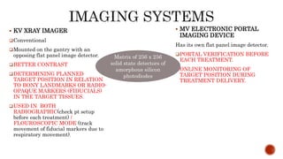
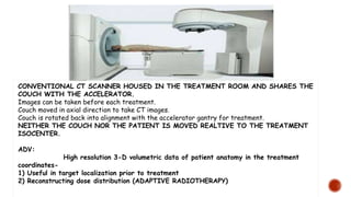
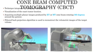
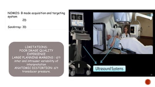
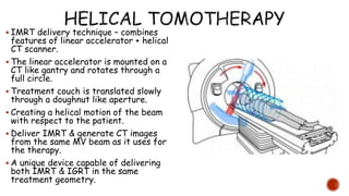
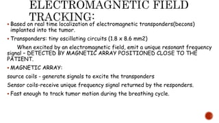
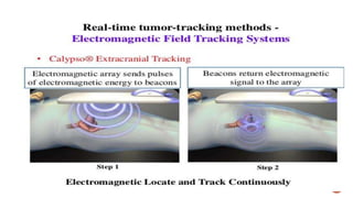
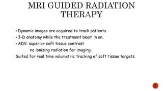
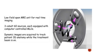
This document discusses image-guided radiation therapy (IGRT) and various IGRT techniques. It describes how IGRT aims to increase the accuracy and precision of radiotherapy delivery by applying image-based target relocalization. Common IGRT techniques mentioned include portal imaging, on-board cone-beam CT (CBCT), in-room CT, ultrasound and real-time tumor tracking. CBCT allows visualization of the tumor location using kilovoltage or megavoltage X-rays rotating around the patient. Real-time tumor tracking involves synchronizing radiation delivery with the respiratory cycle using implanted fiducial markers and fluoroscopy.
1.Aim of Radiotherapy
The goal of radiotherapy is to deliver a prescribed dose of radiation to the Target while sparing surrounding Healthy tissues to the largest extent possible
2.Organ Motion
Intra-fraction motion
during the fraction
Heartbeat
Swallowing
Coughing
Eye movement
Inter-fraction motion
- in between the fractions
Tumour change
Weight gain/loss
Positioning deviation
Breathing
Bowel and rectal filling
Bladder filling
Muscle relaxation/tension
3. Respiratory motion affects:
Respiratory motion affects all tumour sites in the thorax, abdomen and Pelvis. Tumours in the Lung, Liver, Pancreas, Oesophagus, Breast, Kidneys, prostate
Tumour displacement varies depending on the site and organ Location
Lung tumours can move several cm in any direction during irradiation
It is most prevalent and prominent in Lung cancers
4. Problems associated with respiratory motion during RT
Image acquisition limitations
Treatment planning limitations
Radiation delivery limitations
5. Methods to Account for Respiratory Motion
1. Motion encompassing methods
2. Respiratory gating methods
3. Breath hold methods
4. Forced shallow breathing with abdominal compression
5. Real-time tumor tracking methods
Summary:
The management of respiratory motion in radiation oncology is an evolving field
IGRT provides a solution for combating organ motion in radiotherapy
Delivering higher dose to tumor and less dose to normal tissue.
Limited clinical studies, needs to be studied further
IGRT – the future of radiotherapy
Conventional radiotherapy treatments are delivered with radiation beams that are of uniform intensity across the field (within the flatness specification limits). Wedges or compensators are used to modify the intensity profile to offset contour in irregularities and produce more uniform composite dose distributions such as in techniques using wedges. This process of changing beam intensity profile to meet the goals of a composite plan is called intensity modulation
IMRT refers to a radiation therapy technique in which nonuniform fluence is delivered to the patient from any given position of the treatment beam to optimize the composite dose distribution. The optimal fluence profiles for a given set of beam directions are determined through inverse planning. The fluence files thus generated are electronically transmitted to the linear accelerator, which is computer controlled, to deliver intensity modulated beams (IMBs) as calculated.
This seminar is presented as a part of weekly journal club and seminar presented in Apollo Hospital,Kolkata Department of Radiation Oncology.This seminar is moderated by Dr Tanweer Shahid.
The vmat vs other recent radiotherapy techniquesM'dee Phechudi
VMAT is a new type of intensity-modulated radiation therapy (IMRT) treatment technique that uses the same hardware (i.e. a digital linear accelerator) as used for IMRT or conformal treatment, but delivers the radiotherapy treatment using a rotational or arc geometry rather than several static beams.
This technique uses continuous modulation (i.e. moving the collimator leaves) of the multileaf collimator (MLC) fields, continuous change of the fluence rate (the intensity of the X rays) and gantry rotation speed across a single or multiple 360 degree rotations
The vital importance of imaging techniques in radiation oncology now extends beyond diagnostic evaluation and treatment planning. Radiotherapy requires input from imaging for treatment planning and execution, when the treatment target is not located on the surface and, inspection and visual confirmation are not feasible. Traditional radiotherapy practices incorporate use of anatomic surface landmarks as well as radiologic correlation with 2D imaging in the form of port films or fluoroscopic imaging. Targets to be irradiated and normal tissues to be spared are delineated on CT scans in the planning process. Recent technical advances have enabled the integration of various imaging modalities into the everyday practice of radiotherapy directly at the linear accelerator. IGRT seeks to address geometric uncertainties in dose placement for target and normal tissues. It has become a routine part of current RT practice. Safe application of IGRT technology requires additional training and careful integration into the clinical process. IGRT reveals changes in anatomy during treatment which challenges conventional practices. IGRT facilitates the precise application of specialized irradiation techniques with narrow safety margins to radiosensitive organs.
A summary of recent innovations in radiation oncology focussing on the priniciples of different techniques and their application. An overview of clinical results has also been given
Enhancement of clinical outcome using OBI and Cone Beam CT in Radiotherapydrsumandas
Improving the quality of radiation treatment by use of on board image Guidance (OBI) with KV Xray and CBCT. This decreases the variability in daily dose delivery and improves outcome.
enhancement of clinical outcome using On Board Image Guidance Cone Beam CTdrsumandas
Delivery of Radiation therapy has undergone a paradigm shift with the latest technologies. Now with these techniques we can limit daily variation of dose delivery by use of these technology.
A 4 part seminar on 3D cbct technology for seminar presentations. with added technical details and considerations with differences between a CT technology.
Also it features the technical parameters ,uses and how it is considered useful in each departments of medicine and dentistry.
Intensity Modulated Radiation Therapy (IMRT) is an advanced mode of high-precision radiotherapy that uses computer-controlled linear accelerators to deliver precise radiation doses to a malignant tumor or specific areas within the tumor by reducing radiation dose to the nearby normal tissues.
The Roman Empire A Historical Colossus.pdfkaushalkr1407
The Roman Empire, a vast and enduring power, stands as one of history's most remarkable civilizations, leaving an indelible imprint on the world. It emerged from the Roman Republic, transitioning into an imperial powerhouse under the leadership of Augustus Caesar in 27 BCE. This transformation marked the beginning of an era defined by unprecedented territorial expansion, architectural marvels, and profound cultural influence.
The empire's roots lie in the city of Rome, founded, according to legend, by Romulus in 753 BCE. Over centuries, Rome evolved from a small settlement to a formidable republic, characterized by a complex political system with elected officials and checks on power. However, internal strife, class conflicts, and military ambitions paved the way for the end of the Republic. Julius Caesar’s dictatorship and subsequent assassination in 44 BCE created a power vacuum, leading to a civil war. Octavian, later Augustus, emerged victorious, heralding the Roman Empire’s birth.
Under Augustus, the empire experienced the Pax Romana, a 200-year period of relative peace and stability. Augustus reformed the military, established efficient administrative systems, and initiated grand construction projects. The empire's borders expanded, encompassing territories from Britain to Egypt and from Spain to the Euphrates. Roman legions, renowned for their discipline and engineering prowess, secured and maintained these vast territories, building roads, fortifications, and cities that facilitated control and integration.
The Roman Empire’s society was hierarchical, with a rigid class system. At the top were the patricians, wealthy elites who held significant political power. Below them were the plebeians, free citizens with limited political influence, and the vast numbers of slaves who formed the backbone of the economy. The family unit was central, governed by the paterfamilias, the male head who held absolute authority.
Culturally, the Romans were eclectic, absorbing and adapting elements from the civilizations they encountered, particularly the Greeks. Roman art, literature, and philosophy reflected this synthesis, creating a rich cultural tapestry. Latin, the Roman language, became the lingua franca of the Western world, influencing numerous modern languages.
Roman architecture and engineering achievements were monumental. They perfected the arch, vault, and dome, constructing enduring structures like the Colosseum, Pantheon, and aqueducts. These engineering marvels not only showcased Roman ingenuity but also served practical purposes, from public entertainment to water supply.
June 3, 2024 Anti-Semitism Letter Sent to MIT President Kornbluth and MIT Cor...Levi Shapiro
Letter from the Congress of the United States regarding Anti-Semitism sent June 3rd to MIT President Sally Kornbluth, MIT Corp Chair, Mark Gorenberg
Dear Dr. Kornbluth and Mr. Gorenberg,
The US House of Representatives is deeply concerned by ongoing and pervasive acts of antisemitic
harassment and intimidation at the Massachusetts Institute of Technology (MIT). Failing to act decisively to ensure a safe learning environment for all students would be a grave dereliction of your responsibilities as President of MIT and Chair of the MIT Corporation.
This Congress will not stand idly by and allow an environment hostile to Jewish students to persist. The House believes that your institution is in violation of Title VI of the Civil Rights Act, and the inability or
unwillingness to rectify this violation through action requires accountability.
Postsecondary education is a unique opportunity for students to learn and have their ideas and beliefs challenged. However, universities receiving hundreds of millions of federal funds annually have denied
students that opportunity and have been hijacked to become venues for the promotion of terrorism, antisemitic harassment and intimidation, unlawful encampments, and in some cases, assaults and riots.
The House of Representatives will not countenance the use of federal funds to indoctrinate students into hateful, antisemitic, anti-American supporters of terrorism. Investigations into campus antisemitism by the Committee on Education and the Workforce and the Committee on Ways and Means have been expanded into a Congress-wide probe across all relevant jurisdictions to address this national crisis. The undersigned Committees will conduct oversight into the use of federal funds at MIT and its learning environment under authorities granted to each Committee.
• The Committee on Education and the Workforce has been investigating your institution since December 7, 2023. The Committee has broad jurisdiction over postsecondary education, including its compliance with Title VI of the Civil Rights Act, campus safety concerns over disruptions to the learning environment, and the awarding of federal student aid under the Higher Education Act.
• The Committee on Oversight and Accountability is investigating the sources of funding and other support flowing to groups espousing pro-Hamas propaganda and engaged in antisemitic harassment and intimidation of students. The Committee on Oversight and Accountability is the principal oversight committee of the US House of Representatives and has broad authority to investigate “any matter” at “any time” under House Rule X.
• The Committee on Ways and Means has been investigating several universities since November 15, 2023, when the Committee held a hearing entitled From Ivory Towers to Dark Corners: Investigating the Nexus Between Antisemitism, Tax-Exempt Universities, and Terror Financing. The Committee followed the hearing with letters to those institutions on January 10, 202
Macroeconomics- Movie Location
This will be used as part of your Personal Professional Portfolio once graded.
Objective:
Prepare a presentation or a paper using research, basic comparative analysis, data organization and application of economic information. You will make an informed assessment of an economic climate outside of the United States to accomplish an entertainment industry objective.
Operation “Blue Star” is the only event in the history of Independent India where the state went into war with its own people. Even after about 40 years it is not clear if it was culmination of states anger over people of the region, a political game of power or start of dictatorial chapter in the democratic setup.
The people of Punjab felt alienated from main stream due to denial of their just demands during a long democratic struggle since independence. As it happen all over the word, it led to militant struggle with great loss of lives of military, police and civilian personnel. Killing of Indira Gandhi and massacre of innocent Sikhs in Delhi and other India cities was also associated with this movement.
Introduction to AI for Nonprofits with Tapp NetworkTechSoup
Dive into the world of AI! Experts Jon Hill and Tareq Monaur will guide you through AI's role in enhancing nonprofit websites and basic marketing strategies, making it easy to understand and apply.
Biological screening of herbal drugs: Introduction and Need for
Phyto-Pharmacological Screening, New Strategies for evaluating
Natural Products, In vitro evaluation techniques for Antioxidants, Antimicrobial and Anticancer drugs. In vivo evaluation techniques
for Anti-inflammatory, Antiulcer, Anticancer, Wound healing, Antidiabetic, Hepatoprotective, Cardio protective, Diuretics and
Antifertility, Toxicity studies as per OECD guidelines
Acetabularia Information For Class 9 .docxvaibhavrinwa19
Acetabularia acetabulum is a single-celled green alga that in its vegetative state is morphologically differentiated into a basal rhizoid and an axially elongated stalk, which bears whorls of branching hairs. The single diploid nucleus resides in the rhizoid.
Welcome to TechSoup New Member Orientation and Q&A (May 2024).pdfTechSoup
In this webinar you will learn how your organization can access TechSoup's wide variety of product discount and donation programs. From hardware to software, we'll give you a tour of the tools available to help your nonprofit with productivity, collaboration, financial management, donor tracking, security, and more.
Embracing GenAI - A Strategic ImperativePeter Windle
Artificial Intelligence (AI) technologies such as Generative AI, Image Generators and Large Language Models have had a dramatic impact on teaching, learning and assessment over the past 18 months. The most immediate threat AI posed was to Academic Integrity with Higher Education Institutes (HEIs) focusing their efforts on combating the use of GenAI in assessment. Guidelines were developed for staff and students, policies put in place too. Innovative educators have forged paths in the use of Generative AI for teaching, learning and assessments leading to pockets of transformation springing up across HEIs, often with little or no top-down guidance, support or direction.
This Gasta posits a strategic approach to integrating AI into HEIs to prepare staff, students and the curriculum for an evolving world and workplace. We will highlight the advantages of working with these technologies beyond the realm of teaching, learning and assessment by considering prompt engineering skills, industry impact, curriculum changes, and the need for staff upskilling. In contrast, not engaging strategically with Generative AI poses risks, including falling behind peers, missed opportunities and failing to ensure our graduates remain employable. The rapid evolution of AI technologies necessitates a proactive and strategic approach if we are to remain relevant.
2. PROCEDURE THAT REFINES THE DELIVERY OF THERAPEUTIC
RADIATION BY APPLYING IMAGE BASED TARGET
RELOCALISATION TO ENSURE PROPER PATIENT
REPOSITIONING FOR THE PURPOSE OF ENSURING ACCURATE
TREATMENT AND MINIMIZING THE VOLUME OF NORMAL
TISSUE EXPOSED TO IONISING RADIATION.
3. INCREASING THE ACCURACY AND PRECISION OF
RADIOTHERAPY DELIVERY.
INACCURACY:
SYSTEMIC ERRORS-
• IMPROPER TARGET DELINEATION.
• POORLY REPRESENTATIVE
SIMULATION.
• DISSOCIATION B/W SKIN MARK &
INTERNAL ANATOMY.
IMPRECISION:
STOCHASTIC (RANDOM) ERRORS-
PRODUCES VARIANCE IN THE SPATIAL
LOCATION OF THE TREATMENT AROUND
THE TRUE TARGET-
• INEVITABLE FLUCTUATIONS IN THE
DAILY SETUP.
• UNPREDICTABLE TARGET MOTIONS
9. KV XRAY IMAGER
Conventional
Mounted on the gantry with an
opposing flat panel image detector.
BETTER CONTRAST
DETERMINING PLANNED
TARGET POSITION IN RELATION
TO BONY LANDMARKS OR RADIO-
OPAQUE MARKERS (FIDUCIALS)
IN THE TARGET TISSUES.
USED IN BOTH
RADIOGRAPHIC(check pt setup
before each treatment) /
FLOUROSCOPIC MODE (track
movement of fiducial markers due to
respiratory movement).
MV ELECTRONIC PORTAL
IMAGING DEVICE
Has its own flat panel image detector.
PORTAL VERIFICATION BEFORE
EACH TREATMENT.
ONLINE MONITORING OF
TARGET POSITION DURING
TREATMENT DELIVERY.
Matrix of 256 x 256
solid state detectors of
amorphous silicon
photodiodes
12. CONVENTIONAL CT SCANNER HOUSED IN THE TREATMENT ROOM AND SHARES THE
COUCH WITH THE ACCELERATOR.
Images can be taken before each treatment.
Couch moved in axial direction to take CT images.
Couch is rotated back into alignment with the accelerator gantry for treatment.
NEITHER THE COUCH NOR THE PATIENT IS MOVED REALTIVE TO THE TREATMENT
ISOCENTER.
ADV:
High resolution 3-D volumetric data of patient anatomy in the treatment
coordinates-
1) Useful in target localization prior to treatment
2) Reconstructing dose distribution (ADAPTIVE RADIOTHERAPY)
14. Technique integrates CT imaging with LINAC.
Visualization of the exact tumor location.
Acquiring multiple planar images produced by KV or MV cone beam rotating 360 degrees
around the patient.
Filtered back projection algorithm is used to reconstruct the volumetric images of the target
volume.
15. The Xray tube is mounted on a retractable arm at 90 deg with respect to
the central axis of the linear accelerator beam.
Image is generated by the flat panel area detectors mounted opposite the
xray tube.
3D volumetric images are reconstructed by the computer using a filtered
back projection algorithm.
The on board KV imaging system is capable of radiography, fluoroscopy and
cone beam computed tomography.
16. Made possible by the traditional EPID by a Si flat panel
detector.
The Xray beam in this case is the therapy beam of the
accelerator.
Planar projection images are acquired from multiple directions as
the xray source and the detector rotate about the patient.
The soft tissue contrast is reduced in the MV-CBCT.
17. Less susceptibility to imaging artifacts (d/t metallic objects eg-
hip implants, dental fillings and surgical clips).
No need of extrapolating attenuation coeffiecients from KV
beams to therapeutic beams.
The known dose distribution characteristics of the therapeutic
beam allow more accurate calculation of imaging dose in the
MVCBCT acquisition process.
Implementation of MVCBCT does not require extensive
modification of linear accelerator that is already equipped with
EPID.
18. ADVANTAGES OF KV-CBCT OVER MV-CBCT
Better contrast & spatial resolution.
Better soft tissue visibility at much lower doses.
Compatibility of KV-CBCT images with the reference treatment plan images
for patient setup verification and correction.
Combination of Radiography, fluoroscopy, and CBCT capabilities from the
same source and detector which provides great flexibility in implementing
the goals of IGRT.
20. ULTRASOUND: NON INVASIVE, NON RADIOGRAPHIC
REAL TIME IMAGING TECHNIQUE
MOST COMMON IGRT APPROACHES: particularly prostate.
Rationale: emission of high frequency sound waves to produce
images of internal anatomy.
(a transducer is encased in a probe applied to the skin
surface, reflecting sound waves back as echoes when a change in
impedance is encountered due to density differences between
tissues.)
THE TIME AN ECHO TAKES TO RETURN IS USED TO
CALCULATE THE DEPTH OF TISSUE INTERFACE.
3 operational modes: B (brightness) mode is the primary one.
21. NOMOS: B mode acquisition and targeting
system
2D
SonArray: 3D
LIMITATIONS:
POOR IMAGE QUALITY
EXPERIENCE
LARGE PLANNING MARGINS : d/t
inter and intrauser variability of
interpretation
ANATOMIC DISTORTION: d/t
transducer pressure.
22. IMRT delivery technique – combines
features of linear accelerator + helical
CT scanner.
The linear accelerator is mounted on a
CT like gantry and rotates through a
full circle.
Treatment couch is translated slowly
through a doughnut like aperture.
Creating a helical motion of the beam
with respect to the patient.
Deliver IMRT & generate CT images
from the same MV beam as it uses for
the therapy.
A unique device capable of delivering
both IMRT & IGRT in the same
treatment geometry.
23. Detect systemic error.
Position the patient, target or organ at risk.
Modify the treatment plan or choose the appropriate plan.
Detect changes in patient or target size.
Helps in controlling the respiratory motion of the target, most
prominent in lung cancers.
24. Tumor motion in lungs or other sites cannot be predicted with any
degree of accuracy.
Real time tumor tracking or gating process is required to manage
target motion in radiotherapy.
It can be done by -
1) 4D COMPUTED TOMOGRAPHY.
2) REAL TIME TUMOR TRACKING.
25. Acquiring CT scans synchronously with the patient’s respiratory phases.
(4D – being time)
Breathing cycle is divided into 10 respiratory phases and multiple CT volumes
are taken at each phase.
May involve as many as 1500 slices.
One commonly used method – use a reference signal from up and down motion
that could be correlated with the target motion.
eg-Varian Real Time Position Management and Gating System
26. Computer controlled video based system : box with infrared reflectors is
placed on the patient’s surface and the motion of the box is tracked by IR
cameras.
27. The RPM system can be interfaced with a CT or a PET-CT scanner for 4D CT
imaging.
These images are used to design an individualized treatment plan in which
radiation is administered at optimum moments of breathing cycle.
4D CT data acquisition:
2 modes - 1) Prospective gating.
2) Retrospective gating.
Beam is synchronized with the respiratory cycle and switches the beam on
only at selected times of respiration.
Gating threshold is set when the target is in the desired position of the
respiratory cycle .
The gating system turns the beam on or off in accordance with the
programmed gating threshold.
28. prospective
Images are collected only at
one phase of respiratory
cycle.
Eg- end of expiration or end
of inspiration.
retrospective
Scan data for each axial slice
are acquired at all phases of
respiration.
Correlation or registration of
the CT images with the
respiratory phases is
conducted after the data is
acquired.
29. To detect respiratory motion using radiation beam and to follow the
tumor’s changing position.
Surrogate markers used.(identify tumor)
External fiducials on the skin surface and internal fiducials
implanted directly into the tumor.
In order to make this work- the time delay between detection of
motion and the corrective action should be short.(order of 100ms)
2 ways-
1) Flouroscopy based tracking system.
2) Electromagnetic field tracking.
30. Most available tracking system use fluoroscopy to detect metal fiducials
implanted into the tumor.
It continuously images during treatment and the beam is turned on and off
depending on the detected image.
Some are installed in the accelerator gantry while others are installed in the
room.
Eg- 1) Room mounted
2) Gantry mounted
3) Robotic systems
31. By University of Hokkaido.
Dual Xray tube that rotate on a circular track embedded in the floor.
Each tube has a Xray detector that rotates on the ceiling mounted track.
During irradiation the 2 imaging systems continuously track fiducials in the
tumor.
Two fluoroscopic images are combined to construct trajectories of tumor
motion in 3D.
32.
33. Provide IGRT for the delivery of stereotactic radiosurgery and stereotactic
radiotherapy.
Optical + Flouroscopy based tracking are used together.
Optical system consist of IR reflecting markers that are placed on marked
spots on the patients surface or on the immobilization device which is detected
by 2 IR cameras mounted in the ceiling .
Internal target localization : done by stereoscopic Xray imaging device .
2 Xray device on the floor and 2 opposing a Si-detector mounted in the ceiling.
XRAY IMAGING SYSTEM IS FULLY INTEGRTED WITH IR TRACKING
SYSTEM .
TARGET ALIGNMENT IS BASED ON IMPLANTED FIDUCIALS OR
INTERNAL BONY LANMARKS.
34.
35. CYBERKNIFE: image guided frameless stereotactic radiosurgery system for
treating cranial & extracranial lesions.
Two diagnostic xray tube mounted orthogonally in the ceiling and 2 opposing Si-
flat panel detectors.
The robotic arm has 6 degrees of freedom and is capable of pointing the linac
beam almost anywhere in space.
After sensing any target motion, robotic arm moves the beam to the newly
detected target position for alignment.
Not restricted to isocenter geometry, can be directed independently without a
fixed isocenter.
38. Based on real time localization of electromagnetic transponders(becons)
implanted into the tumor.
Transponders: tiny oscillating circuits (1.8 x 8.6 mm2)
When excited by an electromagnetic field, emit a unique resonant frequency
signal – DETECTED BY MAGNETIC ARRAY POSITIONED CLOSE TO THE
PATIENT.
MAGNETIC ARRAY:
source coils - generate signals to excite the transponders
Sensor coils-receive unique frequency signal returned by the responders.
Fast enough to track tumor motion during the breathing cycle.
39.
40. Dynamic images are acquired to track patients.
3-D anatomy while the treatment beam in on.
ADV: superior soft tissue contrast
no ionizing radiation for imaging.
Suited for real time volumetric tracking of soft tissue targets.
41. Low field open MRI unit for real time
imaging
3 cobalt 60 sources, each equipped with
computer controlled MLCs.
Dynamic images are acquired to track
patient 3D anatomy while the treatment
beam is on.
42. POTENTIAL OF EXCESSIVE DOSE TO THE PATIENT DUE TO VARIOUS
RADIOGRAPHIC IMAGING PROCEDURES.
There’s a need to evaluate therapeutic & imaging dose in a much balanced manner.
Editor's Notes
SCANS CAN BE COMPARED TO THE REFERANCE TREATMENT PLAN PERIODICALLY URING THE TREATMENT DURATION…..SETUP CORRCTIONS OR TREATMENT PARAMETERS CAN BE MODIFIED TO MINIMIZE VARIATION B/W THE PLANNE AND THE ACTUAL TREATMENT.