
Recommended
More Related Content
What's hot
What's hot (20)
Similar to Treatment planning in Radiotherapy - field shaping, separation and matching
Similar to Treatment planning in Radiotherapy - field shaping, separation and matching (20)
More from SACHINS700327
More from SACHINS700327 (8)
Recently uploaded
Recently uploaded (20)
Treatment planning in Radiotherapy - field shaping, separation and matching
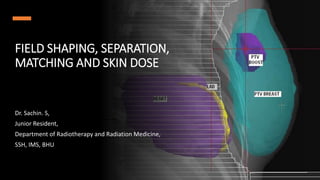
- 1. FIELD SHAPING, SEPARATION, MATCHING AND SKIN DOSE Dr. Sachin. S, Junior Resident, Department of Radiotherapy and Radiation Medicine, SSH, IMS, BHU
- 2. OUTLINE • Field shaping • Custom blocks • Independent Jaws • Multileaf Collimators • Skin dose • Field separation • Geometric • Dosimetric
- 3. WHY TO SHAPE A BEAM? • Collimators provide cuboid field shape • Target can be • regular • Cylindrical • Spherical • Ovoid • cuboid with corners trimmed • irregular
- 4. FIELD SHAPING • Dictated by: • Complete coverage of tumour with prescription dose • Including local and distal disease • Minimal dose to normal tissue • Vital organ tolerance • Primary beam transmission of < 5% through blocked region
- 5. HALF VALUE LAYER n is the number of HVLs
- 6. • High atomic number • High density • Easily available • Less expensive FIELD BLOCKS – IDEAL SHIELD
- 8. DIVERGENT BLOCKS • To reduce transmission penumbra • Uniform penumbra across the beam • Better suited for Linac beams (those with small geometric penumbra)
- 9. CUSTOM BLOCKS PROPERTY LEAD CERROBEND Density (g/𝑐𝑚3) 11.3 9.4 (85% of lead) Melting point 327℃ 70℃ (95℃ for cadmium less) Thickness (4MV) 6cm 7.5cm (1.21 times of Lead)
- 10. CUSTOM BLOCKS
- 11. POSITIVE BLOCKS NEGATIVE BLOCKS
- 12. CUSTOM BLOCKS DISADVANTAGES • Production is time consuming • Cumbersome to use • Increase in treatment and setup time • Toxic fumes while fabricating • Daily variation (manual placement)
- 13. INDEPENDENT JAWS • Asymmetric fields (rectangular blocking) – “beam splitting” • Transmission of 1% (approx.) • Blocks field without changing isocenter • Shift of isodose curves towards blocked edge (elimination of photon and electron scatter from blocked part)
- 15. MULTILEAF COLLIMATORS (MLC) • Large number (60-80) of collimating leaves or blocks • Driven automatically, move independently • Generate a field of any shape • <1cm wide, 6-7.5 cm thick, Tungsten alloy • Transmission: Intra-leaf (<2%), interleaf (<3%)
- 16. MLC - ADVANTAGES • Less time consuming (no need to enter treatment room often) • Less setup and overall treatment time • Automation of field shaping and beam intensity modulation • Suited for treating multiple fields
- 17. MLC - DEMERITS • Island blocking not possible • Larger physical penumbras than Cerrobend blocks and jaws • Jagged boundaries – difficulty in field matching • Radiation leakage between the leaves
- 18. SKIN DOSE • Skin sparing - desirable feature of MV beams • Reduced or lost – secondary electrons contamination or backscattered radiation • 2° electrons from beam shaping devices or air
- 19. SKIN SPARING – FUNCTION OF PHOTON ENERGY
- 20. ABSORBER TO SKIN DISTANCE • Block tray absorbs the 2° electrons produced already but produces own 2° electrons • Skin dose increases with decreased tray to surface distance • Point of maximum dose buildup also moves close to the surface
- 21. EFFECT OF FIELD SIZE • Larger field size – more 2° electrons • Poor skin sparing for large field sizes
- 22. ELECTRON FILTERS • Medium atomic number materials produce better skin sparing • Less forward scattering • Used in large field size and block-tray distance 15-20cms • Thickness equal to 2° electron range: 0.9mm of tin for 𝐶𝑜60
- 23. OBLIQUE INCIDENCE • As the angle of beam incidence increases: • Skin dose increases • Depth of 𝑑𝑚𝑎𝑥 decreases
- 24. OBLIQUITY FACTOR • Obliquity factor (OF) - defined as the dose at a point in phantom on central axis of a beam incident at angle θ, with respect to the perpendicular to the surface, divided by the dose at the same point and depth along central axis with the beam incident at angle 0 degrees • For tangential beam incidence:
- 25. FIELD SEPARATION • Need for treatment with adjacent fields for large areas • Hodgkin’s lymphoma • Medulloblastoma • Some head and neck fields • Divergence of adjacent fields produce • Hotspots at depths • Cold spots at surfaces • Large dosage errors across the junction
- 29. METHODS OF FIELD SEPARATION • Geometric • Fields joined at 50% isodose lines • 100% at the Junction point • Dosimetric • Composite isodose distribution • Uniform at the desired depth
- 30. GEOMETRIC
- 31. IDEAL (NO OVERLAP) MATCHING
- 33. THREE FIELD OVERLAP S1 + S2 + ∆𝑆′
- 38. CRANIOSPINAL FIELDS • More convenient method: • Half beam block • Independent jaws • No couch rotation • Feathering of junction to smear out junctional dose distribution
- 39. FIELD MATCHING GUIDELINES • The site of field matching should be chosen, in so far as possible, over an area that does not contain tumor or a critically sensitive organ • If the tumor is superficial at the junction site, the fields should not be separated because a cold spot on the tumor will risk recurrence • However, if the diverging fields abut on the skin surface, they will overlap at depth
- 40. FIELD MATCHING GUIDELINES • In some cases, this may be clinically acceptable, provided the excessive dosage delivered to the underlying tissues does not exceed their tolerance • In particular, the tolerances of critical structures such as the spinal cord must not be exceeded • In the case of a superficial tumor with a critical organ located at depth, one may abut the fields at the surface but eliminate beam divergence using a beam splitter or by tilting the beams
- 41. FIELD MATCHING GUIDELINES • For deep-seated tumors, the fields may be separated on the skin surface so that the junction point lies at the midline • The line of field matching must be drawn at each treatment session on the basis of the first field treated • It is not necessary anatomically to reproduce this line every day because variation in its location will only smear the junction point, which is desirable • A field-matching technique must be verified by actual isodose distributions before it is adopted for general clinical use
- 42. THANK YOU