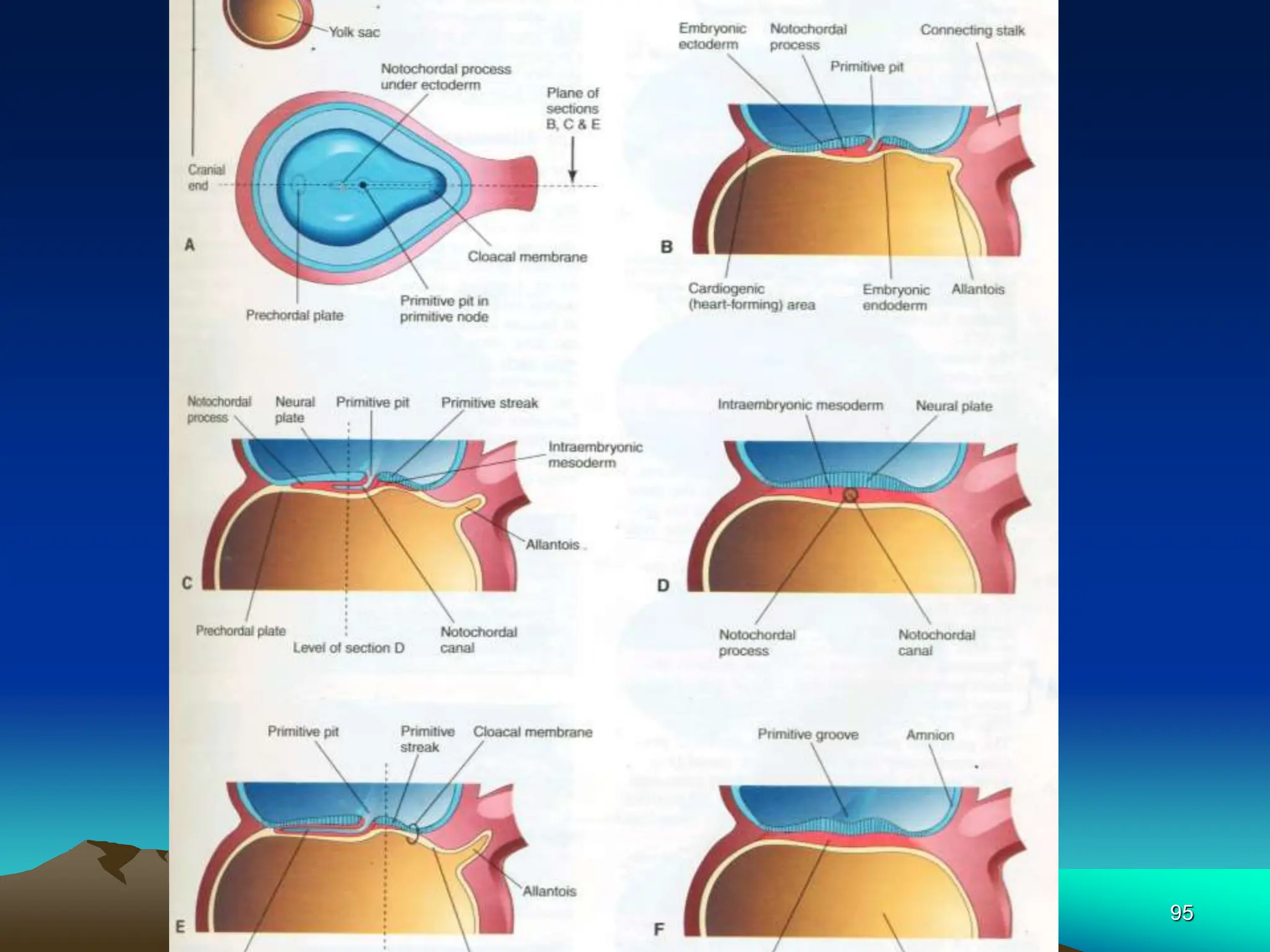
1
2
DEVELOPMENT OF
THE HUMAN BRAIN
Dr. Ademola A. Oremosu
Associate Professor of
Anatomy
ABUAD
3
Objectives/Course Outline
 Introduction
 Primary & secondary brain vesicles
 Embryonic brain flexures
 Development of the hindbrain
 Development of the midbrain
 Development of the forebrain
4
Concerning the development of the brain
• The entire nervous system is of
ectodermal origin
• By end of 3rd week, neural folds have begun to
move together & fuse, converting the neural
plate into neural tube, the primordium of the
CNS.
• Primitive streak results from proliferation &
migration of cells of the epiblast to the
median plane of the embryonic disc
• By the 18th day floor of the notochordal process
fuses with underlying endoderm.
5
Secondary brain vesicles include
• Telecenphalon
• Diencephalon
• Metencephalon
• Myelencephalon
• rhombencephalon
6
INTRODUCTION
Divisions of the Brain
• Forebrain, midbrain, hindbrain
• 5 major subdivisions
• Forebrain
1. Telencephalon
2. Diencephalon
7
• Midbrain
3. Mesencephalon
• Hindbrain
4. Metencephalon
5. Myelencephalon
8
• The entire nervous system is of
ectodermal origin, and its first rudiment
is seen in the neural groove which
extends along the dorsal aspect of the
embryo
9
 Gastrulation is the process by
which the bilaminar embryonic
disc is converted into a trilaminar
embryonic disc.
 Begins with formation of the
primitive streak at the caudal end
of the embryo.
10
 Primitive streak results from proliferation
& migration of cells of the epiblast to the
median plane of the embryonic disc.
 Cranial end proliferates to form primitive
node.
 Primitive groove develops in the primitive
streak.
 Primitive pit – small depression in
primitive node.
 Primitive groove & pit result from
invagination of epiblastic cells.
11
16-day
12
13
• Some mesenchymal cells migrate cranially from
primitive node & pit, forming a median cellular
cord- notochordal/head process.
• By the 18th day floor of the notochordal process
fuses with underlying endoderm.
• Gradually the lumen of notochordal process
disappears completely.
• With further development the notochordal cells
proliferate & form a solid cord- the definitive
notochord/chordamesoderm (end of 3rd wk).
• Primitive streak undergoes degenerative
changes & disappears by the end of the 4th
week (!Sacrococcygeal teratoma ~1/35000 newborns).
14
15
16
• As the notochord develops, the embryonic
ectoderm over it thickens to form an elongated,
slipper-shaped plate- neural plate.
• The ectoderm of the neural plate forms the
CNS.
• At first the neural plate corresponds precisely in
length to the underlying notochord.
• Neural plate appears cranial to the primitive
node & dorsal to the notochord & mesoderm
adjacent to it.
• As the notochord elongates, the neural plate
broadens & eventually extends cranially as far
as the oropharyngeal membrane.
17
18
• Eventually the neural plate extends beyond the
notochord.
• ~ 18th day, neural plate invaginates along its
central axis to form a longitudinal median-
neural groove, that has neural folds on each
side.
• Neural folds become particularly prominent at
the cranial end of embryo & are first signs of
brain development.
• By end of 3rd week, neural folds have begun to
move together & fuse, converting the neural
plate into neural tube, the primordium of the
CNS.
19
20
21
22
Regarding the notochord
• The notochordal process and adjacent
mesoderm induce the overlying embyonic
ectoderm to form the neural plate
• As the notochordal process elongates, the
primitive streak shortens
• At end of the 3rd week the notochordal process
is transformed into notochord
• Embryonic disc is originally egg-shaped but
soon becomes pear-shaped & then slipperlike
• The notochord functions as the primary inductor
in the early embryo
23
• It is a prime mover in a sries of signal-
calling episodes that ultimately transform
unspecialized embyonic cells into definitive
adult tissues & organs
• The notochord is an intricate structure
around which the vertebral column forms.
• It extends from oropharyngeal membrane
to the primitive node
• Notochord degenerates & disappears as
the bodies of the vertebrae form, but
persists as the nucleus pulposus of each IV
disc.
• Neurulation is completed during the 4th week.
24
NEURAL CREST FORMATION
• As the neural folds fuse to form neural tube,
some neuroectodermal cells on the crest of
each fold lose their epithelial affinities &
attachments to neighbouring cells.
• As the neural tube separates from surface
ectoderm, neural crest cells migrate
dorsolaterally on each side of the neural
tube.
• The neural crest is eventually formed
between the neural tube & the overlying
surface ectoderm.
25
26
27
28
Derivatives of Neural Crest Cells
 Spinal ganglia (dorsal ganglia)
 Ganglia of ANS
 Ganglia of cranial nerves V, VII, IX &
X (in part)
 Neurolemmal sheaths of peripheral
nerves
 Meninges of brain & spinal cord
29
Embryological Origin of the Brain
 The brain develops from the anterior end of
the neural tube (cranial to the 4th pair of
somites), which at an early period becomes
expanded into 3 vesicles, the primary
cerebral vesicles.
 These are marked off from each other by
intervening constrictions, and are named the
fore-brain or prosencephalon, the mid-
brain or mesencephalon, and the hind-
brain or rhombencephalon—the last being
continuous with the medulla spinalis.
30
Secondary Brain Vesicles
 During the 5th week
forebrain partly divides into 2 secondary
vesicles- telecenphalon &
diencephalon;
Midbrain/ mesencephalon does not
divide;
hindbrain partly divides into
metencephalon & myelencephalon;
hence 5 secondary brain vesicles
31
 As a result of unequal growth of these
different parts 3 flexures are formed
and the embryonic brain becomes
bent on itself in a somewhat zigzag
fashion
 The 2 earliest flexures are concave
ventrally and are associated with
corresponding flexures of the whole
head
32
The first flexure appears in the
region of the mid-brain, and is
named the ventral cephalic
flexure
33
 By means of the midbrain flexure, fore-
brain is bent in a ventral direction around
the anterior end of the notochord & fore-
gut, with the result that the floor of the
fore-brain comes to lie almost parallel with
that of the hind-brain.
 This flexure causes the mid-brain to
become, for a time, the most prominent
part of the brain, since its dorsal surface
corresponds with the convexity of the
curve.
34
 The 2nd bend appears at the junction of
the hind-brain and medulla spinalis -
cervical flexure, and increases from 3rd
to the end of 5th week, when the hind-
brain forms nearly a right angle with the
medulla spinalis
 After the 5th week, erection of the head
takes place & the cervical flexure
diminishes & disappears.
35
5th week
36
 The 3rd bend is named the pontine
flexure
 Located in future pontine region; divides
hindbrain into caudal- myelencephalon &
rostral metencephalon
 It differs from the other 2 in that (a) its
convexity is forward, and (b) it does not
affect the head
37
 The lateral walls of the brain-
tube, like those of the medulla
spinalis, are divided by internal
furrows into alar/dorsal &
basal/ventral laminæ
38
The Hind-brain or Rhombencephalon
The cavity of the hind-brain becomes the
4th ventricle.
At the time when the ventral cephalic
flexure makes its appearance, the length
of the hind-brain exceeds the combined
lengths of the other 2 vesicles.
Immediately behind the mid-brain it
exhibits a marked constriction, the
isthmus rhombencephali.
39
40
From the isthmus the anterior medullary
velum & the superior peduncle of the
cerebellum are formed.
The cerebellum is developed by a
thickening of roof & pons by a
thickening in the floor & lateral walls of the
metencephalon.
41
The floor & lateral walls of the
myelencephalon are thickened to form
the medulla oblongata.
42
43
The development of the medulla
oblongata
• On transverse section the
myelencephalon at an early stage is seen
to consist of 2 lateral walls, connected
across the middle line by floor- and roof-
plates
• Each lateral wall consists of an alar and a
basal lamina, separated by an internal
furrow, the remains of which are
represented in the adult brain by the
sulcus limitans on the rhomboid fossa.
44
5th Week
45
 The contained cavity is more or less
triangular in outline, the base being
formed by the roof-plate, which is
thin and greatly expanded
transversely.
 Neuroblasts are developed in the
alar & basal laminæ & their narrow
stalks are elongated to form the
axis-cylinders of the nerve
fibres.
46
Opposite the furrow or boundary between
the alar & basal laminæ a bundle of nerve
fibres attaches itself to the outer surface of
the alar lamina -tractus solitarius, & is
formed by the sensory fibres of the
glossopharyngeal & vagus n.
 It is developed by an ingrowth of fibres
from the ganglia of the neural crest.
47
48
• Within a few days the rhomboid lip
becomes applied to, and unites with, the
outer surface of the main part of the alar
lamina, and so covers in the tractus
solitarius & also the spinal root of the
trigeminal nerve;
• The nodulus & flocculus of the
cerebellum are developed from the
rhombic lip.
49
• From the alar lamina and its rhombic lip,
neuroblasts migrate into the basal lamina, and
become aggregated to form the olivary nuclei,
while many send their axis-cylinders thru the
floor-plate to the opposite side, and thus
constitute the rudiment of the raphé of the
medulla oblongata.
• By means of this thickening of the ventral
portion, the motor nuclei are buried deeply in
the interior, and, in the adult, are found close to
the rhomboid fossa.
• This is still further accentuated: (a) by the development of
the pyramids, which are formed about the 4th month by
the downward growth of the motor fibres from the
cerebral cortex; and (b) by the fibres which pass to & from
the cerebellum.
50
• On the rhomboid fossa a series of 6
temporary furrows appears- rhombic
grooves.
•They bear a definite relationship to
some of the cranial nerves; thus, from
before backward the 1st & 2nd
grooves overlie the nucleus of the
trigeminal; the 3rd, the nucleus of the
facial; the 4th, that of the abducent;
the 5th, that of the glossopharyngeal;
and the 6th, that of the vagus.
51
Development of the Pons
 The pons is developed from the
ventro-lateral wall of the
metencephalon.
 The pontine flexure causes
divergence of the lateral walls of
the pons, which spreads the
grey matter in floor of 4th
ventricle.
52
Development of the Cerebellum
 The cerebellum is developed in the roof
of the anterior part of the hind-brain
 The alar laminæ of this region become
thickened to form two lateral plates which
soon fuse in the middle line & produce a
thick lamina which roofs in the upper part
of the cavity of the hind-brain vesicle; the
outer surface of which is originally smooth
and convex.
53
 The fissures of the cerebellum appear first
in the vermis & floccular region, and traces
of them are found during the 3rd month;
 Fissures on the cerebellar hemispheres do not
appear until the 5th month.
 The primitive fissures are not developed in the
order of their relative size in the adult—thus the
horizontal sulcus in the fifth month is merely a
shallow groove.
 The best marked of the early fissures are: (a)
the fissura prima between the developing
culmen and declive, and (b) the fissura
secunda between the future pyramid and
uvula.
54
 The flocculus & nodule are developed from
the rhombic lip, and are therefore recognizable
as separate portions before any of the other
cerebellar lobules.
55
 On the ventricular surface of the cerebellar
lamina a transverse furrow, the incisura
fastigii, appears, and deepens to form the tent-
like recess of the roof of the 4th ventricle
 As the cerebellar rudiments enlarge & fuse in
the median plane, they overgrow the rostral half
of 4th ventricle & overlap pons & medulla
 Some neuroblasts in intermediate zone of the
alar plates migrate to marginal zone &
differentiate into neurons of the cerebellar
cortex
 Other neuroblats from these plates give rise to
the central nuclei
56
Regarding the metencephalon
 Cells from the alar plates give rise to
the pontine nuclei, cochlear &
vestibular nuclei, sensory nuclei of
trigeminal nerve
57
The Mid-brain or Mesencephalon
• The mid-brain exists for a time as a thin-
walled cavity of some size, separated from
isthmus rhombencephali behind, and from
the fore-brain in front, by slight constrictions.
• Its cavity becomes relatively reduced in
diameter, and forms the cerebral aqueduct
of the adult brain.
• Its basal laminæ increase in thickness to
form the cerebral peduncles, which are at
first of small size, but rapidly enlarge after
the 4th month.
58
• The neuroblasts of these laminæ are
grouped in relation to sides & floor of
cerebral aqueduct, & constitute the nuclei
of the III & trochlear n. & of the
mesencephalic root of the trigeminal n.
• By a similar thickening process its alar
laminæ are developed into the
quadrigeminal lamina.
59
• Neuroblasts migrate from the alar plates
of midbrain into the tectum & aggregate to
form 4 large groups of neurons- sup & inf
colliculi
• Neuroblasts from basal plates may give
rise to groups of neurons in the
tegmentum (red nuclei, nuclei of 3rd & 4th
cranial nerves, & reticular nuclei)
60
The Fore-brain/Prosencephalon
At a very early period 2 lateral diverticula,
the optic vesicles, appear, one on either
side of the fore-brain; for a time they
communicate with the cavity of the fore-
brain by relatively wide openings.
The peripheral parts of the vesicles
expand, while the proximal parts are
reduced to tubular stalks, the optic
stalks.
61
5th Week
62
The optic vesicle gives rise to the retina
and the epithelium on the back of the
ciliary body and iris.
The optic stalk is invaded by nerve fibres
to form the optic nerve.
63
A 2nd pair of diverticula soon arise more
dorsally & rostrally- cerebral/telencephalic
vesicles- primordia of cerebral
hemispheres.
The cavities of these diverticula are the
rudiments of the lateral ventricles; they
communicate with the median part of the
fore-brain cavity by relatively wide
openings, which ultimately form the
interventricular foramen.
64
The median portion of the wall of the fore-
brain vesicle consists of a thin lamina, the
lamina terminalis, which stretches from
the interventricular foramen to the recess
at the base of the optic stalk.
The anterior part of the fore-brain,
including the rudiments of the cerebral
hemispheres, is named the
telencephalon, & its posterior portion is
termed the diencephalon; both of these
contribute to the formation of the third
ventricle.
65
Diencephalon
 From the alar lamina of the
diencephalon, the thalamus,
metathalamus, and epithalamus are
developed.
 Thalamus separated from
epithalamus by epithalamic sulcus &
from hypothalamus by hypothalamic
sulcus
66
Thalamus
 Develops rapidly on each side &
bulges into cavity of 3rd ventricle,
reducing it to a narrow cleft
 Thalami meet & fuse in midline in
~70% of brains, forming a bridge of
gray matter across the 3rd ventricle-
interthalamic adhesion (massa
intermedia)
67
Metathalamus
The metathalamus comprises the
geniculate bodies which originate as
slight outward bulgings of the alar
lamina.
68
Epithamus
 The epithalamus includes the pineal
body, the posterior commissure, & the
trigonum habenulæ.
 The pineal body arises as an upward
evagination of the caudal part of roof-plate
of diencephalon, immediately in front of
the midbrian;
this evagination becomes solid with the
exception of its proximal part, which
persists as the recessus pinealis.
69
In lizards the pineal evagination is elongated
into a stalk, and its peripheral extremity is
expanded into a vesicle, in which a
rudimentary lens and retina are formed; the
stalk becomes solid and nerve fibres make
their appearance in it, so that in these
animals the pineal body forms a rudimentary
eye.
The posterior commissure is formed by
the ingrowth of fibres into the depression
behind & below the pineal evagination, &
the trigonum habenulæ is developed in
front of the pineal recess.
70
Hypothalamus
 From the basal laminæ of the
diencephalon the pars mamillaris
hypothalami is developed; this
comprises the corpora mamillaria &
the posterior part of the tuber
cinereum.
71
The corpora mamillaria arise as a single
thickening, which becomes divided into 2
by a median furrow during the 3rd month.
The roof-plate of the diencephalon, in front
of the pineal body, remains thin and
epithelial in character, and is subsequently
invaginated by the choroid plexuses of the
3rd ventricle.
72
Concerning the development of the hind-
brain in the 4th and 5th week of foetal life
• The cavity of the hind-brain becomes the
4th ventricle.
• The length of the hind-brain exceeds the
combined lengths of fore- and mid-brain.
• The anterior medullary velum and the
superior peduncle of the cerebellum are
formed from the isthmus
• The isthmus rhombencephali is located
Immediately behind the mid-brain
• Olivary nuclei are formed from neuroblasts from the
alar lamina and its rhombic lip.
73
Telecephalon
• Consists of a median portion & 2 lateral
diverticulae. The median portion forms
the anterior part of the cavity of the 3rd
ventricle, and is closed below and in front
by the lamina terminalis.
• The lateral diverticulae consist of outward
pouchings of the alar laminæ; the cavities
represent the lateral ventricles, and their
walls become thickened to form the
nervous matter of the cerebral
hemispheres.
74
Pituitary Gland
• The hypophysis cerebri is ectodermal in
origin
• Develops from 2 sources
• An upgrowth from ectodermal roof of
stomodeum
• A downgrowth from neuroectoderm of
diencephalon- the neurohypophysial bud
75
Rhinencephalon, Corpus
triatum & Neopallium
Elliott-Smith divides each
cerebral hemisphere into 3
fundamental parts, viz., the
rhinencephalon, the corpus
striatum, and the neopallium.
76
The Rhinencephalon
 The rhinencephalon represents the
oldest part of the telencephalon, and
forms almost the whole of the hemisphere in
fishes, amphibians, and reptiles.
 In man it is feebly developed in comparison with
the rest of the hemisphere, and comprises the
following parts, viz., the olfactory lobe
(consisting of the olfactory tract and bulb and
the trigonum olfactorium), the anterior
perforated substance, the septum pellucidum,
the subcallosal, supracallosal, and dentate gyri,
the fornix, the hippocampus, and the uncus.
77
Corpus Striatum
 The corpus striatum appears in the 4th
week as a triangular thickening of the floor
of the telencephalon between the optic
recess and the interventricular
foramen, and continuous behind with the
thalamic part of the diencephalon
 It increases in size, and by the 2nd month is
seen as a swelling in the floor of the future
lateral ventricle
78
The neopallium
 The neopallium forms the remaining, and by
far the greater, part of the cerebral
hemisphere.
 Consists, at an early stage, of a relatively large,
more or less hemispherical cavity—the primitive
lateral ventricle—enclosed by a thin wall from
which the cortex of the hemisphere is developed.
 The vesicle expands in all directions, but more
especially upward and backward, so that by the
3rd month the hemispheres cover the
diencephalon, by the 6th they overlap the mid-
brain, and by the 8th the hind-brain
79
Congenital Anomalies of the Brain
• Abnormal development of the brain is
common- 3 per 1000 births
•Because of the complexity of its
embryological history
80
Neural Tube Defects: Rostral
• Neural tube fails to close
• Exencephaly
• Meroanencephaly -1 /1000 births; 2-4 x in
females
– skull & brain partially or totally absent
– perinatal mortality
• Meningocele/Meningocencephalocele/Meningo
hydroencephalocele
– parts of meninges/brain/ventricular system
protrude outside skull
– mental disability depends on extent
Causative factors of NTDs
• Genetic, Nutritional, Environmental
81
• Microcephaly- calvaria & brain are small;
grossly mentally retarded
• Agenesis of corpus callosum- may be
asymptomatic; seizures & mental def
common
• Hydrocephalus
• Holoprosencephaly- small forebrain, lat
ventricles often merge to 1 large ventricle
• Hydranencephaly- cerebral hemispheres
are absent/represented by membranous
sacs
• Arnold-Chiari Malformation- 1/1000 births
82
• Mental Retardation- from chromosomal
abnormalities/ mutant gene
•Maternal alcohol abuse is the
commonest cause of mental
retardation
• Congenital anomalies of brain may be
caused by alterations in morphogenesis or
histogenesis of the nervous tissue or from
developmental failures
83
• Abnormal histogenesis of cerebral cortex
can result in seizures & various degrees of
mental retardation
• Submental intellectual development may
result from exposure of embyo/foetus
during 8- to 16-week period of
development to certain viruses & high
levels of radiation
• Prenatal factors may be involved in the
development of cerebral palsy
84
85
Essay 1
• A) What is the incidence of congenital
abnormalities of the brain and why are
they common.
• B) Discuss the morphogenesis and
implications of 6 congenital abnormalities
of the brain.
86
Essay 2
• With the aid of well-labeled diagrams,
describe the embyology of the fore-brain
87
Stages of Cellular Activity
• 6 distinct stages
1. Neurogenesis
2. Cell migration
3. Differentiation
4. Synaptogenesis
5. Neuronal cell death
6. Synaptic rearrangement ~
88
Stages of Cellular Activity
1. Neurogenesis
– mitosis
– nonneural cells
neurons do not divide
– develop into neurons or glia
glia produced throughout life ~
89
Stages of Cellular Activity
2. Cell migration
– to specific “brain”
locations
– along radial glia
– later: along other
neurons ~
90
Stages of Cellular Activity
3. Differentiation
– into specific neuron types
– by induction
influenced by surrounding cells
– or cell-autonomous ~
91
Stages of Cellular Activity
4. Synaptogenesis
– formation of synapses
– growth of axons &
dendrites
growth cones
– neurotrophic factors~
92
Stages of Cellular Activity
5. Neuronal cell death
– apoptosis: programmed cell
death
– 20-80% of neurons in a
region
– genetically programmed
– lack of neurotrophic factors ~
93
Stages of Cellular Activity
6. synaptic rearrangement
– elimination of synapse
– formation of new synapses
– dependent on neural activity ~
94
95
96

BRAIN-EMBRYOLOGY-2-2.ppt.. .

  • 1.
  • 2.
    2 DEVELOPMENT OF THE HUMANBRAIN Dr. Ademola A. Oremosu Associate Professor of Anatomy ABUAD
  • 3.
    3 Objectives/Course Outline  Introduction Primary & secondary brain vesicles  Embryonic brain flexures  Development of the hindbrain  Development of the midbrain  Development of the forebrain
  • 4.
    4 Concerning the developmentof the brain • The entire nervous system is of ectodermal origin • By end of 3rd week, neural folds have begun to move together & fuse, converting the neural plate into neural tube, the primordium of the CNS. • Primitive streak results from proliferation & migration of cells of the epiblast to the median plane of the embryonic disc • By the 18th day floor of the notochordal process fuses with underlying endoderm.
  • 5.
    5 Secondary brain vesiclesinclude • Telecenphalon • Diencephalon • Metencephalon • Myelencephalon • rhombencephalon
  • 6.
    6 INTRODUCTION Divisions of theBrain • Forebrain, midbrain, hindbrain • 5 major subdivisions • Forebrain 1. Telencephalon 2. Diencephalon
  • 7.
    7 • Midbrain 3. Mesencephalon •Hindbrain 4. Metencephalon 5. Myelencephalon
  • 8.
    8 • The entirenervous system is of ectodermal origin, and its first rudiment is seen in the neural groove which extends along the dorsal aspect of the embryo
  • 9.
    9  Gastrulation isthe process by which the bilaminar embryonic disc is converted into a trilaminar embryonic disc.  Begins with formation of the primitive streak at the caudal end of the embryo.
  • 10.
    10  Primitive streakresults from proliferation & migration of cells of the epiblast to the median plane of the embryonic disc.  Cranial end proliferates to form primitive node.  Primitive groove develops in the primitive streak.  Primitive pit – small depression in primitive node.  Primitive groove & pit result from invagination of epiblastic cells.
  • 11.
  • 12.
  • 13.
    13 • Some mesenchymalcells migrate cranially from primitive node & pit, forming a median cellular cord- notochordal/head process. • By the 18th day floor of the notochordal process fuses with underlying endoderm. • Gradually the lumen of notochordal process disappears completely. • With further development the notochordal cells proliferate & form a solid cord- the definitive notochord/chordamesoderm (end of 3rd wk). • Primitive streak undergoes degenerative changes & disappears by the end of the 4th week (!Sacrococcygeal teratoma ~1/35000 newborns).
  • 14.
  • 15.
  • 16.
    16 • As thenotochord develops, the embryonic ectoderm over it thickens to form an elongated, slipper-shaped plate- neural plate. • The ectoderm of the neural plate forms the CNS. • At first the neural plate corresponds precisely in length to the underlying notochord. • Neural plate appears cranial to the primitive node & dorsal to the notochord & mesoderm adjacent to it. • As the notochord elongates, the neural plate broadens & eventually extends cranially as far as the oropharyngeal membrane.
  • 17.
  • 18.
    18 • Eventually theneural plate extends beyond the notochord. • ~ 18th day, neural plate invaginates along its central axis to form a longitudinal median- neural groove, that has neural folds on each side. • Neural folds become particularly prominent at the cranial end of embryo & are first signs of brain development. • By end of 3rd week, neural folds have begun to move together & fuse, converting the neural plate into neural tube, the primordium of the CNS.
  • 19.
  • 20.
  • 21.
  • 22.
    22 Regarding the notochord •The notochordal process and adjacent mesoderm induce the overlying embyonic ectoderm to form the neural plate • As the notochordal process elongates, the primitive streak shortens • At end of the 3rd week the notochordal process is transformed into notochord • Embryonic disc is originally egg-shaped but soon becomes pear-shaped & then slipperlike • The notochord functions as the primary inductor in the early embryo
  • 23.
    23 • It isa prime mover in a sries of signal- calling episodes that ultimately transform unspecialized embyonic cells into definitive adult tissues & organs • The notochord is an intricate structure around which the vertebral column forms. • It extends from oropharyngeal membrane to the primitive node • Notochord degenerates & disappears as the bodies of the vertebrae form, but persists as the nucleus pulposus of each IV disc. • Neurulation is completed during the 4th week.
  • 24.
    24 NEURAL CREST FORMATION •As the neural folds fuse to form neural tube, some neuroectodermal cells on the crest of each fold lose their epithelial affinities & attachments to neighbouring cells. • As the neural tube separates from surface ectoderm, neural crest cells migrate dorsolaterally on each side of the neural tube. • The neural crest is eventually formed between the neural tube & the overlying surface ectoderm.
  • 25.
  • 26.
  • 27.
  • 28.
    28 Derivatives of NeuralCrest Cells  Spinal ganglia (dorsal ganglia)  Ganglia of ANS  Ganglia of cranial nerves V, VII, IX & X (in part)  Neurolemmal sheaths of peripheral nerves  Meninges of brain & spinal cord
  • 29.
    29 Embryological Origin ofthe Brain  The brain develops from the anterior end of the neural tube (cranial to the 4th pair of somites), which at an early period becomes expanded into 3 vesicles, the primary cerebral vesicles.  These are marked off from each other by intervening constrictions, and are named the fore-brain or prosencephalon, the mid- brain or mesencephalon, and the hind- brain or rhombencephalon—the last being continuous with the medulla spinalis.
  • 30.
    30 Secondary Brain Vesicles During the 5th week forebrain partly divides into 2 secondary vesicles- telecenphalon & diencephalon; Midbrain/ mesencephalon does not divide; hindbrain partly divides into metencephalon & myelencephalon; hence 5 secondary brain vesicles
  • 31.
    31  As aresult of unequal growth of these different parts 3 flexures are formed and the embryonic brain becomes bent on itself in a somewhat zigzag fashion  The 2 earliest flexures are concave ventrally and are associated with corresponding flexures of the whole head
  • 32.
    32 The first flexureappears in the region of the mid-brain, and is named the ventral cephalic flexure
  • 33.
    33  By meansof the midbrain flexure, fore- brain is bent in a ventral direction around the anterior end of the notochord & fore- gut, with the result that the floor of the fore-brain comes to lie almost parallel with that of the hind-brain.  This flexure causes the mid-brain to become, for a time, the most prominent part of the brain, since its dorsal surface corresponds with the convexity of the curve.
  • 34.
    34  The 2ndbend appears at the junction of the hind-brain and medulla spinalis - cervical flexure, and increases from 3rd to the end of 5th week, when the hind- brain forms nearly a right angle with the medulla spinalis  After the 5th week, erection of the head takes place & the cervical flexure diminishes & disappears.
  • 35.
  • 36.
    36  The 3rdbend is named the pontine flexure  Located in future pontine region; divides hindbrain into caudal- myelencephalon & rostral metencephalon  It differs from the other 2 in that (a) its convexity is forward, and (b) it does not affect the head
  • 37.
    37  The lateralwalls of the brain- tube, like those of the medulla spinalis, are divided by internal furrows into alar/dorsal & basal/ventral laminæ
  • 38.
    38 The Hind-brain orRhombencephalon The cavity of the hind-brain becomes the 4th ventricle. At the time when the ventral cephalic flexure makes its appearance, the length of the hind-brain exceeds the combined lengths of the other 2 vesicles. Immediately behind the mid-brain it exhibits a marked constriction, the isthmus rhombencephali.
  • 39.
  • 40.
    40 From the isthmusthe anterior medullary velum & the superior peduncle of the cerebellum are formed. The cerebellum is developed by a thickening of roof & pons by a thickening in the floor & lateral walls of the metencephalon.
  • 41.
    41 The floor &lateral walls of the myelencephalon are thickened to form the medulla oblongata.
  • 42.
  • 43.
    43 The development ofthe medulla oblongata • On transverse section the myelencephalon at an early stage is seen to consist of 2 lateral walls, connected across the middle line by floor- and roof- plates • Each lateral wall consists of an alar and a basal lamina, separated by an internal furrow, the remains of which are represented in the adult brain by the sulcus limitans on the rhomboid fossa.
  • 44.
  • 45.
    45  The containedcavity is more or less triangular in outline, the base being formed by the roof-plate, which is thin and greatly expanded transversely.  Neuroblasts are developed in the alar & basal laminæ & their narrow stalks are elongated to form the axis-cylinders of the nerve fibres.
  • 46.
    46 Opposite the furrowor boundary between the alar & basal laminæ a bundle of nerve fibres attaches itself to the outer surface of the alar lamina -tractus solitarius, & is formed by the sensory fibres of the glossopharyngeal & vagus n.  It is developed by an ingrowth of fibres from the ganglia of the neural crest.
  • 47.
  • 48.
    48 • Within afew days the rhomboid lip becomes applied to, and unites with, the outer surface of the main part of the alar lamina, and so covers in the tractus solitarius & also the spinal root of the trigeminal nerve; • The nodulus & flocculus of the cerebellum are developed from the rhombic lip.
  • 49.
    49 • From thealar lamina and its rhombic lip, neuroblasts migrate into the basal lamina, and become aggregated to form the olivary nuclei, while many send their axis-cylinders thru the floor-plate to the opposite side, and thus constitute the rudiment of the raphé of the medulla oblongata. • By means of this thickening of the ventral portion, the motor nuclei are buried deeply in the interior, and, in the adult, are found close to the rhomboid fossa. • This is still further accentuated: (a) by the development of the pyramids, which are formed about the 4th month by the downward growth of the motor fibres from the cerebral cortex; and (b) by the fibres which pass to & from the cerebellum.
  • 50.
    50 • On therhomboid fossa a series of 6 temporary furrows appears- rhombic grooves. •They bear a definite relationship to some of the cranial nerves; thus, from before backward the 1st & 2nd grooves overlie the nucleus of the trigeminal; the 3rd, the nucleus of the facial; the 4th, that of the abducent; the 5th, that of the glossopharyngeal; and the 6th, that of the vagus.
  • 51.
    51 Development of thePons  The pons is developed from the ventro-lateral wall of the metencephalon.  The pontine flexure causes divergence of the lateral walls of the pons, which spreads the grey matter in floor of 4th ventricle.
  • 52.
    52 Development of theCerebellum  The cerebellum is developed in the roof of the anterior part of the hind-brain  The alar laminæ of this region become thickened to form two lateral plates which soon fuse in the middle line & produce a thick lamina which roofs in the upper part of the cavity of the hind-brain vesicle; the outer surface of which is originally smooth and convex.
  • 53.
    53  The fissuresof the cerebellum appear first in the vermis & floccular region, and traces of them are found during the 3rd month;  Fissures on the cerebellar hemispheres do not appear until the 5th month.  The primitive fissures are not developed in the order of their relative size in the adult—thus the horizontal sulcus in the fifth month is merely a shallow groove.  The best marked of the early fissures are: (a) the fissura prima between the developing culmen and declive, and (b) the fissura secunda between the future pyramid and uvula.
  • 54.
    54  The flocculus& nodule are developed from the rhombic lip, and are therefore recognizable as separate portions before any of the other cerebellar lobules.
  • 55.
    55  On theventricular surface of the cerebellar lamina a transverse furrow, the incisura fastigii, appears, and deepens to form the tent- like recess of the roof of the 4th ventricle  As the cerebellar rudiments enlarge & fuse in the median plane, they overgrow the rostral half of 4th ventricle & overlap pons & medulla  Some neuroblasts in intermediate zone of the alar plates migrate to marginal zone & differentiate into neurons of the cerebellar cortex  Other neuroblats from these plates give rise to the central nuclei
  • 56.
    56 Regarding the metencephalon Cells from the alar plates give rise to the pontine nuclei, cochlear & vestibular nuclei, sensory nuclei of trigeminal nerve
  • 57.
    57 The Mid-brain orMesencephalon • The mid-brain exists for a time as a thin- walled cavity of some size, separated from isthmus rhombencephali behind, and from the fore-brain in front, by slight constrictions. • Its cavity becomes relatively reduced in diameter, and forms the cerebral aqueduct of the adult brain. • Its basal laminæ increase in thickness to form the cerebral peduncles, which are at first of small size, but rapidly enlarge after the 4th month.
  • 58.
    58 • The neuroblastsof these laminæ are grouped in relation to sides & floor of cerebral aqueduct, & constitute the nuclei of the III & trochlear n. & of the mesencephalic root of the trigeminal n. • By a similar thickening process its alar laminæ are developed into the quadrigeminal lamina.
  • 59.
    59 • Neuroblasts migratefrom the alar plates of midbrain into the tectum & aggregate to form 4 large groups of neurons- sup & inf colliculi • Neuroblasts from basal plates may give rise to groups of neurons in the tegmentum (red nuclei, nuclei of 3rd & 4th cranial nerves, & reticular nuclei)
  • 60.
    60 The Fore-brain/Prosencephalon At avery early period 2 lateral diverticula, the optic vesicles, appear, one on either side of the fore-brain; for a time they communicate with the cavity of the fore- brain by relatively wide openings. The peripheral parts of the vesicles expand, while the proximal parts are reduced to tubular stalks, the optic stalks.
  • 61.
  • 62.
    62 The optic vesiclegives rise to the retina and the epithelium on the back of the ciliary body and iris. The optic stalk is invaded by nerve fibres to form the optic nerve.
  • 63.
    63 A 2nd pairof diverticula soon arise more dorsally & rostrally- cerebral/telencephalic vesicles- primordia of cerebral hemispheres. The cavities of these diverticula are the rudiments of the lateral ventricles; they communicate with the median part of the fore-brain cavity by relatively wide openings, which ultimately form the interventricular foramen.
  • 64.
    64 The median portionof the wall of the fore- brain vesicle consists of a thin lamina, the lamina terminalis, which stretches from the interventricular foramen to the recess at the base of the optic stalk. The anterior part of the fore-brain, including the rudiments of the cerebral hemispheres, is named the telencephalon, & its posterior portion is termed the diencephalon; both of these contribute to the formation of the third ventricle.
  • 65.
    65 Diencephalon  From thealar lamina of the diencephalon, the thalamus, metathalamus, and epithalamus are developed.  Thalamus separated from epithalamus by epithalamic sulcus & from hypothalamus by hypothalamic sulcus
  • 66.
    66 Thalamus  Develops rapidlyon each side & bulges into cavity of 3rd ventricle, reducing it to a narrow cleft  Thalami meet & fuse in midline in ~70% of brains, forming a bridge of gray matter across the 3rd ventricle- interthalamic adhesion (massa intermedia)
  • 67.
    67 Metathalamus The metathalamus comprisesthe geniculate bodies which originate as slight outward bulgings of the alar lamina.
  • 68.
    68 Epithamus  The epithalamusincludes the pineal body, the posterior commissure, & the trigonum habenulæ.  The pineal body arises as an upward evagination of the caudal part of roof-plate of diencephalon, immediately in front of the midbrian; this evagination becomes solid with the exception of its proximal part, which persists as the recessus pinealis.
  • 69.
    69 In lizards thepineal evagination is elongated into a stalk, and its peripheral extremity is expanded into a vesicle, in which a rudimentary lens and retina are formed; the stalk becomes solid and nerve fibres make their appearance in it, so that in these animals the pineal body forms a rudimentary eye. The posterior commissure is formed by the ingrowth of fibres into the depression behind & below the pineal evagination, & the trigonum habenulæ is developed in front of the pineal recess.
  • 70.
    70 Hypothalamus  From thebasal laminæ of the diencephalon the pars mamillaris hypothalami is developed; this comprises the corpora mamillaria & the posterior part of the tuber cinereum.
  • 71.
    71 The corpora mamillariaarise as a single thickening, which becomes divided into 2 by a median furrow during the 3rd month. The roof-plate of the diencephalon, in front of the pineal body, remains thin and epithelial in character, and is subsequently invaginated by the choroid plexuses of the 3rd ventricle.
  • 72.
    72 Concerning the developmentof the hind- brain in the 4th and 5th week of foetal life • The cavity of the hind-brain becomes the 4th ventricle. • The length of the hind-brain exceeds the combined lengths of fore- and mid-brain. • The anterior medullary velum and the superior peduncle of the cerebellum are formed from the isthmus • The isthmus rhombencephali is located Immediately behind the mid-brain • Olivary nuclei are formed from neuroblasts from the alar lamina and its rhombic lip.
  • 73.
    73 Telecephalon • Consists ofa median portion & 2 lateral diverticulae. The median portion forms the anterior part of the cavity of the 3rd ventricle, and is closed below and in front by the lamina terminalis. • The lateral diverticulae consist of outward pouchings of the alar laminæ; the cavities represent the lateral ventricles, and their walls become thickened to form the nervous matter of the cerebral hemispheres.
  • 74.
    74 Pituitary Gland • Thehypophysis cerebri is ectodermal in origin • Develops from 2 sources • An upgrowth from ectodermal roof of stomodeum • A downgrowth from neuroectoderm of diencephalon- the neurohypophysial bud
  • 75.
    75 Rhinencephalon, Corpus triatum &Neopallium Elliott-Smith divides each cerebral hemisphere into 3 fundamental parts, viz., the rhinencephalon, the corpus striatum, and the neopallium.
  • 76.
    76 The Rhinencephalon  Therhinencephalon represents the oldest part of the telencephalon, and forms almost the whole of the hemisphere in fishes, amphibians, and reptiles.  In man it is feebly developed in comparison with the rest of the hemisphere, and comprises the following parts, viz., the olfactory lobe (consisting of the olfactory tract and bulb and the trigonum olfactorium), the anterior perforated substance, the septum pellucidum, the subcallosal, supracallosal, and dentate gyri, the fornix, the hippocampus, and the uncus.
  • 77.
    77 Corpus Striatum  Thecorpus striatum appears in the 4th week as a triangular thickening of the floor of the telencephalon between the optic recess and the interventricular foramen, and continuous behind with the thalamic part of the diencephalon  It increases in size, and by the 2nd month is seen as a swelling in the floor of the future lateral ventricle
  • 78.
    78 The neopallium  Theneopallium forms the remaining, and by far the greater, part of the cerebral hemisphere.  Consists, at an early stage, of a relatively large, more or less hemispherical cavity—the primitive lateral ventricle—enclosed by a thin wall from which the cortex of the hemisphere is developed.  The vesicle expands in all directions, but more especially upward and backward, so that by the 3rd month the hemispheres cover the diencephalon, by the 6th they overlap the mid- brain, and by the 8th the hind-brain
  • 79.
    79 Congenital Anomalies ofthe Brain • Abnormal development of the brain is common- 3 per 1000 births •Because of the complexity of its embryological history
  • 80.
    80 Neural Tube Defects:Rostral • Neural tube fails to close • Exencephaly • Meroanencephaly -1 /1000 births; 2-4 x in females – skull & brain partially or totally absent – perinatal mortality • Meningocele/Meningocencephalocele/Meningo hydroencephalocele – parts of meninges/brain/ventricular system protrude outside skull – mental disability depends on extent Causative factors of NTDs • Genetic, Nutritional, Environmental
  • 81.
    81 • Microcephaly- calvaria& brain are small; grossly mentally retarded • Agenesis of corpus callosum- may be asymptomatic; seizures & mental def common • Hydrocephalus • Holoprosencephaly- small forebrain, lat ventricles often merge to 1 large ventricle • Hydranencephaly- cerebral hemispheres are absent/represented by membranous sacs • Arnold-Chiari Malformation- 1/1000 births
  • 82.
    82 • Mental Retardation-from chromosomal abnormalities/ mutant gene •Maternal alcohol abuse is the commonest cause of mental retardation • Congenital anomalies of brain may be caused by alterations in morphogenesis or histogenesis of the nervous tissue or from developmental failures
  • 83.
    83 • Abnormal histogenesisof cerebral cortex can result in seizures & various degrees of mental retardation • Submental intellectual development may result from exposure of embyo/foetus during 8- to 16-week period of development to certain viruses & high levels of radiation • Prenatal factors may be involved in the development of cerebral palsy
  • 84.
  • 85.
    85 Essay 1 • A)What is the incidence of congenital abnormalities of the brain and why are they common. • B) Discuss the morphogenesis and implications of 6 congenital abnormalities of the brain.
  • 86.
    86 Essay 2 • Withthe aid of well-labeled diagrams, describe the embyology of the fore-brain
  • 87.
    87 Stages of CellularActivity • 6 distinct stages 1. Neurogenesis 2. Cell migration 3. Differentiation 4. Synaptogenesis 5. Neuronal cell death 6. Synaptic rearrangement ~
  • 88.
    88 Stages of CellularActivity 1. Neurogenesis – mitosis – nonneural cells neurons do not divide – develop into neurons or glia glia produced throughout life ~
  • 89.
    89 Stages of CellularActivity 2. Cell migration – to specific “brain” locations – along radial glia – later: along other neurons ~
  • 90.
    90 Stages of CellularActivity 3. Differentiation – into specific neuron types – by induction influenced by surrounding cells – or cell-autonomous ~
  • 91.
    91 Stages of CellularActivity 4. Synaptogenesis – formation of synapses – growth of axons & dendrites growth cones – neurotrophic factors~
  • 92.
    92 Stages of CellularActivity 5. Neuronal cell death – apoptosis: programmed cell death – 20-80% of neurons in a region – genetically programmed – lack of neurotrophic factors ~
  • 93.
    93 Stages of CellularActivity 6. synaptic rearrangement – elimination of synapse – formation of new synapses – dependent on neural activity ~
  • 94.
  • 95.
  • 96.