Cholera
Dr. Aswartha Harinatha Reddy
Assistant Professor
Department of Biotechnology
Bangalore
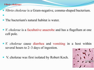
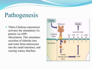
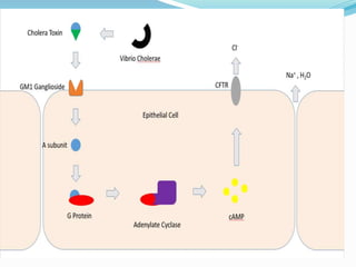
 Vibrio cholerae is a Gram-negative, comma-shaped bacterium.

 The bacterium's natural habitat is water.
 V. cholerae is a facultative anaerobe and has a flagellum at one
cell pole.
 V. cholerae cause diarrhea and vomiting in a host within
several hours to 2–3 days of ingestion.
 V. cholerae was first isolated by Robert Koch.
Vibrio cholerae:
Pathogenesis:
 Cholera toxin (also known as choleragen and sometimes
abbreviated to CTX, Ctx or CT) is protein complex secreted by the
bacterium Vibrio cholerae.
 The heat-labile enterotoxin is inactivated at high temperatures.
 The cholera toxin is an oligomeric complex made up of six protein
subunits:
 A single copy of the Alpha subunit (part A, enzymatic), and five
copies of the Beta subunit (part B, receptor binding), denoted as
AB5.
 Subunit B binds while subunit A activates the G protein which
activates adenylate cyclase.
 Vibrio Cholerae enterotoxin
activates the stimulatory Gs
protein via ADP-
ribosylation. This stimulates
secretion of chloride ions
and water from enterocytes
into the small intestines, and
causing watery diarrhea.
Pathogenesis
 CT’s bind to the GM1 gangliosides .
 Once the Beta subunits bind GM1 molecules (located in the
plasma membrane of the mucosal cells).
 The toxin complex gets endocytosed and transported to the
endoplasmic reticulum.
 Once in the ER, the disulfide bond in the A subunit then gets
reduced, causing the A subunit to dissociate from the complex
and enter the cytoplasm.

 Where it binds an ADP ribosylation factor.
 This process causes the activation of adenylate cyclase,
which produces cyclic adenosine monophosphate (cAMP)
from ATP.
 The increase in cAMP changes the transmural ion flow,
causing the secretion of Cl-, K+, Na+ H2O2 in to
intestine.
Symptoms of Cholera:
 Watery diarrhea with mucus, which is called rice-water
stool and is the hallmark of cholera.
 In very severe cases, a person can lose 1 L of fluid an hour!
This intense diarrhea leads to other complications, including
 Dehydration
 Muscle cramps due to the loss of potassium ions
 Low blood pH (acidosis)
 Sunken eyes and skin
 Loss of blood volume, leading to very low blood pressure
(hypotension)
 Coma in young children
 Very fast heart rate.
Transmission:
 The bacteria causing cholera is present in faeces or other
effluent that may seep into and contaminate waterways, soil or
sources of drinking water.
 Drinking infected water or even just using it to wash foods,
kitchen utensils or culinary items can lead to transmission of
the infection.
Treatment of Cholera:
Fluids:
 In most cases, cholera can be successfully treated with
oral rehydration therapy, which is highly effective, safe,
and simple to administer.
 Rice-based solutions are preferred to glucose-based ones
due to greater efficiency.

Electrolytes:
 The potassium levels may decrease rapidly in cholera, and
thus need to be replaced.
 This may be done by eating foods high in potassium like
bananas or green coconut water.
Antibiotics:
 Doxycycline is typically used first line, although some strains
of V. cholerae have shown resistance.
 Other antibiotics proven to be effective include erythromycin,
tetracycline and chloramphenicol.
Streptococcus pneumoniae
Pathogenesis:
 Pneumonia caused by Streptococcus pneumoniae, Gram positive,
facultative anaerobic member of the genus Streptococcus.
 S. pneumoniae was recognized as a major cause of pneumonia.
 S. pneumoniae typically colonizing the respiratory tract and
nasal cavity.
 However, in individuals with weaker immune systems and
young children, the bacterium may become pathogenic and
spread to other locations to cause disease.
 The capsulated, a polysaccharide capsule that acts as a
virulence factor.
 Usually they are found in pairs of cocci, or diplococci or in
short chains or singly.
 When cultured on blood agar they demonstrate alpha
hemolysis.
 They are non motile organisms.
Pathogenesis:
 S. pneumoniae colonization of the nasopharynx is
facilitated by mucus degradation by the enzymes NanA,
BgaA, StrH, and NanB. Ply decreases epithelial cell
ciliary beating, enhancing bacterial adherence.
 Pneumococcal cell wall peptidoglycans may be destroyed
by lysozyme.
 PdgA and Adr deacetylate pneumococcal cell surface
petidoglycan molecules, rendering them resistant to
lysozyme.
Symptoms:
Pneumonia may begin suddenly.
 High fever
 Cough
 Shortness of breath
 Rapid breathing
 Chest pains
Treatment:
Antibiotics:
 The most commonly used antibiotics, including penicillins,,
cephalosporins, rifampin and vancomycin.
Vaccine:
 There are two types of pneumococcal vaccines: conjugate
vaccines and polysaccharide vaccines.
 They are given by injection either into a muscle or just under
the skin.
THANK YOU

Cholera and streptococcus pneumoniae

  • 1.
    Cholera Dr. Aswartha HarinathaReddy Assistant Professor Department of Biotechnology Bangalore
  • 2.
     Vibrio choleraeis a Gram-negative, comma-shaped bacterium.   The bacterium's natural habitat is water.  V. cholerae is a facultative anaerobe and has a flagellum at one cell pole.  V. cholerae cause diarrhea and vomiting in a host within several hours to 2–3 days of ingestion.  V. cholerae was first isolated by Robert Koch. Vibrio cholerae:
  • 3.
    Pathogenesis:  Cholera toxin(also known as choleragen and sometimes abbreviated to CTX, Ctx or CT) is protein complex secreted by the bacterium Vibrio cholerae.  The heat-labile enterotoxin is inactivated at high temperatures.  The cholera toxin is an oligomeric complex made up of six protein subunits:  A single copy of the Alpha subunit (part A, enzymatic), and five copies of the Beta subunit (part B, receptor binding), denoted as AB5.  Subunit B binds while subunit A activates the G protein which activates adenylate cyclase.
  • 4.
     Vibrio Choleraeenterotoxin activates the stimulatory Gs protein via ADP- ribosylation. This stimulates secretion of chloride ions and water from enterocytes into the small intestines, and causing watery diarrhea. Pathogenesis
  • 5.
     CT’s bindto the GM1 gangliosides .  Once the Beta subunits bind GM1 molecules (located in the plasma membrane of the mucosal cells).  The toxin complex gets endocytosed and transported to the endoplasmic reticulum.  Once in the ER, the disulfide bond in the A subunit then gets reduced, causing the A subunit to dissociate from the complex and enter the cytoplasm. 
  • 6.
     Where itbinds an ADP ribosylation factor.  This process causes the activation of adenylate cyclase, which produces cyclic adenosine monophosphate (cAMP) from ATP.  The increase in cAMP changes the transmural ion flow, causing the secretion of Cl-, K+, Na+ H2O2 in to intestine.
  • 8.
    Symptoms of Cholera: Watery diarrhea with mucus, which is called rice-water stool and is the hallmark of cholera.  In very severe cases, a person can lose 1 L of fluid an hour! This intense diarrhea leads to other complications, including  Dehydration  Muscle cramps due to the loss of potassium ions  Low blood pH (acidosis)  Sunken eyes and skin  Loss of blood volume, leading to very low blood pressure (hypotension)  Coma in young children  Very fast heart rate.
  • 9.
    Transmission:  The bacteriacausing cholera is present in faeces or other effluent that may seep into and contaminate waterways, soil or sources of drinking water.  Drinking infected water or even just using it to wash foods, kitchen utensils or culinary items can lead to transmission of the infection.
  • 10.
    Treatment of Cholera: Fluids: In most cases, cholera can be successfully treated with oral rehydration therapy, which is highly effective, safe, and simple to administer.  Rice-based solutions are preferred to glucose-based ones due to greater efficiency. 
  • 11.
    Electrolytes:  The potassiumlevels may decrease rapidly in cholera, and thus need to be replaced.  This may be done by eating foods high in potassium like bananas or green coconut water.
  • 12.
    Antibiotics:  Doxycycline istypically used first line, although some strains of V. cholerae have shown resistance.  Other antibiotics proven to be effective include erythromycin, tetracycline and chloramphenicol.
  • 13.
  • 14.
    Pathogenesis:  Pneumonia causedby Streptococcus pneumoniae, Gram positive, facultative anaerobic member of the genus Streptococcus.  S. pneumoniae was recognized as a major cause of pneumonia.  S. pneumoniae typically colonizing the respiratory tract and nasal cavity.  However, in individuals with weaker immune systems and young children, the bacterium may become pathogenic and spread to other locations to cause disease.
  • 15.
     The capsulated,a polysaccharide capsule that acts as a virulence factor.  Usually they are found in pairs of cocci, or diplococci or in short chains or singly.  When cultured on blood agar they demonstrate alpha hemolysis.  They are non motile organisms.
  • 16.
    Pathogenesis:  S. pneumoniaecolonization of the nasopharynx is facilitated by mucus degradation by the enzymes NanA, BgaA, StrH, and NanB. Ply decreases epithelial cell ciliary beating, enhancing bacterial adherence.  Pneumococcal cell wall peptidoglycans may be destroyed by lysozyme.  PdgA and Adr deacetylate pneumococcal cell surface petidoglycan molecules, rendering them resistant to lysozyme.
  • 17.
    Symptoms: Pneumonia may beginsuddenly.  High fever  Cough  Shortness of breath  Rapid breathing  Chest pains
  • 18.
    Treatment: Antibiotics:  The mostcommonly used antibiotics, including penicillins,, cephalosporins, rifampin and vancomycin. Vaccine:  There are two types of pneumococcal vaccines: conjugate vaccines and polysaccharide vaccines.  They are given by injection either into a muscle or just under the skin.
  • 19.