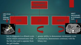
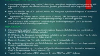
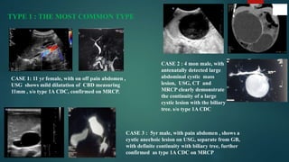
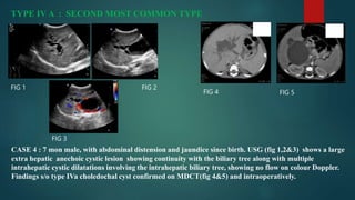
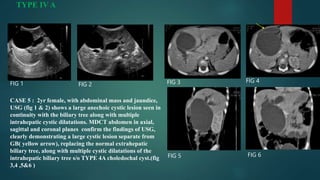
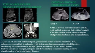
TITLE: EVALUATION OF CHOLEDOCHAL
CYSTS USING ULTRASONOGRAPHY IN A
SPECTRUM OF CASES
GUIDE :
DR. ALKA AGRAWAL
HEAD OF DEPARTMENT
DEPARTMENT OF RADIODIAGNOSIS
MGMMC INDORE
COGUIDE :
DR. GAURAV BHANDARI
ASSISTANT PROFESSOR
DEPARTMENT OF
RADIODIAGNOSIS
MGMMC INDORE
PRESENTER :
DR. SUDHA CHANDANA
PG RESIDENT
DEPARTMENT OF RADIODIAGNOSIS
MGMMC INDORE
INTRODUCTION
 Choledochal cysts are cystic dilatation of the biliary tree, with an incidence of
1:100,000-150,000, an uncommon but serious condition that requires surgical
treatment. (1)
 Although choledochal cysts are generally considered a disorder of childhood and
infancy, the ages in reported cases have ranged from newly born to 80 years.
however, 60% of such cysts are diagnosed in patients less than 10 years old . (1)
 PATHOGENESIS:
 Close association with the anomalous formation of the pancreaticobiliary ductal
junction is reported in some subtypes .(2)
CLASSIFICATION
 Commonly accepted classification is one devised by Todani et al. (3)
 There are five main and several subtypes
 type I: most common, accounting for 80-90% ( can present in utero)
 Ia: dilatation of extrahepatic bile duct (entire)
 Ib: dilatation of extrahepatic bile duct (focal segment)
 Ic: dilatation of the CBD portion of extrahepatic bile duct
 type II: true diverticulum from extrahepatic bile duct
 type III: dilatation of extrahepatic bile duct within the duodenal wall (choledochocoele)
 type IV: next most common
 IVa: cysts involving both intra and extrahepatic ducts
 IVb: multiple dilatations/cysts of extrahepatic ducts only
 type V: multiple dilatations/cysts of intrahepatic ducts only (Caroli disease)
 Recently a type VI cyst as been identified as cystic dilatation involving cystic duct (only a
few case reports are available in literature). (4)
IMAGING
MODALITIES
USG CT MRCP
key to diagnosis is a dilated cystic
lesion which communicates with
the bile duct and is separate from
the gall bladder
• greater ability to demonstrate intrahepatic cysts
• conclusively demonstrates continuity with the
biliary tree
GB
CDC
MDCT
demonstratin
g intrahepatic
cysts
USG shows
CDC
separate
From GB
MATERIALS AND METHODS
 Ultrasonography was done using convex( 2-5MHz) and linear (3-9MHz) probe in patients presenting with
a spectrum of symptoms like neonatal cholestasis, jaundice, pain abdomen and or palpable abdominal
mass.
 Study was done in a total of 20 pediatric patients who were given a provisional diagnosis of choledochal
cyst on ultrasonography.
 The patients were followed up for a variable period of time and USG findings were evaluated using
MRCP, MDCT and/or per operative and histopathology findings as and when applicable.
 USG was done to make a diagnosis of choledochal cyst, determining the type of cyst as per Todani’s
classification and also evaluating complications.
DISCUSSION
 Ultrasonography was found 100 % sensitive in making a diagnosis of choledochal cyst (confirmed per
operatively/MRCP/MDCT/histopathologically )
 16 (80%) out of the 20 cases of choledochal cysts included in our study were found to be of type 1 , which
is also the most common type as per Todani et al. (3)
 There were 3 cases of type IV a , and a single case of type V choledochal cyst.
 The most common presentation was that of abdominal pain and jaundice. 6 of them were large enough to
present as palpable abdominal mass.
 11 of the 20 cases underwent cyst excision and hepaticojejunostomy under GA for curative management
which is the recommended form of treatment . (5)
 9 out of 20 had confirmation of USG diagnosis by either MRCP or MDCT abdomen
TYPE 1 : THE MOST COMMON TYPE
CASE 1: 11 yr female, with on off pain abdomen ,
USG shows mild dilatation of CBD measuring
11mm , s/o type 1A CDC, confirmed on MRCP.
CASE 2 : 4 mon male, with
antenatally detected large
abdominal cystic mass
lesion, USG, CT and
MRCP clearly demonstrate
the continuity of a large
cystic lesion with the biliary
tree. s/o type 1A CDC
CASE 3 : 5yr male, with pain abdomen , shows a
cystic anechoic lesion on USG, separate from GB,
with definite continuity with biliary tree, further
confirmed as type 1A CDC on MRCP
TYPE IV A : SECOND MOST COMMON TYPE
CASE 4 : 7 mon male, with abdominal distension and jaundice since birth. USG (fig 1,2&3) shows a large
extra hepatic anechoic cystic lesion showing continuity with the biliary tree along with multiple
intrahepatic cystic dilatations involving the intrahepatic biliary tree, showing no flow on colour Doppler.
Findings s/o type IVa choledochal cyst confirmed on MDCT(fig 4&5) and intraoperatively.
FIG 1 FIG 2
FIG 4 FIG 5
FIG 3
TYPE IV A
FIG 1 FIG 2 FIG 3 FIG 4
FIG 5 FIG 6
CASE 5 : 2yr female, with abdominal mass and jaundice,
USG (fig 1 & 2) shows a large anechoic cystic lesion seen in
continuity with the biliary tree along with multiple
intrahepatic cystic dilatations. MDCT abdomen in axial,
sagittal and coronal planes confirm the findings of USG,
clearly demonstrating a large cystic lesion separate from
GB( yellow arrow), replacing the normal extrahepatic
biliary tree, along with multiple cystic dilatations of the
intrahepatic biliary tree s/o TYPE 4A choledochal cyst.(fig
3,4 ,5&6 )
TYPE 5 CAROLI’S WITH
INFANTILE POLYCYSTIC KIDNEY
DISEASE
CASE 6 : 2.5 Y old, with abdominal pain , jaundice and failure to thrive since birth. USG
abdomen reveals multiple intrahepatic cystic lesions showing communication with biliary tree
and showing the classical central dot sign ( yellow arrows) on CT s/o Caroli’s disease .
Bilateral kidneys are enlarged, echogenic and shows multiple non communicating cysts s/o
infantile variety of polycystic kidney disease .
COMPLICATIONS OF
CHOLEDOCHAL CYSTS
CASE 8
CASE 7
CASE 7 shows a fusiform dilatation of CBD
with multiple echogenic foci within s/o calculi
Case 8 in another patient, shows echogenic
sludge within the lumen of a choledochal cyst.
CONCLUSION
 Ultrasonography is an excellent modality for initial evaluation of pediatric patients with
choledochal cysts. It not only helps establish a provisional diagnosis of choledochal cyst, but
also helps us decide the subtype of cyst, presence of complications and other syndromic
associations. MR is mostly used for confirmation of diagnosis in complex cases and also for
pre-operative evaluation for ductal anatomy.
 REFERENCES :
 1: Kim OH, Chung HJ, Choi BG. Imaging of the choledochal cyst. Radiographics. 1995;15 (1): 69-88.
 2: Babbitt DP. Congenital choledochal cyst: New etiologic concept based on anomalous relationships of common bile duct and
pancreatic bulb. Ann Radiol 1969; 12:231-40
 3: Todani T, Watanabe Y, Narusue M, et al. Congenital bile duct cysts. Am J Surg 1977; 134:263-69
 4: Bode WE, Aust JB. Isolated cystic dilatation of the cystic duct. Am J Surg. 1983;145:828–9.
 5: Liu CL, Fan ST, Lo CM, Lam CM, Poon RT, Wang J. Choledochal cysts in adults. Arch Surg.2002;137:465–468

Choledochal Cyst.pptx

  • 1.
    TITLE: EVALUATION OFCHOLEDOCHAL CYSTS USING ULTRASONOGRAPHY IN A SPECTRUM OF CASES GUIDE : DR. ALKA AGRAWAL HEAD OF DEPARTMENT DEPARTMENT OF RADIODIAGNOSIS MGMMC INDORE COGUIDE : DR. GAURAV BHANDARI ASSISTANT PROFESSOR DEPARTMENT OF RADIODIAGNOSIS MGMMC INDORE PRESENTER : DR. SUDHA CHANDANA PG RESIDENT DEPARTMENT OF RADIODIAGNOSIS MGMMC INDORE
  • 2.
    INTRODUCTION  Choledochal cystsare cystic dilatation of the biliary tree, with an incidence of 1:100,000-150,000, an uncommon but serious condition that requires surgical treatment. (1)  Although choledochal cysts are generally considered a disorder of childhood and infancy, the ages in reported cases have ranged from newly born to 80 years. however, 60% of such cysts are diagnosed in patients less than 10 years old . (1)  PATHOGENESIS:  Close association with the anomalous formation of the pancreaticobiliary ductal junction is reported in some subtypes .(2)
  • 3.
    CLASSIFICATION  Commonly acceptedclassification is one devised by Todani et al. (3)  There are five main and several subtypes  type I: most common, accounting for 80-90% ( can present in utero)  Ia: dilatation of extrahepatic bile duct (entire)  Ib: dilatation of extrahepatic bile duct (focal segment)  Ic: dilatation of the CBD portion of extrahepatic bile duct  type II: true diverticulum from extrahepatic bile duct  type III: dilatation of extrahepatic bile duct within the duodenal wall (choledochocoele)  type IV: next most common  IVa: cysts involving both intra and extrahepatic ducts  IVb: multiple dilatations/cysts of extrahepatic ducts only  type V: multiple dilatations/cysts of intrahepatic ducts only (Caroli disease)  Recently a type VI cyst as been identified as cystic dilatation involving cystic duct (only a few case reports are available in literature). (4)
  • 4.
    IMAGING MODALITIES USG CT MRCP keyto diagnosis is a dilated cystic lesion which communicates with the bile duct and is separate from the gall bladder • greater ability to demonstrate intrahepatic cysts • conclusively demonstrates continuity with the biliary tree GB CDC MDCT demonstratin g intrahepatic cysts USG shows CDC separate From GB
  • 5.
    MATERIALS AND METHODS Ultrasonography was done using convex( 2-5MHz) and linear (3-9MHz) probe in patients presenting with a spectrum of symptoms like neonatal cholestasis, jaundice, pain abdomen and or palpable abdominal mass.  Study was done in a total of 20 pediatric patients who were given a provisional diagnosis of choledochal cyst on ultrasonography.  The patients were followed up for a variable period of time and USG findings were evaluated using MRCP, MDCT and/or per operative and histopathology findings as and when applicable.  USG was done to make a diagnosis of choledochal cyst, determining the type of cyst as per Todani’s classification and also evaluating complications. DISCUSSION  Ultrasonography was found 100 % sensitive in making a diagnosis of choledochal cyst (confirmed per operatively/MRCP/MDCT/histopathologically )  16 (80%) out of the 20 cases of choledochal cysts included in our study were found to be of type 1 , which is also the most common type as per Todani et al. (3)  There were 3 cases of type IV a , and a single case of type V choledochal cyst.  The most common presentation was that of abdominal pain and jaundice. 6 of them were large enough to present as palpable abdominal mass.  11 of the 20 cases underwent cyst excision and hepaticojejunostomy under GA for curative management which is the recommended form of treatment . (5)  9 out of 20 had confirmation of USG diagnosis by either MRCP or MDCT abdomen
  • 6.
    TYPE 1 :THE MOST COMMON TYPE CASE 1: 11 yr female, with on off pain abdomen , USG shows mild dilatation of CBD measuring 11mm , s/o type 1A CDC, confirmed on MRCP. CASE 2 : 4 mon male, with antenatally detected large abdominal cystic mass lesion, USG, CT and MRCP clearly demonstrate the continuity of a large cystic lesion with the biliary tree. s/o type 1A CDC CASE 3 : 5yr male, with pain abdomen , shows a cystic anechoic lesion on USG, separate from GB, with definite continuity with biliary tree, further confirmed as type 1A CDC on MRCP
  • 7.
    TYPE IV A: SECOND MOST COMMON TYPE CASE 4 : 7 mon male, with abdominal distension and jaundice since birth. USG (fig 1,2&3) shows a large extra hepatic anechoic cystic lesion showing continuity with the biliary tree along with multiple intrahepatic cystic dilatations involving the intrahepatic biliary tree, showing no flow on colour Doppler. Findings s/o type IVa choledochal cyst confirmed on MDCT(fig 4&5) and intraoperatively. FIG 1 FIG 2 FIG 4 FIG 5 FIG 3
  • 8.
    TYPE IV A FIG1 FIG 2 FIG 3 FIG 4 FIG 5 FIG 6 CASE 5 : 2yr female, with abdominal mass and jaundice, USG (fig 1 & 2) shows a large anechoic cystic lesion seen in continuity with the biliary tree along with multiple intrahepatic cystic dilatations. MDCT abdomen in axial, sagittal and coronal planes confirm the findings of USG, clearly demonstrating a large cystic lesion separate from GB( yellow arrow), replacing the normal extrahepatic biliary tree, along with multiple cystic dilatations of the intrahepatic biliary tree s/o TYPE 4A choledochal cyst.(fig 3,4 ,5&6 )
  • 9.
    TYPE 5 CAROLI’SWITH INFANTILE POLYCYSTIC KIDNEY DISEASE CASE 6 : 2.5 Y old, with abdominal pain , jaundice and failure to thrive since birth. USG abdomen reveals multiple intrahepatic cystic lesions showing communication with biliary tree and showing the classical central dot sign ( yellow arrows) on CT s/o Caroli’s disease . Bilateral kidneys are enlarged, echogenic and shows multiple non communicating cysts s/o infantile variety of polycystic kidney disease . COMPLICATIONS OF CHOLEDOCHAL CYSTS CASE 8 CASE 7 CASE 7 shows a fusiform dilatation of CBD with multiple echogenic foci within s/o calculi Case 8 in another patient, shows echogenic sludge within the lumen of a choledochal cyst.
  • 10.
    CONCLUSION  Ultrasonography isan excellent modality for initial evaluation of pediatric patients with choledochal cysts. It not only helps establish a provisional diagnosis of choledochal cyst, but also helps us decide the subtype of cyst, presence of complications and other syndromic associations. MR is mostly used for confirmation of diagnosis in complex cases and also for pre-operative evaluation for ductal anatomy.  REFERENCES :  1: Kim OH, Chung HJ, Choi BG. Imaging of the choledochal cyst. Radiographics. 1995;15 (1): 69-88.  2: Babbitt DP. Congenital choledochal cyst: New etiologic concept based on anomalous relationships of common bile duct and pancreatic bulb. Ann Radiol 1969; 12:231-40  3: Todani T, Watanabe Y, Narusue M, et al. Congenital bile duct cysts. Am J Surg 1977; 134:263-69  4: Bode WE, Aust JB. Isolated cystic dilatation of the cystic duct. Am J Surg. 1983;145:828–9.  5: Liu CL, Fan ST, Lo CM, Lam CM, Poon RT, Wang J. Choledochal cysts in adults. Arch Surg.2002;137:465–468