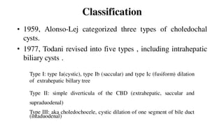
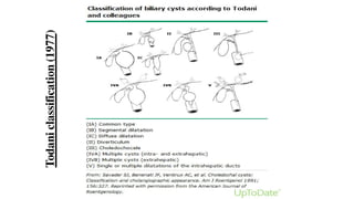
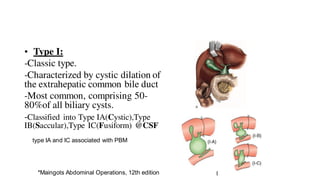
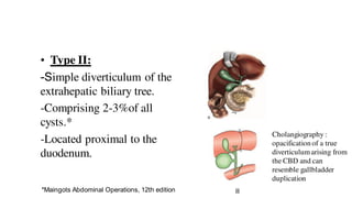
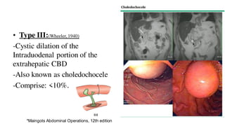
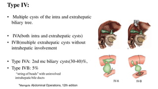
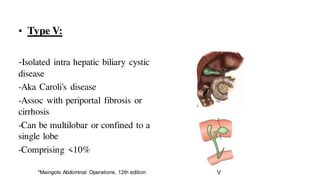
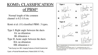
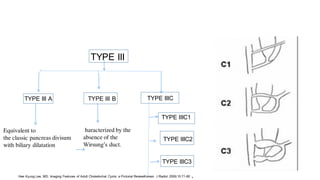
CHOLEDOCHAL CYSTS
Introduction:
• Focal or diffuse pathological dilatation of the biliary system.
• Rare, usually diagnosed in children (15%antenatally, 60% 1st
decade) and 25% in adults. (Y.Parikh, Maingots abdominal operations.12th edition,pn 1029)
• A premalignant condition .
• Treatment depend upon type , S/S, potential long term sequelae.
Demographics
• Uncommon finding.
• Incidence: 1 in 100000 to 1in 150000*, in Asia (1 in 13000)*
• One-half to two-thirds of the reported cases occurring in Japan#
• Frequency of bile duct cyst: type I(79%), followed by type IV (13%)
Then type III(4%) and type II(2.6%).
• Female preponderance (4:1).
*Lipsett PA et al,Choledochal cyst disease. A changing pattern of presentation,Annals of surg,1998
#O'Neill JA Jr et al, Choledochal cyst,Curr Probl Surg. 1992;29(6):361
Pathogenesis
Congenital :
• Defect in maturation with ductal plate malformation
• Defective bile duct plate remodelling
• Fetal viral infection(reovirus)* *Tyler KL et al,Hepatology. 1998;27(6):1475.
• Bile duct obstruction: sec to web, stricture, sphincter of oddi
dysfunction
• Fewer ganglion cells in distal CBD in CCs pt resulting in proximal
dilatation .Kusunoki et al,1998
Pancreatobiliary maljunction: (Babbit, 1969)
•Extramural junction of the pancreatic and biliary ducts in the
duodenum beyond the intramural sphincter
•PD and BD connects 1-2cm proximal to the sphincter of Oddi.
•Allow reflux of pancreatic juice into the biliary tree
•Proteolysis within the CBD ductal dilation.
•High trypsinogen and phospholipase A2 levels in bile.
•Present in 50 to 80 percent of patients with biliary cysts.
*Maingots Abdominal Operations, 12th edition
• Biliary epithelium dysplastic
changes.
• Insertion of the CBD farther than
15mm from the ampulla of Vater*
• Failure of the embryological ducts
to migrate fully into the duodenum.
• Bile amylase concentration of
>8,000 IU/L.
*Funabiki T et al, Langenbecks Arch Surg. 2009
Classification
• 1959, Alonso-Lej categorized three types of choledochal
cysts.
• 1977, Todani revised into five types , including intrahepatic
biliary cysts .
Type I: type Ia(cystic), type Ib (saccular) and type Ic (fusiform) dilation
of extrahepatic billary tree
Type II: simple diverticula of the CBD (extrahepatic, saccular and
supraduodenal)
Type III: aka choledochocele, cystic dilation of one segment of bile duct
(intaduodenal)
Todaniclassification(1977)
type IA and IC associated with PBM
• Type I:
-Classic type.
-Characterized by cystic dilation of
the extrahepatic common bile duct
-Most common, comprising 50-
80%of all biliary cysts.
-Classified into Type IA(Cystic),Type
IB(Saccular),Type IC(Fusiform) @CSF
*Maingots Abdominal Operations, 12th edition
Cholangiography :
opacification of a true
diverticulumarising from
the CBD and can
resemble gallbladder
duplication
• Type II:
-Simple diverticulum of the
extrahepatic biliary tree.
-Comprising 2-3%of all
cysts.*
-Located proximal to the
duodenum.
*Maingots Abdominal Operations, 12th edition
• Type III:(Wheeler, 1940)
-Cystic dilation of the
Intraduodenal portion of the
extrahepatic CBD
-Also known as choledochocele
-Comprise: <10%.
*Maingots Abdominal Operations, 12th edition
Type IV:
“string of beads” with uninvolved
intrahepaticbile ducts
• Multiple cysts of the intra and extrahepatic
biliary tree.
• IVA(both intra and extrahepatic cysts)
• IVB(multiple extrahepatic cysts without
intrahepatic involvement
• Type IVA: 2nd mc biliary cysts(30-40)%,
• Type IVB: 5%
*Maingots Abdominal Operations, 12th edition
• Type V:
-Isolated intra hepatic biliary cystic
disease
-Aka Caroli's disease
-Assoc with periportal fibrosis or
cirrhosis
-Can be multilobar or confined to a
single lobe
-Comprising <10%
*Maingots Abdominal Operations, 12th edition
KOMI's CLASSIFICATION
of PBM*
Normal length of the common
channel is 0.2-1.0 cm.
Komi et al. (11) classified PBM : 3 types.
Type I: Right angle between the ducts
IA: no dilatation
IB: dilatation +
Type II: Acute angle between the ducts
IIA: no dilatation
IIB: dilatation +
*Hae Kyung Lee, MD, Imaging Features of Adult Choledochal
Cysts: a Pictorial ReviewKoreanJ Radiol 2009;10:71-80 ,
TYPE III A TYPE III B TYPE IIIC
Equivalent to
the classic pancreas divisum
with biliary dilatation
haracterized by the
absence of the
Wirsung’s duct.
TYPE IIIC2
TYPE IIIC3
Hae Kyung Lee, MD, Imaging Features of Adult Choledochal Cysts: a Pictorial ReviewKorean J Radiol 2009;10:71-80 ,
TYPE III
TYPE IIIC1
Acquired cyst:
Rarely Type 1 biliary cysts can occur after
cholecystectomy
Sphincter of Oddi stenosis might contribute to
cyst formation
Pathology:
Histological apperance of thick and dense
fibrotic cystic wall with inflammation
sparceness of mucin glands and metaplasia
Hisami et al, cystic disorders of bile ducts, Shackelford's surgery of the alimentary tract,20th edition
Developmental anomalies associated with biliary cysts include:
• Abnormal pancreatobiliary junction
• Biliary atresia
• Duodenal atresia
• Pancreatic arteriovenous malformation
• Multiseptate gallbladder
• Hemifacial macrosomia with extracranial anomalies.
• Polycystic kidney disease
Presentation
• May remain asymptomatic
• Classic triad : female child with jaundice, abdominal pain and RUQ
mass
• Infants : elevated conjugated bilirubin , failure to thrive or an
abdominal mass
• Adults : mass rare, abdominal pain and recurrent cholangitis
• Type III cyst : pancreatitis (30%)
• Symptoms mimic calculous biliary cholecystitis
Hisami et al, cystic disorders of bile ducts, Shackelford's surgery of the alimentary tract,20th edition
Difference between Adult and Pediatric patients
ADULTCHOLEDOCHALCYST
• F:M 1.5:1
• Per abdominal mass>abdominal
pain
• More frequently associated with
APBJ(59%)
• Malignancy risk: less
common(21.2%)
PEDIATRIC CHOLEDOCHALCYST
• 4:1
• Abdominal pain> per abdominal
mass
• Less frequently assoc with
APBJ(85.7%)
• Malignancy risk: more
common(0.7%)
(Ching Shui et al, Choledochal Cysts: Differences Between Pediatric and Adult Patients,Journal of GI Surgery,2010)
Approach to Diagnosis:
• Requires high level of suspicion.
• Ductal dilation : Biliary obstruction vs choledochal cyst ?
• Absence of biochemical, radiographic, or endoscopic
evidence of obstruction.
• USG: most common first line imaging tool.
 Irregular hypoechoic segmental dilation of extrahepatic bile
duct
 Absence of septationsdistinguish from extrahepatic biliary
tumors
 Limited in identifying choledochoceles
(A CBD measuring greater than 10 mm)
• CECT :
• MRCP: Non invasive procedure of choice for diagnosis of choledochal cyst
.(pancreatic, intrahepatic and extrahepatic ductal anatomy).
ADDITIONAL TESTS:
•Types I and IVA cysts:
 MRCP-obstruction (ERCP or EUS performed)
 Direct visualization of the ampulla as well as the peri-ampullary bile duct and
pancreatic duct
Hepatobiliaryand pancreaticanatomy
Biliary malignancy
Matastaticdisease
Vascular encasement
relation of cyst with adjacent structures
• Types II and IVB cysts:
 Hepatobiliary scintigraphy( communication with biliary tree)*
 ERCP/ EUS. EUS :distinguish a pancreatic head cyst from a Type II
biliary cyst
• Type III cysts:
ERCP: Endoscopic therapy and cyst mucosal biopsy.
*(Lambie el,Tc99m-hepatobiliary iminodiacetic acid(HIDA)scintigraphyin clinical practice,clin radiology,2011)
Cholangiography: (endoscopic, percutaneous or
intraoperative)
sensitivity of up to 100 percent for diagnosing biliary cysts
-Preoperative evaluation of the biliary anatomy
-Detect filling defects due to stones or malignancy
-Primarily therapeutic procedure to place stents or to obtain brushing
for cytology.
-Gold standard for preoperative diagnosis of an abnormal
pancreatobiliary junction.
• ERCP:
- Better visualization of PBM,
- Biopsy, brush cytology(can be taken), extraction of intracystic stones
after papillotomy.
- Exclude signs of portal hypertension.
- Procedure of choice for type III cysts.
•PTC:
- Patients with previous Roux-en-Y cystoenterostomy or type IV cysts
with ductal stricture/tumors.
- Percutaneous billary drainage for control of bilary sepsis.
• EUS:
-Demonstrate extrahepatic biliary cysts
-Detailed image of the cyst wall and PBM
-Evidence of biliary obstruction
•Hepatobiliary scintigraphy:
-Demonstrate continuity of cysts with the bile ducts.
- HIDA scan:Selectively taken up by hepatocytes.
Differential Diagnosis:
• Pancreatic, mesenteric, and hepatic cysts:
- Do not communicate with the biliary tree
-Confirmed by biliary scintigraphy or ERCP.
•Acute or chronic biliary obstruction :
- Marked biliary dilation that mimics a type I cyst.
- Jaundice or elevated serum liver tests
- Identifiable obstructing lesion such as a stone, stricture, or mass.
-Biliary dilation often improves after appropriate treatment
Associated Hepatobiliary Pathologies:
• Cystolithiasis (most frequent)
- Thick viscous bile
-Anastomotic strictures after previous cystoenterostomies
• Hepatolithiasis
-Proximal bile stasis or migration of intracystic stones
• Calculous Cholecystitis
• Pancreatitis
- Prevalance 2% to 70% (Lipsett.et.al, 1994)
- Mild, acute often relapsing
- Obstruction of the pranceatic duct at the PBJ or distally in the ampulla.
Kristopher et al, Bile Duct Cysts In Adults,Blumgart's Surgery of the Liver, Biliary Tract, and Pancreas,6th edition
• Cholangiocarinoma
• Intrahepatic abscess
- Recurrent cholangitis and billary obstruction.
• Cirrhosis with portal hypertension
- Secondary billary cirrhosis, portal vein thrombosis or Caroli
disease with congenital hepatic fibrosis.
• Spontaneous perforation of bile duct cysts
- Reported in infants and children, not in adults (Ando.et.al 1998)
Malignancy and bile duct cysts
- Incidence 7.5% (Sastry. et.al 2015)
- Cholangiocarcinoma 70%
Ca GB 20%
- More common in older age (0.4% in 1st decade to 14% after age 2, 38% after age 60. (Sastry.
et.al 2015)
- Malignancy may occur after cyst excision
- Significantly greater in type I and IV cyst (7.6% and 9.2% respectively)
- 20 to 30 fold increased risk of cholangiocarcinoma in biliary cysts.*
*Søreide K, et al,Ann Surg Oncol. 2007
} of associated malignancy
Abnormal pancreatobiliary junction and cancer:
-Increases the risk of malignancy
-Gallbladder cancer who do not have a biliary cyst
-Prophylactic cholecystectomy in patients with APBJ has been advised
Etiology:
-K-ras and P-53 mutations seen in more than 60% carcinomas.(Shimatoke et al, 2003)
- Unconjugated Deoxycolate and lithocolate → Billary metaplasia
- Reported survival: 6 to 21 months (Mabrut.et.al 2013)
Kristopheret al, Bile Duct Cysts In Adults,Blumgart's Surgery of the Liver, Biliary Tract, and Pancreas,6th edition
Treatment
• Surgical management: based on cyst type , associated hepatobiliary
pathology
• Type I, II, or IV: Surgical resection of the cysts( risk of malignancy).
• Aims of preop management:
 Complete cholangiographic extent of cystic process.
 Control of biliary infections.
 Percutaneous or endoscopic drainage of infected bile duct cysts.
 Evaluation for anastomotic stricture, ductal stones, biliary tract malignancy,
cirrhosis and portal hypertension in prior cyst related surgery.
• Definitive treatment : Surgical
• Historically: Cystoenterostomy.
• Recommended management : Complete Cyst Excision
• Goal: prevent malignant degeneration of the cyst
• Mucosa to mucosa bilioenteric anastomosis.
• External drainage alone has no role in definitive management
• Long term follow up : age related risk of malignancy and late
anastomotic strictures.
Principle of Surgery:
•Cholecystectomy and complete Choledochal Cyst excision.
•Bile duct reconstruction ( Roux-en-Y Hepaticojejunostomy )
•Enteric interposition grafts : jejunal and appendiceal between
duodenum and bile duct.*
*Blumgart's Surgery of the Liver, Biliary tract and Pancreas,20th edition
Type I Cysts:
• Total cystectomy down to the level
of communication with the
pancreatic duct and Roux -en-Y HJ
(Wiseman et al,2005)
• Reduction in risk of malignancy
• Roux -en-Y jejunal limb approx
60cm in length, standard retrocolic
end -to-side Roux -en-Y HJ .
• Difficult dissection from portal vein or hepatic artery: post wall left intact
+mucosectomy (removal of the epithelial lining of the cyst)
• Intrapancreatic portion of type I cysts: intramural dissection of the bile
duct.
• Complication: in long term , stenosis of HJ leading to recurrent cholangitis.
-up to 30 percent of patients over time*
-monitored with annual LFTs
*Röthlin MA et al, Am J Surg. 1998;
Type II cysts:
• Complete cyst excision
• CBD wall defect closed transversely with or without a T-tube.
• Cholecystectomy same setting.
Type III cyst (Choledochocele)
• Typically small (<= 2cm)
• Biliary colic, cholangitis, pancreatitis: more common.
• Previously transduodenal cyst excision +/- sphincterotomy
prefered.
• Endoscopy sphincterotomy and cyst unroofing at present
• Diagnostic advantage of ERCP with endoscopy treatment
Type IV cyst
• Excision of extra hepatic cysts, cholecystectomy, Roux-en-Y HJ
• Extrahepatic component of type IVa and type IVb cyst is
approached as type I bile duct cyst.
• Lower hepatic resection usually required for intrahepatic cystic
component.
• Both lobe of liver: Transhepatic drainge catheters with Roux-en-Y
HJ .
Caroli Disease
• 1958, Caroli
- Segmental cystic dilation of the intrahepatic ducts
- Increased incidence of the biliary lithiasis, cholangitis, and abscesses
- Absence of cirrhosis and portal hypertension
- Association of renal tubular ectasia or similar renal cystic disease.
• Autosomal Recessive
• M:F; 3:2
• CT scan of liver show tiny dots with strong contrast inhancement
with in dilated IHBDs (central dot sign)
• Long term prognosis poor
• Treatment depends on distribution of intrahepatic bile duct cysts,
presence of congenital hepatic fibrosis, cirrhosis or carcinoma
• May present as localized form or diffuse form
• In the absence of cirrhosis or malignancy, Roux-en-Y HJ with
bilateral transhepatic silastic stents indicated to improve the bilary
drainage.
• Caroli disease with progressive liver disease : Liver Transplantation.
Laparoscopic Advances in Surgical
Management
• 1st reported in pediatric patient with type I cyst (Farello et al 1995)
• Major advantage:
- Superior visualization of structures around the cyst and the hepatic
ileum.
• Bile leakage rate 1.6 to 8.1 % (Liem collegues 2001)
• No difference in overall post operative complication between lap and
the open groups.
Follow up:
• Malignancy : 2 percent of patients
-higher in partial cyst resection.
• Portions of cysts that were left behind at surgery, at the
anastomotic site, or in the pancreas
• Yearly serum liver tests: screen for anastomotic biliary stenosis.
• Yearly imaging studies: for detecting malignant change
• Type III cyst treated with sphincterotomy: endoscopic biopsy after
1 yr to assess for dysplasia.
Type A: stenotic type
Type B: non stenotic type
Type C: dilated channel typ
Type D: complex type
References
• Mastery of Surgery , 7th edition
• Blumgart's Surgery fo the Liver, Biliary tract and Pancreas,20th
edition
• Maingots Abdominal Operations, 12th edition
• Shackelford's Surgery Of The Alimentary Tract, 20th edition
• Schwartz Principles Of Surgery,11th Edition
THANK YOU

Choledochal cyst

  • 1.
  • 2.
    Introduction: • Focal ordiffuse pathological dilatation of the biliary system. • Rare, usually diagnosed in children (15%antenatally, 60% 1st decade) and 25% in adults. (Y.Parikh, Maingots abdominal operations.12th edition,pn 1029) • A premalignant condition . • Treatment depend upon type , S/S, potential long term sequelae.
  • 3.
    Demographics • Uncommon finding. •Incidence: 1 in 100000 to 1in 150000*, in Asia (1 in 13000)* • One-half to two-thirds of the reported cases occurring in Japan# • Frequency of bile duct cyst: type I(79%), followed by type IV (13%) Then type III(4%) and type II(2.6%). • Female preponderance (4:1). *Lipsett PA et al,Choledochal cyst disease. A changing pattern of presentation,Annals of surg,1998 #O'Neill JA Jr et al, Choledochal cyst,Curr Probl Surg. 1992;29(6):361
  • 4.
    Pathogenesis Congenital : • Defectin maturation with ductal plate malformation • Defective bile duct plate remodelling • Fetal viral infection(reovirus)* *Tyler KL et al,Hepatology. 1998;27(6):1475. • Bile duct obstruction: sec to web, stricture, sphincter of oddi dysfunction • Fewer ganglion cells in distal CBD in CCs pt resulting in proximal dilatation .Kusunoki et al,1998
  • 5.
    Pancreatobiliary maljunction: (Babbit,1969) •Extramural junction of the pancreatic and biliary ducts in the duodenum beyond the intramural sphincter •PD and BD connects 1-2cm proximal to the sphincter of Oddi. •Allow reflux of pancreatic juice into the biliary tree •Proteolysis within the CBD ductal dilation. •High trypsinogen and phospholipase A2 levels in bile. •Present in 50 to 80 percent of patients with biliary cysts. *Maingots Abdominal Operations, 12th edition
  • 6.
    • Biliary epitheliumdysplastic changes. • Insertion of the CBD farther than 15mm from the ampulla of Vater* • Failure of the embryological ducts to migrate fully into the duodenum. • Bile amylase concentration of >8,000 IU/L. *Funabiki T et al, Langenbecks Arch Surg. 2009
  • 7.
    Classification • 1959, Alonso-Lejcategorized three types of choledochal cysts. • 1977, Todani revised into five types , including intrahepatic biliary cysts . Type I: type Ia(cystic), type Ib (saccular) and type Ic (fusiform) dilation of extrahepatic billary tree Type II: simple diverticula of the CBD (extrahepatic, saccular and supraduodenal) Type III: aka choledochocele, cystic dilation of one segment of bile duct (intaduodenal)
  • 8.
  • 9.
    type IA andIC associated with PBM • Type I: -Classic type. -Characterized by cystic dilation of the extrahepatic common bile duct -Most common, comprising 50- 80%of all biliary cysts. -Classified into Type IA(Cystic),Type IB(Saccular),Type IC(Fusiform) @CSF *Maingots Abdominal Operations, 12th edition
  • 10.
    Cholangiography : opacification ofa true diverticulumarising from the CBD and can resemble gallbladder duplication • Type II: -Simple diverticulum of the extrahepatic biliary tree. -Comprising 2-3%of all cysts.* -Located proximal to the duodenum. *Maingots Abdominal Operations, 12th edition
  • 11.
    • Type III:(Wheeler,1940) -Cystic dilation of the Intraduodenal portion of the extrahepatic CBD -Also known as choledochocele -Comprise: <10%. *Maingots Abdominal Operations, 12th edition
  • 12.
    Type IV: “string ofbeads” with uninvolved intrahepaticbile ducts • Multiple cysts of the intra and extrahepatic biliary tree. • IVA(both intra and extrahepatic cysts) • IVB(multiple extrahepatic cysts without intrahepatic involvement • Type IVA: 2nd mc biliary cysts(30-40)%, • Type IVB: 5% *Maingots Abdominal Operations, 12th edition
  • 13.
    • Type V: -Isolatedintra hepatic biliary cystic disease -Aka Caroli's disease -Assoc with periportal fibrosis or cirrhosis -Can be multilobar or confined to a single lobe -Comprising <10% *Maingots Abdominal Operations, 12th edition
  • 14.
    KOMI's CLASSIFICATION of PBM* Normallength of the common channel is 0.2-1.0 cm. Komi et al. (11) classified PBM : 3 types. Type I: Right angle between the ducts IA: no dilatation IB: dilatation + Type II: Acute angle between the ducts IIA: no dilatation IIB: dilatation + *Hae Kyung Lee, MD, Imaging Features of Adult Choledochal Cysts: a Pictorial ReviewKoreanJ Radiol 2009;10:71-80 ,
  • 15.
    TYPE III ATYPE III B TYPE IIIC Equivalent to the classic pancreas divisum with biliary dilatation haracterized by the absence of the Wirsung’s duct. TYPE IIIC2 TYPE IIIC3 Hae Kyung Lee, MD, Imaging Features of Adult Choledochal Cysts: a Pictorial ReviewKorean J Radiol 2009;10:71-80 , TYPE III TYPE IIIC1
  • 16.
    Acquired cyst: Rarely Type1 biliary cysts can occur after cholecystectomy Sphincter of Oddi stenosis might contribute to cyst formation Pathology: Histological apperance of thick and dense fibrotic cystic wall with inflammation sparceness of mucin glands and metaplasia Hisami et al, cystic disorders of bile ducts, Shackelford's surgery of the alimentary tract,20th edition
  • 17.
    Developmental anomalies associatedwith biliary cysts include: • Abnormal pancreatobiliary junction • Biliary atresia • Duodenal atresia • Pancreatic arteriovenous malformation • Multiseptate gallbladder • Hemifacial macrosomia with extracranial anomalies. • Polycystic kidney disease
  • 18.
    Presentation • May remainasymptomatic • Classic triad : female child with jaundice, abdominal pain and RUQ mass • Infants : elevated conjugated bilirubin , failure to thrive or an abdominal mass • Adults : mass rare, abdominal pain and recurrent cholangitis • Type III cyst : pancreatitis (30%) • Symptoms mimic calculous biliary cholecystitis Hisami et al, cystic disorders of bile ducts, Shackelford's surgery of the alimentary tract,20th edition
  • 19.
    Difference between Adultand Pediatric patients ADULTCHOLEDOCHALCYST • F:M 1.5:1 • Per abdominal mass>abdominal pain • More frequently associated with APBJ(59%) • Malignancy risk: less common(21.2%) PEDIATRIC CHOLEDOCHALCYST • 4:1 • Abdominal pain> per abdominal mass • Less frequently assoc with APBJ(85.7%) • Malignancy risk: more common(0.7%) (Ching Shui et al, Choledochal Cysts: Differences Between Pediatric and Adult Patients,Journal of GI Surgery,2010)
  • 20.
    Approach to Diagnosis: •Requires high level of suspicion. • Ductal dilation : Biliary obstruction vs choledochal cyst ? • Absence of biochemical, radiographic, or endoscopic evidence of obstruction. • USG: most common first line imaging tool.  Irregular hypoechoic segmental dilation of extrahepatic bile duct  Absence of septationsdistinguish from extrahepatic biliary tumors  Limited in identifying choledochoceles (A CBD measuring greater than 10 mm)
  • 21.
    • CECT : •MRCP: Non invasive procedure of choice for diagnosis of choledochal cyst .(pancreatic, intrahepatic and extrahepatic ductal anatomy). ADDITIONAL TESTS: •Types I and IVA cysts:  MRCP-obstruction (ERCP or EUS performed)  Direct visualization of the ampulla as well as the peri-ampullary bile duct and pancreatic duct Hepatobiliaryand pancreaticanatomy Biliary malignancy Matastaticdisease Vascular encasement relation of cyst with adjacent structures
  • 22.
    • Types IIand IVB cysts:  Hepatobiliary scintigraphy( communication with biliary tree)*  ERCP/ EUS. EUS :distinguish a pancreatic head cyst from a Type II biliary cyst • Type III cysts: ERCP: Endoscopic therapy and cyst mucosal biopsy. *(Lambie el,Tc99m-hepatobiliary iminodiacetic acid(HIDA)scintigraphyin clinical practice,clin radiology,2011)
  • 23.
    Cholangiography: (endoscopic, percutaneousor intraoperative) sensitivity of up to 100 percent for diagnosing biliary cysts -Preoperative evaluation of the biliary anatomy -Detect filling defects due to stones or malignancy -Primarily therapeutic procedure to place stents or to obtain brushing for cytology. -Gold standard for preoperative diagnosis of an abnormal pancreatobiliary junction.
  • 24.
    • ERCP: - Bettervisualization of PBM, - Biopsy, brush cytology(can be taken), extraction of intracystic stones after papillotomy. - Exclude signs of portal hypertension. - Procedure of choice for type III cysts. •PTC: - Patients with previous Roux-en-Y cystoenterostomy or type IV cysts with ductal stricture/tumors. - Percutaneous billary drainage for control of bilary sepsis.
  • 25.
    • EUS: -Demonstrate extrahepaticbiliary cysts -Detailed image of the cyst wall and PBM -Evidence of biliary obstruction •Hepatobiliary scintigraphy: -Demonstrate continuity of cysts with the bile ducts. - HIDA scan:Selectively taken up by hepatocytes.
  • 26.
    Differential Diagnosis: • Pancreatic,mesenteric, and hepatic cysts: - Do not communicate with the biliary tree -Confirmed by biliary scintigraphy or ERCP. •Acute or chronic biliary obstruction : - Marked biliary dilation that mimics a type I cyst. - Jaundice or elevated serum liver tests - Identifiable obstructing lesion such as a stone, stricture, or mass. -Biliary dilation often improves after appropriate treatment
  • 27.
    Associated Hepatobiliary Pathologies: •Cystolithiasis (most frequent) - Thick viscous bile -Anastomotic strictures after previous cystoenterostomies • Hepatolithiasis -Proximal bile stasis or migration of intracystic stones • Calculous Cholecystitis • Pancreatitis - Prevalance 2% to 70% (Lipsett.et.al, 1994) - Mild, acute often relapsing - Obstruction of the pranceatic duct at the PBJ or distally in the ampulla. Kristopher et al, Bile Duct Cysts In Adults,Blumgart's Surgery of the Liver, Biliary Tract, and Pancreas,6th edition
  • 28.
    • Cholangiocarinoma • Intrahepaticabscess - Recurrent cholangitis and billary obstruction. • Cirrhosis with portal hypertension - Secondary billary cirrhosis, portal vein thrombosis or Caroli disease with congenital hepatic fibrosis. • Spontaneous perforation of bile duct cysts - Reported in infants and children, not in adults (Ando.et.al 1998)
  • 29.
    Malignancy and bileduct cysts - Incidence 7.5% (Sastry. et.al 2015) - Cholangiocarcinoma 70% Ca GB 20% - More common in older age (0.4% in 1st decade to 14% after age 2, 38% after age 60. (Sastry. et.al 2015) - Malignancy may occur after cyst excision - Significantly greater in type I and IV cyst (7.6% and 9.2% respectively) - 20 to 30 fold increased risk of cholangiocarcinoma in biliary cysts.* *Søreide K, et al,Ann Surg Oncol. 2007 } of associated malignancy
  • 30.
    Abnormal pancreatobiliary junctionand cancer: -Increases the risk of malignancy -Gallbladder cancer who do not have a biliary cyst -Prophylactic cholecystectomy in patients with APBJ has been advised Etiology: -K-ras and P-53 mutations seen in more than 60% carcinomas.(Shimatoke et al, 2003) - Unconjugated Deoxycolate and lithocolate → Billary metaplasia - Reported survival: 6 to 21 months (Mabrut.et.al 2013) Kristopheret al, Bile Duct Cysts In Adults,Blumgart's Surgery of the Liver, Biliary Tract, and Pancreas,6th edition
  • 31.
    Treatment • Surgical management:based on cyst type , associated hepatobiliary pathology • Type I, II, or IV: Surgical resection of the cysts( risk of malignancy). • Aims of preop management:  Complete cholangiographic extent of cystic process.  Control of biliary infections.  Percutaneous or endoscopic drainage of infected bile duct cysts.  Evaluation for anastomotic stricture, ductal stones, biliary tract malignancy, cirrhosis and portal hypertension in prior cyst related surgery.
  • 32.
    • Definitive treatment: Surgical • Historically: Cystoenterostomy. • Recommended management : Complete Cyst Excision • Goal: prevent malignant degeneration of the cyst • Mucosa to mucosa bilioenteric anastomosis. • External drainage alone has no role in definitive management • Long term follow up : age related risk of malignancy and late anastomotic strictures.
  • 33.
    Principle of Surgery: •Cholecystectomyand complete Choledochal Cyst excision. •Bile duct reconstruction ( Roux-en-Y Hepaticojejunostomy ) •Enteric interposition grafts : jejunal and appendiceal between duodenum and bile duct.* *Blumgart's Surgery of the Liver, Biliary tract and Pancreas,20th edition
  • 34.
    Type I Cysts: •Total cystectomy down to the level of communication with the pancreatic duct and Roux -en-Y HJ (Wiseman et al,2005) • Reduction in risk of malignancy • Roux -en-Y jejunal limb approx 60cm in length, standard retrocolic end -to-side Roux -en-Y HJ .
  • 35.
    • Difficult dissectionfrom portal vein or hepatic artery: post wall left intact +mucosectomy (removal of the epithelial lining of the cyst) • Intrapancreatic portion of type I cysts: intramural dissection of the bile duct. • Complication: in long term , stenosis of HJ leading to recurrent cholangitis. -up to 30 percent of patients over time* -monitored with annual LFTs *Röthlin MA et al, Am J Surg. 1998;
  • 36.
    Type II cysts: •Complete cyst excision • CBD wall defect closed transversely with or without a T-tube. • Cholecystectomy same setting.
  • 37.
    Type III cyst(Choledochocele) • Typically small (<= 2cm) • Biliary colic, cholangitis, pancreatitis: more common. • Previously transduodenal cyst excision +/- sphincterotomy prefered. • Endoscopy sphincterotomy and cyst unroofing at present • Diagnostic advantage of ERCP with endoscopy treatment
  • 38.
    Type IV cyst •Excision of extra hepatic cysts, cholecystectomy, Roux-en-Y HJ • Extrahepatic component of type IVa and type IVb cyst is approached as type I bile duct cyst. • Lower hepatic resection usually required for intrahepatic cystic component. • Both lobe of liver: Transhepatic drainge catheters with Roux-en-Y HJ .
  • 39.
    Caroli Disease • 1958,Caroli - Segmental cystic dilation of the intrahepatic ducts - Increased incidence of the biliary lithiasis, cholangitis, and abscesses - Absence of cirrhosis and portal hypertension - Association of renal tubular ectasia or similar renal cystic disease. • Autosomal Recessive • M:F; 3:2 • CT scan of liver show tiny dots with strong contrast inhancement with in dilated IHBDs (central dot sign) • Long term prognosis poor
  • 40.
    • Treatment dependson distribution of intrahepatic bile duct cysts, presence of congenital hepatic fibrosis, cirrhosis or carcinoma • May present as localized form or diffuse form • In the absence of cirrhosis or malignancy, Roux-en-Y HJ with bilateral transhepatic silastic stents indicated to improve the bilary drainage. • Caroli disease with progressive liver disease : Liver Transplantation.
  • 41.
    Laparoscopic Advances inSurgical Management • 1st reported in pediatric patient with type I cyst (Farello et al 1995) • Major advantage: - Superior visualization of structures around the cyst and the hepatic ileum. • Bile leakage rate 1.6 to 8.1 % (Liem collegues 2001) • No difference in overall post operative complication between lap and the open groups.
  • 42.
    Follow up: • Malignancy: 2 percent of patients -higher in partial cyst resection. • Portions of cysts that were left behind at surgery, at the anastomotic site, or in the pancreas • Yearly serum liver tests: screen for anastomotic biliary stenosis. • Yearly imaging studies: for detecting malignant change • Type III cyst treated with sphincterotomy: endoscopic biopsy after 1 yr to assess for dysplasia.
  • 43.
    Type A: stenotictype Type B: non stenotic type Type C: dilated channel typ Type D: complex type
  • 47.
    References • Mastery ofSurgery , 7th edition • Blumgart's Surgery fo the Liver, Biliary tract and Pancreas,20th edition • Maingots Abdominal Operations, 12th edition • Shackelford's Surgery Of The Alimentary Tract, 20th edition • Schwartz Principles Of Surgery,11th Edition
  • 48.