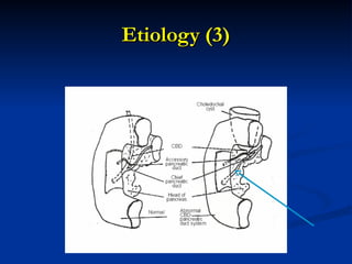
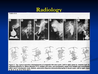
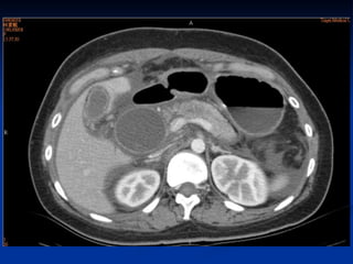
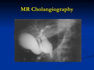
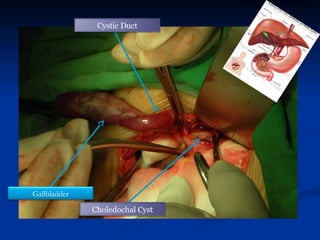
CHOLEDOCHAL CYST
CHOLEDOCHAL CYST
HAMED RASHAD
HAMED RASHAD
Professor of surgery Banha
faculty of medicine - Egypt
Background
Background
•
• Congenital anomalies of the bile ducts.
Congenital anomalies of the bile ducts.
•
• First found western literature 1723
First found western literature 1723 (Vater and
(Vater and
Ezler).
Ezler).
•
• First systematic description of 96 cases in
First systematic description of 96 cases in 1959.
1959.
•
• Classification of 5 types
Classification of 5 types (Todani 1977)
(Todani 1977)
Definition
Definition
 Cyst of the choledocal duct
Cyst of the choledocal duct
 Approximately 1 in 10.000-13.000 live births
Approximately 1 in 10.000-13.000 live births
 More common in women (3-4:1)
More common in women (3-4:1)
 More common in Asian (up to 1:1000) ,
More common in Asian (up to 1:1000) ,
especially in Japan
especially in Japan
Etiology
Etiology
• Long common channel of the terminal bile duct
Long common channel of the terminal bile duct
and pancreatic duct (more than 1 cm from the
sphincter)
sphincter)
• Reflux of the pancreatic enzyms
Reflux of the pancreatic enzyms
• Possible genetic influence, common with other
Possible genetic influence, common with other
anomalies (congenital hepatic fibrosis, intrahepatic
biliary cysts, and polycystic kidney disease)
• Possible congenital duct wall anomaly
Possible congenital duct wall anomaly
Etiology (2)
Etiology (2)
• Weakness of the wall of the bile duct (Babbitt,
Weakness of the wall of the bile duct (Babbitt,
1973; Bismuth, 1999; Flanigan, 1975)
• Obstruction of the distal choledochus (Spitz, 1977)
Obstruction of the distal choledochus (Spitz, 1977)
• Combination of obstruction and weakness (Kato,
Combination of obstruction and weakness (Kato,
1974)
1974)
• Reflux of pancreatic enzymes into the CBD
Reflux of pancreatic enzymes into the CBD
secondary to an anomaly of the pancreaticobiliary
junction (Babbitt, 1969; Kato, 1981; Miyano, 1979;
Okada, 1990; Todani, 1984, Yamashiro, 1984)
Etiology (3)
Etiology (3)
Mortality/morbidity
Mortality/morbidity
•
• Infants and children – pancreatitis,
Infants and children – pancreatitis,
cholangitis and histological evidence
of hepatocellular inflammation
and damage.
and damage.
•
• cholangiocarcinoma 9-28%.
cholangiocarcinoma 9-28%.
Clinical Presentation
 Jaundice
Jaundice
 Pain in RUQ
Pain in RUQ
 Mass (rare)
Mass (rare)
Symptoms
Symptoms
•
• Jaundice
Jaundice
•
• Pale stools (putty colour)
Pale stools (putty colour)
•
• Palpable mass right upper abdominal quadrant
Palpable mass right upper abdominal quadrant
•
• Hepatomegaly
Hepatomegaly
•
• Acute pancreatitis
Acute pancreatitis
Classification
Classification
(Todani 1977)
(Todani 1977)
•
• Type I
Type I 80-90%
80-90%
•
• Type II
Type II
•
• Type III
Type III
•
• Type IV
Type IV
•
• Type V
Type V
Todani Classification
• Type I - Cystic or fusiform dilatation of the CBD; most
Type I - Cystic or fusiform dilatation of the CBD; most
frequent type (90-95% of the cases).
• Type II - Diverticulum of the CBD, with normal size CBD
Type II - Diverticulum of the CBD, with normal size CBD
• Type III - Choledochocele, a cystic dilatation of the distal
Type III - Choledochocele, a cystic dilatation of the distal
intramural portion of the CBD, typically protruding into the
second portion of the duodenum
• Type IV - Cystic or fusiform dilatation of the CBD associated
Type IV - Cystic or fusiform dilatation of the CBD associated
with cystic, fusiform, or saccular dilatation of intrahepatic bile
ducts, also termed form fruste
• Type V - Cystic, fusiform, or saccular dilatation of the
Type V - Cystic, fusiform, or saccular dilatation of the
intrahepatic bile ducts associated with a normal CBD; may be
associated with hepatic fibrosis (referred to as Caroli disease)
Type I choledochal cyst
•
• Dilation of entire common hapatic
Dilation of entire common hapatic
and common bile duct or segments
of each.
of each.
•
• Sacular or fusiform.
Sacular or fusiform.
Type II choledochal cyst
•
• Relatively isolated protrusion that
Relatively isolated protrusion that
project from common bile duct wall.
•
• Connected by narrow stalk.
Connected by narrow stalk.
Type III choledochal cyst
•
• Found in intraduodenal portion of the
Found in intraduodenal portion of the
common bile duct (choledochocade).
Type IV choledochal cyst
•
• Multiple dilations of the
Multiple dilations of the
Intrahepatic and extrahepatic
biliary tree.
biliary tree.
•
• Large extrahepatic cyst with
Large extrahepatic cyst with
multiple intrahepatic cysts.
Type v choledochal cysts
•
• Dilation of intraphepatic biliary
Dilation of intraphepatic biliary radicles.
radicles.
•
• Numerous cysts with structures
Numerous cysts with structures
causing intraphepatic stone
formation, obstruction and cholangitis.
Types of choledocal cysts
Investigations
Investigations
•
• USS abdomen
USS abdomen
•
• CT (93%) or MRI (100%) + cholangiogram
CT (93%) or MRI (100%) + cholangiogram
•
• HIDA scan
HIDA scan
•
• Plain AXR do not identify choledochal cysts
Plain AXR do not identify choledochal cysts
well.
well.
Radiology
Radiology
US
US
CT : choledocal cyst and pancreatitis
MR Cholangiography
MR Cholangiography
Therapy
Therapy
Type I-IV
Type I-IV
• Cystojejunostomy
Cystojejunostomy
• Total Excision and Cystojejunostomy
Total Excision and Cystojejunostomy
• Biliary reconstruction can be performed with a
Biliary reconstruction can be performed with a
Roux-en-Y hepaticojejunostomy as high as
possible, near the hilum of the liver (Hata, 1993;
Joseph, 1990; Kasai, 1970; Miyano, 1996)
Type V
Type V
• Hepatic Lobectomy
Hepatic Lobectomy
Treatment Type I
Treatment Type I
•
• Surgery
Surgery
•
• Complete excision with Rovx-en-y
Complete excision with Rovx-en-y
biliary end to side anastromosis to
biliary end to side anastromosis to restore
restore
biliary continuity to
biliary continuity to gastrointestinal track.
gastrointestinal track.
Treatment Type II
Treatment Type II
•
• Total excision
Total excision
Treatment Type III
Treatment Type III
•
• 3 cm or less endoscopic removal.
3 cm or less endoscopic removal.
•
• >3 cm surgical excised.
>3 cm surgical excised.
Treatment Type IV
Treatment Type IV
•
• Dilated extraphepatic
Dilated extraphepatic
duct removed.
duct removed.
Treatment Type V
Treatment Type V
•
• Liver Transplantation
Liver Transplantation
Surgery
Surgery
Gallbladder
Choledochal Cyst
Cystic Duct
Gallbladder
Choledochal Cyst
Hepaticojejunostomy Roux-en-Y
Complications
Complications
 Cholangitis
Cholangitis
 Biliary stone formation
Biliary stone formation
 Anastomotic stricture
Anastomotic stricture
 Residual debris in the intrahepatic bile ducts
Residual debris in the intrahepatic bile ducts
 Intrahepatic bile duct dilatation
Intrahepatic bile duct dilatation
 Malignancy
Malignancy
References
References
 Btaiche I and Khalidi N (2002). Parenteral Nutrition - Associated
Btaiche I and Khalidi N (2002). Parenteral Nutrition - Associated
Liver Complications in Children.
Liver Complications in Children. Pharmacotherapy.
Pharmacotherapy. 22(2): 188- 211
22(2): 188- 211
 Howard E, Stringer M and Colombani P (2002) (2ND Ed).
Howard E, Stringer M and Colombani P (2002) (2ND Ed).
Surgery of the Liver, bile ducts and Pancreas in Children.
Surgery of the Liver, bile ducts and Pancreas in Children. Arnold.
Arnold.
London
London
 Piwko J, Caty M, Ryan R (2003). Neonatal considerations for the
Piwko J, Caty M, Ryan R (2003). Neonatal considerations for the
Paediatric Surgeon.
Paediatric Surgeon. www.emedicine.com/ped/topic2982.htm
 Puri P (2003) Newborn Surgery (2nd Ed).
Puri P (2003) Newborn Surgery (2nd Ed). Arnold.
Arnold. London
London
 Sadiq J, Nondi B, Lakhoo K (2009). An unusual variant of
Sadiq J, Nondi B, Lakhoo K (2009). An unusual variant of
choledochal cyst : a case report.
choledochal cyst : a case report. Journal of Medical Case Reports.
Journal of Medical Case Reports. 3 :
3 :
54
54
References Cont.
References Cont.
 Sawyer M, Varma M, Murphy T (2009). Choledochal Cyst.
Sawyer M, Varma M, Murphy T (2009). Choledochal Cyst.
http://emedicine.medscape.com/article/366004-overview
 Strodtbeck F(2003) The pathophysiology of prolonged periods of
Strodtbeck F(2003) The pathophysiology of prolonged periods of
No Enteral Nutrition or Nothing by Mouth. Newborn and Infant
Nursing Reviews.
Nursing Reviews. www.medscape.com/viewarticle/458473
 Subramanian K, Yoon H and Toral J (2002). Extremely Low Birth
Subramanian K, Yoon H and Toral J (2002). Extremely Low Birth
Weight Infant.
Weight Infant. www.emedicine.com/ped/topic2784.htm
 Tortora G and Grabowski S (1993) (7TH Ed). Principles of
Tortora G and Grabowski S (1993) (7TH Ed). Principles of
Anatomy and
Anatomy and Physiology
Physiology.
. Harper Collins College Publishers.
Harper Collins College Publishers. New
New
York Pp790
York Pp790
THAK YOU
THAK YOU

Choledochal cyst the lect. types and management

  • 1.
    CHOLEDOCHAL CYST CHOLEDOCHAL CYST HAMEDRASHAD HAMED RASHAD Professor of surgery Banha Professor of surgery Banha faculty of medicine - Egypt faculty of medicine - Egypt
  • 2.
    Background Background • • Congenital anomaliesof the bile ducts. Congenital anomalies of the bile ducts. • • First found western literature 1723 First found western literature 1723 (Vater and (Vater and Ezler). Ezler). • • First systematic description of 96 cases in First systematic description of 96 cases in 1959. 1959. • • Classification of 5 types Classification of 5 types (Todani 1977) (Todani 1977)
  • 3.
    Definition Definition  Cyst ofthe choledocal duct Cyst of the choledocal duct  Approximately 1 in 10.000-13.000 live births Approximately 1 in 10.000-13.000 live births  More common in women (3-4:1) More common in women (3-4:1)  More common in Asian (up to 1:1000) , More common in Asian (up to 1:1000) , especially in Japan especially in Japan
  • 4.
    Etiology Etiology • Long commonchannel of the terminal bile duct Long common channel of the terminal bile duct and pancreatic duct (more than 1 cm from the and pancreatic duct (more than 1 cm from the sphincter) sphincter) • Reflux of the pancreatic enzyms Reflux of the pancreatic enzyms • Possible genetic influence, common with other Possible genetic influence, common with other anomalies (congenital hepatic fibrosis, intrahepatic anomalies (congenital hepatic fibrosis, intrahepatic biliary cysts, and polycystic kidney disease) biliary cysts, and polycystic kidney disease) • Possible congenital duct wall anomaly Possible congenital duct wall anomaly
  • 5.
    Etiology (2) Etiology (2) •Weakness of the wall of the bile duct (Babbitt, Weakness of the wall of the bile duct (Babbitt, 1973; Bismuth, 1999; Flanigan, 1975) 1973; Bismuth, 1999; Flanigan, 1975) • Obstruction of the distal choledochus (Spitz, 1977) Obstruction of the distal choledochus (Spitz, 1977) • Combination of obstruction and weakness (Kato, Combination of obstruction and weakness (Kato, 1974) 1974) • Reflux of pancreatic enzymes into the CBD Reflux of pancreatic enzymes into the CBD secondary to an anomaly of the pancreaticobiliary secondary to an anomaly of the pancreaticobiliary junction (Babbitt, 1969; Kato, 1981; Miyano, 1979; junction (Babbitt, 1969; Kato, 1981; Miyano, 1979; Okada, 1990; Todani, 1984, Yamashiro, 1984) Okada, 1990; Todani, 1984, Yamashiro, 1984)
  • 6.
  • 7.
    Mortality/morbidity Mortality/morbidity • • Infants andchildren – pancreatitis, Infants and children – pancreatitis, cholangitis and histological evidence cholangitis and histological evidence of hepatocellular inflammation of hepatocellular inflammation and damage. and damage. • • cholangiocarcinoma 9-28%. cholangiocarcinoma 9-28%.
  • 8.
    Clinical Presentation Clinical Presentation Jaundice Jaundice  Pain in RUQ Pain in RUQ  Mass (rare) Mass (rare)
  • 9.
    Symptoms Symptoms • • Jaundice Jaundice • • Palestools (putty colour) Pale stools (putty colour) • • Palpable mass right upper abdominal quadrant Palpable mass right upper abdominal quadrant • • Hepatomegaly Hepatomegaly • • Acute pancreatitis Acute pancreatitis
  • 10.
    Classification Classification (Todani 1977) (Todani 1977) • •Type I Type I 80-90% 80-90% • • Type II Type II • • Type III Type III • • Type IV Type IV • • Type V Type V
  • 11.
    Todani Classification Todani Classification •Type I - Cystic or fusiform dilatation of the CBD; most Type I - Cystic or fusiform dilatation of the CBD; most frequent type (90-95% of the cases). frequent type (90-95% of the cases). • Type II - Diverticulum of the CBD, with normal size CBD Type II - Diverticulum of the CBD, with normal size CBD • Type III - Choledochocele, a cystic dilatation of the distal Type III - Choledochocele, a cystic dilatation of the distal intramural portion of the CBD, typically protruding into the intramural portion of the CBD, typically protruding into the second portion of the duodenum second portion of the duodenum • Type IV - Cystic or fusiform dilatation of the CBD associated Type IV - Cystic or fusiform dilatation of the CBD associated with cystic, fusiform, or saccular dilatation of intrahepatic bile with cystic, fusiform, or saccular dilatation of intrahepatic bile ducts, also termed form fruste ducts, also termed form fruste • Type V - Cystic, fusiform, or saccular dilatation of the Type V - Cystic, fusiform, or saccular dilatation of the intrahepatic bile ducts associated with a normal CBD; may be intrahepatic bile ducts associated with a normal CBD; may be associated with hepatic fibrosis (referred to as Caroli disease) associated with hepatic fibrosis (referred to as Caroli disease)
  • 12.
    Type I choledochalcyst Type I choledochal cyst • • Dilation of entire common hapatic Dilation of entire common hapatic and common bile duct or segments and common bile duct or segments of each. of each. • • Sacular or fusiform. Sacular or fusiform.
  • 13.
    Type II choledochalcyst Type II choledochal cyst • • Relatively isolated protrusion that Relatively isolated protrusion that project from common bile duct wall. project from common bile duct wall. • • Connected by narrow stalk. Connected by narrow stalk.
  • 14.
    Type III choledochalcyst Type III choledochal cyst • • Found in intraduodenal portion of the Found in intraduodenal portion of the common bile duct (choledochocade). common bile duct (choledochocade).
  • 15.
    Type IV choledochalcyst Type IV choledochal cyst • • Multiple dilations of the Multiple dilations of the Intrahepatic and extrahepatic Intrahepatic and extrahepatic biliary tree. biliary tree. • • Large extrahepatic cyst with Large extrahepatic cyst with multiple intrahepatic cysts. multiple intrahepatic cysts.
  • 16.
    Type v choledochalcysts Type v choledochal cysts • • Dilation of intraphepatic biliary Dilation of intraphepatic biliary radicles. radicles. • • Numerous cysts with structures Numerous cysts with structures causing intraphepatic stone causing intraphepatic stone formation, obstruction and cholangitis. formation, obstruction and cholangitis.
  • 17.
    Types of choledocalcysts Types of choledocal cysts
  • 18.
    Investigations Investigations • • USS abdomen USSabdomen • • CT (93%) or MRI (100%) + cholangiogram CT (93%) or MRI (100%) + cholangiogram • • HIDA scan HIDA scan • • Plain AXR do not identify choledochal cysts Plain AXR do not identify choledochal cysts well. well.
  • 19.
  • 20.
  • 21.
    CT : choledocalcyst and pancreatitis CT : choledocal cyst and pancreatitis
  • 23.
  • 24.
    Therapy Therapy Type I-IV Type I-IV •Cystojejunostomy Cystojejunostomy • Total Excision and Cystojejunostomy Total Excision and Cystojejunostomy • Biliary reconstruction can be performed with a Biliary reconstruction can be performed with a Roux-en-Y hepaticojejunostomy as high as Roux-en-Y hepaticojejunostomy as high as possible, near the hilum of the liver (Hata, 1993; possible, near the hilum of the liver (Hata, 1993; Joseph, 1990; Kasai, 1970; Miyano, 1996) Joseph, 1990; Kasai, 1970; Miyano, 1996) Type V Type V • Hepatic Lobectomy Hepatic Lobectomy
  • 25.
    Treatment Type I TreatmentType I • • Surgery Surgery • • Complete excision with Rovx-en-y Complete excision with Rovx-en-y biliary end to side anastromosis to biliary end to side anastromosis to restore restore biliary continuity to biliary continuity to gastrointestinal track. gastrointestinal track.
  • 26.
    Treatment Type II TreatmentType II • • Total excision Total excision
  • 27.
    Treatment Type III TreatmentType III • • 3 cm or less endoscopic removal. 3 cm or less endoscopic removal. • • >3 cm surgical excised. >3 cm surgical excised.
  • 28.
    Treatment Type IV TreatmentType IV • • Dilated extraphepatic Dilated extraphepatic duct removed. duct removed.
  • 29.
    Treatment Type V TreatmentType V • • Liver Transplantation Liver Transplantation
  • 30.
  • 31.
  • 32.
  • 33.
    Complications Complications  Cholangitis Cholangitis  Biliarystone formation Biliary stone formation  Anastomotic stricture Anastomotic stricture  Residual debris in the intrahepatic bile ducts Residual debris in the intrahepatic bile ducts  Intrahepatic bile duct dilatation Intrahepatic bile duct dilatation  Malignancy Malignancy
  • 34.
    References References  Btaiche Iand Khalidi N (2002). Parenteral Nutrition - Associated Btaiche I and Khalidi N (2002). Parenteral Nutrition - Associated Liver Complications in Children. Liver Complications in Children. Pharmacotherapy. Pharmacotherapy. 22(2): 188- 211 22(2): 188- 211  Howard E, Stringer M and Colombani P (2002) (2ND Ed). Howard E, Stringer M and Colombani P (2002) (2ND Ed). Surgery of the Liver, bile ducts and Pancreas in Children. Surgery of the Liver, bile ducts and Pancreas in Children. Arnold. Arnold. London London  Piwko J, Caty M, Ryan R (2003). Neonatal considerations for the Piwko J, Caty M, Ryan R (2003). Neonatal considerations for the Paediatric Surgeon. Paediatric Surgeon. www.emedicine.com/ped/topic2982.htm  Puri P (2003) Newborn Surgery (2nd Ed). Puri P (2003) Newborn Surgery (2nd Ed). Arnold. Arnold. London London  Sadiq J, Nondi B, Lakhoo K (2009). An unusual variant of Sadiq J, Nondi B, Lakhoo K (2009). An unusual variant of choledochal cyst : a case report. choledochal cyst : a case report. Journal of Medical Case Reports. Journal of Medical Case Reports. 3 : 3 : 54 54
  • 35.
    References Cont. References Cont. Sawyer M, Varma M, Murphy T (2009). Choledochal Cyst. Sawyer M, Varma M, Murphy T (2009). Choledochal Cyst. http://emedicine.medscape.com/article/366004-overview  Strodtbeck F(2003) The pathophysiology of prolonged periods of Strodtbeck F(2003) The pathophysiology of prolonged periods of No Enteral Nutrition or Nothing by Mouth. Newborn and Infant No Enteral Nutrition or Nothing by Mouth. Newborn and Infant Nursing Reviews. Nursing Reviews. www.medscape.com/viewarticle/458473  Subramanian K, Yoon H and Toral J (2002). Extremely Low Birth Subramanian K, Yoon H and Toral J (2002). Extremely Low Birth Weight Infant. Weight Infant. www.emedicine.com/ped/topic2784.htm  Tortora G and Grabowski S (1993) (7TH Ed). Principles of Tortora G and Grabowski S (1993) (7TH Ed). Principles of Anatomy and Anatomy and Physiology Physiology. . Harper Collins College Publishers. Harper Collins College Publishers. New New York Pp790 York Pp790
  • 36.