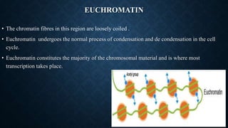
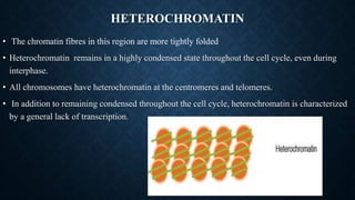
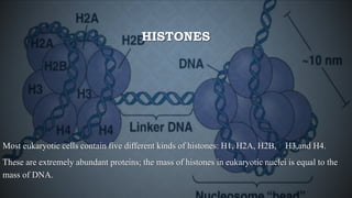
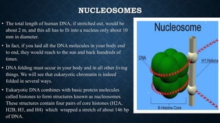
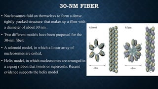
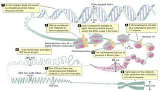
CHROMATIN STRUCTURE
• Chromatin is composed of DNA and proteins, mostly basic proteins called histones
• That help chromatin fold so it can pack into the tiny volume of a cell’s nucleus.
• The two basic types of chromatin are
1. Euchromatin
2. Heterochromatin
EUCHROMATIN
• The chromatin fibres in this region are loosely coiled .
• Euchromatin undergoes the normal process of condensation and de condensation in the cell
cycle.
• Euchromatin constitutes the majority of the chromosomal material and is where most
transcription takes place.
HETEROCHROMATIN
• The chromatin fibres in this region are more tightly folded
• Heterochromatin remains in a highly condensed state throughout the cell cycle, even during
interphase.
• All chromosomes have heterochromatin at the centromeres and telomeres.
• In addition to remaining condensed throughout the cell cycle, heterochromatin is characterized
by a general lack of transcription.
HISTONES
Most eukaryotic cells contain five different kinds of histones: H1, H2A, H2B, H3,and H4.
These are extremely abundant proteins; the mass of histones in eukaryotic nuclei is equal to the
mass of DNA.
NUCLEOSOMES
• The total length of human DNA, if stretched out, would be
about 2 m, and this all has to fit into a nucleus only about 10
mm in diameter.
• In fact, if you laid all the DNA molecules in your body end
to end, they would reach to the sun and back hundreds of
times.
• DNA folding must occur in your body and in all other living
things. We will see that eukaryotic chromatin is indeed
folded in several ways.
• Eukaryotic DNA combines with basic protein molecules
called histones to form structures known as nucleosomes.
These structures contain four pairs of core histones (H2A,
H2B, H3, and H4) which wrapped a stretch of about 146 bp
of DNA.
30-NM FIBER
• Nucleosomes fold on themselves to form a dense,
tightly packed structure that makes up a fiber with
a diameter of about 30 nm .
• Two different models have been proposed for the
30-nm fiber:
• A solenoid model, in which a linear array of
nucleosomes are coiled,
• Helix model, in which nucleosomes are arranged in
a zigzag ribbon that twists or supercoils. Recent
evidence supports the helix model
HIGHER-ORDER CHROMATIN FOLDING
• The next-higher level of chromatin structure is a series of loops of 30-nm fibers.
• On average, each loop encompasses some 20,000 to 100,000 bp of DNA and is about 300 nm in
length.
• The 300-nm loops are packed and folded to produce a 250-nm-wide fiber.
• Tight helical coiling of the 250-nm fiber in turn produces the structure that appears in metaphase
individual chromatids approximately 700 nm in width.
Chromatin structure

Chromatin structure

  • 1.
    CHROMATIN STRUCTURE • Chromatinis composed of DNA and proteins, mostly basic proteins called histones • That help chromatin fold so it can pack into the tiny volume of a cell’s nucleus. • The two basic types of chromatin are 1. Euchromatin 2. Heterochromatin
  • 2.
    EUCHROMATIN • The chromatinfibres in this region are loosely coiled . • Euchromatin undergoes the normal process of condensation and de condensation in the cell cycle. • Euchromatin constitutes the majority of the chromosomal material and is where most transcription takes place.
  • 3.
    HETEROCHROMATIN • The chromatinfibres in this region are more tightly folded • Heterochromatin remains in a highly condensed state throughout the cell cycle, even during interphase. • All chromosomes have heterochromatin at the centromeres and telomeres. • In addition to remaining condensed throughout the cell cycle, heterochromatin is characterized by a general lack of transcription.
  • 4.
    HISTONES Most eukaryotic cellscontain five different kinds of histones: H1, H2A, H2B, H3,and H4. These are extremely abundant proteins; the mass of histones in eukaryotic nuclei is equal to the mass of DNA.
  • 5.
    NUCLEOSOMES • The totallength of human DNA, if stretched out, would be about 2 m, and this all has to fit into a nucleus only about 10 mm in diameter. • In fact, if you laid all the DNA molecules in your body end to end, they would reach to the sun and back hundreds of times. • DNA folding must occur in your body and in all other living things. We will see that eukaryotic chromatin is indeed folded in several ways. • Eukaryotic DNA combines with basic protein molecules called histones to form structures known as nucleosomes. These structures contain four pairs of core histones (H2A, H2B, H3, and H4) which wrapped a stretch of about 146 bp of DNA.
  • 6.
    30-NM FIBER • Nucleosomesfold on themselves to form a dense, tightly packed structure that makes up a fiber with a diameter of about 30 nm . • Two different models have been proposed for the 30-nm fiber: • A solenoid model, in which a linear array of nucleosomes are coiled, • Helix model, in which nucleosomes are arranged in a zigzag ribbon that twists or supercoils. Recent evidence supports the helix model
  • 7.
    HIGHER-ORDER CHROMATIN FOLDING •The next-higher level of chromatin structure is a series of loops of 30-nm fibers. • On average, each loop encompasses some 20,000 to 100,000 bp of DNA and is about 300 nm in length. • The 300-nm loops are packed and folded to produce a 250-nm-wide fiber. • Tight helical coiling of the 250-nm fiber in turn produces the structure that appears in metaphase individual chromatids approximately 700 nm in width.