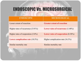
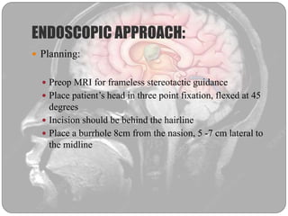
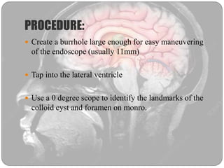
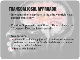
Dr. Rabail Akbar Qazi
Neurosurgery Resident
COLLOID CYST
 Benign tumors of the neuroectodermal origin
constituting 0.5% to 1% of all intracranial neoplasms
 They arise from the anterior aspect of velum
interpositum or the choroid plexus of the third
ventricle, in close proximity with the foramen of
monro
 Slow growing
ANATOMY
PATHOGENESIS
 Origin: Unknown
 Implicated structures include:
 Paraphysis
 Diencephalic ependyma in the recess of postvelar arch
 Ventricular neuroepithelium
 Comprised of fibrous epithelial-lined wall filled with
either mucoid or dense hyaloid substance
 Can occur in any age with peak between the 2nd and 4th
decades of life with no sex predilection.
 Cyst wall is lined with simple or pseudostratified
cuboidal or low columnar ciliated epithelial cells
 Stain positive for PAS & S100 and negative for GFAP,
vimentin and neurofilament.
SYMPTOMS
 In 75% of the patients, the initial symptom is headache
with variable intensity.
 Other symptoms include nausea, vomiting, gait
disturbance and blurred vision.
 A sudden increase in ICP from rapid onset
hydrocephalus can lead to sudden death
 By compression of the surrounding anatomic
structures, the patient can experience thermal
dysregulation, electrolyte and hormonal imbalances,
endocrine dysfunction , altered personality, memory
loss or visual changes.
 Most cysts <1cm donot produce hydrocephalus and are
asymptomatic.
 Colloid cysts may cause chronic, acute or intermittent
hydrocephalus .
 Intermittent symptoms are attributed to the movement
on the cyst on its pedicle causing intermittent
obstruction to CSF flow.
EVALUATION
 Imaging usually demonstrates the tumor located in the
anterior third ventricle.
 If there is obstruction to CSF flow due to the cyst, it
manifests as pathognomonic hydrocephalus involving
only the lateral ventricles.
 MRI is usually the imaging modality of choice.
CT SCAN:
 Most are hyperdense
 Density may correlate with
viscosity of the contents
 Typically homogenous, with
presence of calcifications being
an exception.
MRI :
 Appearance variable
 T1WI: Hyperintense
 T2WI: Hypointense
 No or minimal contrast enhancement, sometimes only
involving the capsule
 Symptomatic patients are more likely to display T2
hyperintense cysts
TREATMENT
 Conservative
 Shunting procedure
 Surgical
 Microsurgical
 Endoscopic
RECURSIVE PARTITIONING ANALYSIS
DIVERSION PROCEDURES:
 Bilateral ventriculoperitoneal shunts
 Unilateral shunt with septum pellucidotomy
 However, direct surgical treatment is usually
recommended due to:
 To prevent shunt dependency
 To reduce the possibility of tumor progression
 Risk of cardiovascular instability
SURGICAL MANAGEMENT:
 Transcallosal approach : Not dependent on
ventriculomegaly
 Transcortical approach: In patients with HCP
 Stereotactic drainage
 Ventriculoscopic removal
STEREOTACTIC DRAINAGE:
 Maybe useful in patients with normal ventricles
 High recurrence rate
 Short hospital stay and early recovery time
 Viscous contents and tough capsule lead to failure of
procedure
 Two features correlating with unsuccesful aspiration:
 High viscosity: Correlates with hyperdensity on CT
 Deflection of the cyst from tip of the aspiration needle
due to small size
Technique:
 Insertion point of the needle is just anterior to the right
coronal suture
 Start with sharp-tipped 1.8mm probe, and advance to
3–5mm beyond target site (to accommodate for
displacement of cyst wall
 Use a 10ml syringe and apply 6–8ml of negative
aspiration pressure
 If this does not yield any material, repeat with a
2.1mm probe.
 Although complete cyst evacuation is desirable, if this
cannot be accomplished an acceptable goal of
aspiration is re-establishment of patency of the
ventricular pathways (may be verified by injecting 1–2
cc of iohexol)
ENDOSCOPIC Vs. MICROSURGICAL
ENDOSCOPIC MICROSURGICAL
Lesser extent of resection Greater extent of resection
Higher rates of recurrence (3.91%) Lower rates of recurrence (1.48%)
Higher rates of reoperation (3.0%) Lower rates of reoperation (0.38%)
Lower complication rate (10.5%) Higher complication rate (16.3%)
Similar mortality rate Similar mortality rate
ENDOSCOPIC APPROACH:
 Planning:
 Preop MRI for frameless stereotactic guidance
 Place patient’s head in three point fixation, flexed at 45
degrees
 Incision should be behind the hairline
 Place a burrhole 8cm from the nasion, 5 -7 cm lateral to
the midline
PROCEDURE:
 Create a burrhole large enough for easy maneuvering
of the endoscope (usually 11mm)
 Tap into the lateral ventricle
 Use a 0 degree scope to identify the landmarks of the
colloid cyst and foramen on monro.
 Change to a 30 degree scope
 Colloid cyst can be identified by its greenish grey
membrane
 Resection technique for the cyst depends on it’s size
and consistency
 If the contents are liquid, cyst can be punctured and
contents aspirated through an appropriate suction tube.
 Sometimes, cyst has to be opened with microscissors
and contents removed in a piecemeal fashion
TRANSCALLOSAL APPROACH:
 Interhemispheric approach to the third ventricle via a
parietal craniotomy
 Position: Supine with neck flexed. Thorax elevated at
20 degrees. Keep the head vertical
 Skin Incision:
 Inverted U with the top just left of midline, 6cm anterior
to the coronal suture, to 2 cm behind the coronal suture,
taking the sides for 7-8cm
 Souttar skin incision
Craniotomy:
 The bone flap is either trapezoid or triangular
 For adequate exposure, it is critical to go all the way to
the superior sagittal sinus
 To stay away from the motor strip and to keep sagittal
sinus exposure as anterior as possible:
 2/3rd of the craniotomy anterior to the coronal suture
 1/3rd posterior to the coronal suture
 Total craniotomy size should be 6cm
 4cm anterior to the coronal suture
 2 cm posterior to the coronal suture
 4cm to the right of midline
 The dural flap is based towards the sagittal sinus
COMPLICATIONS:
 Venous infarction
 Sacrifice of critical cortical draining veins
 SSS thrombosis
 Transient Mutism due to rough retraction of cingulate gyrus
 Decrease in spontaneous speech due to resection of anterior
portion of CC
 Damage to fornix resulting in temporary or persistent
amnestic syndromes
 Intraventricular bleeding
 CSF leak
TRANSCORTICAL APPROACH:
 Patient supine with three pin fixation, head flexed at
30 degrees, 10 to 50 degree rotation to the contralateral
side
 3x4 cm bone flap placed over the location of the
middle frontal gyrus
 Medial border should just cross the midline and the
anterior border 2cm anterior to the coronal suture and
posterior border 2 cm behind.
Colloid cyst
Colloid cyst
Colloid cyst

Colloid cyst

  • 1.
    Dr. Rabail AkbarQazi Neurosurgery Resident COLLOID CYST
  • 2.
     Benign tumorsof the neuroectodermal origin constituting 0.5% to 1% of all intracranial neoplasms  They arise from the anterior aspect of velum interpositum or the choroid plexus of the third ventricle, in close proximity with the foramen of monro  Slow growing
  • 3.
  • 13.
    PATHOGENESIS  Origin: Unknown Implicated structures include:  Paraphysis  Diencephalic ependyma in the recess of postvelar arch  Ventricular neuroepithelium  Comprised of fibrous epithelial-lined wall filled with either mucoid or dense hyaloid substance
  • 15.
     Can occurin any age with peak between the 2nd and 4th decades of life with no sex predilection.  Cyst wall is lined with simple or pseudostratified cuboidal or low columnar ciliated epithelial cells  Stain positive for PAS & S100 and negative for GFAP, vimentin and neurofilament.
  • 16.
    SYMPTOMS  In 75%of the patients, the initial symptom is headache with variable intensity.  Other symptoms include nausea, vomiting, gait disturbance and blurred vision.  A sudden increase in ICP from rapid onset hydrocephalus can lead to sudden death
  • 17.
     By compressionof the surrounding anatomic structures, the patient can experience thermal dysregulation, electrolyte and hormonal imbalances, endocrine dysfunction , altered personality, memory loss or visual changes.
  • 20.
     Most cysts<1cm donot produce hydrocephalus and are asymptomatic.  Colloid cysts may cause chronic, acute or intermittent hydrocephalus .  Intermittent symptoms are attributed to the movement on the cyst on its pedicle causing intermittent obstruction to CSF flow.
  • 21.
    EVALUATION  Imaging usuallydemonstrates the tumor located in the anterior third ventricle.  If there is obstruction to CSF flow due to the cyst, it manifests as pathognomonic hydrocephalus involving only the lateral ventricles.  MRI is usually the imaging modality of choice.
  • 22.
    CT SCAN:  Mostare hyperdense  Density may correlate with viscosity of the contents  Typically homogenous, with presence of calcifications being an exception.
  • 23.
    MRI :  Appearancevariable  T1WI: Hyperintense  T2WI: Hypointense  No or minimal contrast enhancement, sometimes only involving the capsule  Symptomatic patients are more likely to display T2 hyperintense cysts
  • 27.
    TREATMENT  Conservative  Shuntingprocedure  Surgical  Microsurgical  Endoscopic
  • 28.
  • 30.
    DIVERSION PROCEDURES:  Bilateralventriculoperitoneal shunts  Unilateral shunt with septum pellucidotomy  However, direct surgical treatment is usually recommended due to:  To prevent shunt dependency  To reduce the possibility of tumor progression  Risk of cardiovascular instability
  • 31.
    SURGICAL MANAGEMENT:  Transcallosalapproach : Not dependent on ventriculomegaly  Transcortical approach: In patients with HCP  Stereotactic drainage  Ventriculoscopic removal
  • 32.
    STEREOTACTIC DRAINAGE:  Maybeuseful in patients with normal ventricles  High recurrence rate  Short hospital stay and early recovery time  Viscous contents and tough capsule lead to failure of procedure
  • 33.
     Two featurescorrelating with unsuccesful aspiration:  High viscosity: Correlates with hyperdensity on CT  Deflection of the cyst from tip of the aspiration needle due to small size
  • 34.
    Technique:  Insertion pointof the needle is just anterior to the right coronal suture
  • 35.
     Start withsharp-tipped 1.8mm probe, and advance to 3–5mm beyond target site (to accommodate for displacement of cyst wall
  • 36.
     Use a10ml syringe and apply 6–8ml of negative aspiration pressure
  • 37.
     If thisdoes not yield any material, repeat with a 2.1mm probe.  Although complete cyst evacuation is desirable, if this cannot be accomplished an acceptable goal of aspiration is re-establishment of patency of the ventricular pathways (may be verified by injecting 1–2 cc of iohexol)
  • 38.
    ENDOSCOPIC Vs. MICROSURGICAL ENDOSCOPICMICROSURGICAL Lesser extent of resection Greater extent of resection Higher rates of recurrence (3.91%) Lower rates of recurrence (1.48%) Higher rates of reoperation (3.0%) Lower rates of reoperation (0.38%) Lower complication rate (10.5%) Higher complication rate (16.3%) Similar mortality rate Similar mortality rate
  • 39.
    ENDOSCOPIC APPROACH:  Planning: Preop MRI for frameless stereotactic guidance  Place patient’s head in three point fixation, flexed at 45 degrees  Incision should be behind the hairline  Place a burrhole 8cm from the nasion, 5 -7 cm lateral to the midline
  • 40.
    PROCEDURE:  Create aburrhole large enough for easy maneuvering of the endoscope (usually 11mm)  Tap into the lateral ventricle  Use a 0 degree scope to identify the landmarks of the colloid cyst and foramen on monro.
  • 43.
     Change toa 30 degree scope  Colloid cyst can be identified by its greenish grey membrane  Resection technique for the cyst depends on it’s size and consistency  If the contents are liquid, cyst can be punctured and contents aspirated through an appropriate suction tube.  Sometimes, cyst has to be opened with microscissors and contents removed in a piecemeal fashion
  • 45.
    TRANSCALLOSAL APPROACH:  Interhemisphericapproach to the third ventricle via a parietal craniotomy  Position: Supine with neck flexed. Thorax elevated at 20 degrees. Keep the head vertical  Skin Incision:  Inverted U with the top just left of midline, 6cm anterior to the coronal suture, to 2 cm behind the coronal suture, taking the sides for 7-8cm  Souttar skin incision
  • 47.
    Craniotomy:  The boneflap is either trapezoid or triangular  For adequate exposure, it is critical to go all the way to the superior sagittal sinus  To stay away from the motor strip and to keep sagittal sinus exposure as anterior as possible:  2/3rd of the craniotomy anterior to the coronal suture  1/3rd posterior to the coronal suture
  • 48.
     Total craniotomysize should be 6cm  4cm anterior to the coronal suture  2 cm posterior to the coronal suture  4cm to the right of midline  The dural flap is based towards the sagittal sinus
  • 54.
    COMPLICATIONS:  Venous infarction Sacrifice of critical cortical draining veins  SSS thrombosis  Transient Mutism due to rough retraction of cingulate gyrus  Decrease in spontaneous speech due to resection of anterior portion of CC  Damage to fornix resulting in temporary or persistent amnestic syndromes  Intraventricular bleeding  CSF leak
  • 55.
    TRANSCORTICAL APPROACH:  Patientsupine with three pin fixation, head flexed at 30 degrees, 10 to 50 degree rotation to the contralateral side  3x4 cm bone flap placed over the location of the middle frontal gyrus  Medial border should just cross the midline and the anterior border 2cm anterior to the coronal suture and posterior border 2 cm behind.

Editor's Notes

  • #14 Paraphysis : Evagination of the roof of the third ventricle
  • #54 Mid and posterior third ventricle