Downloaded 534 times


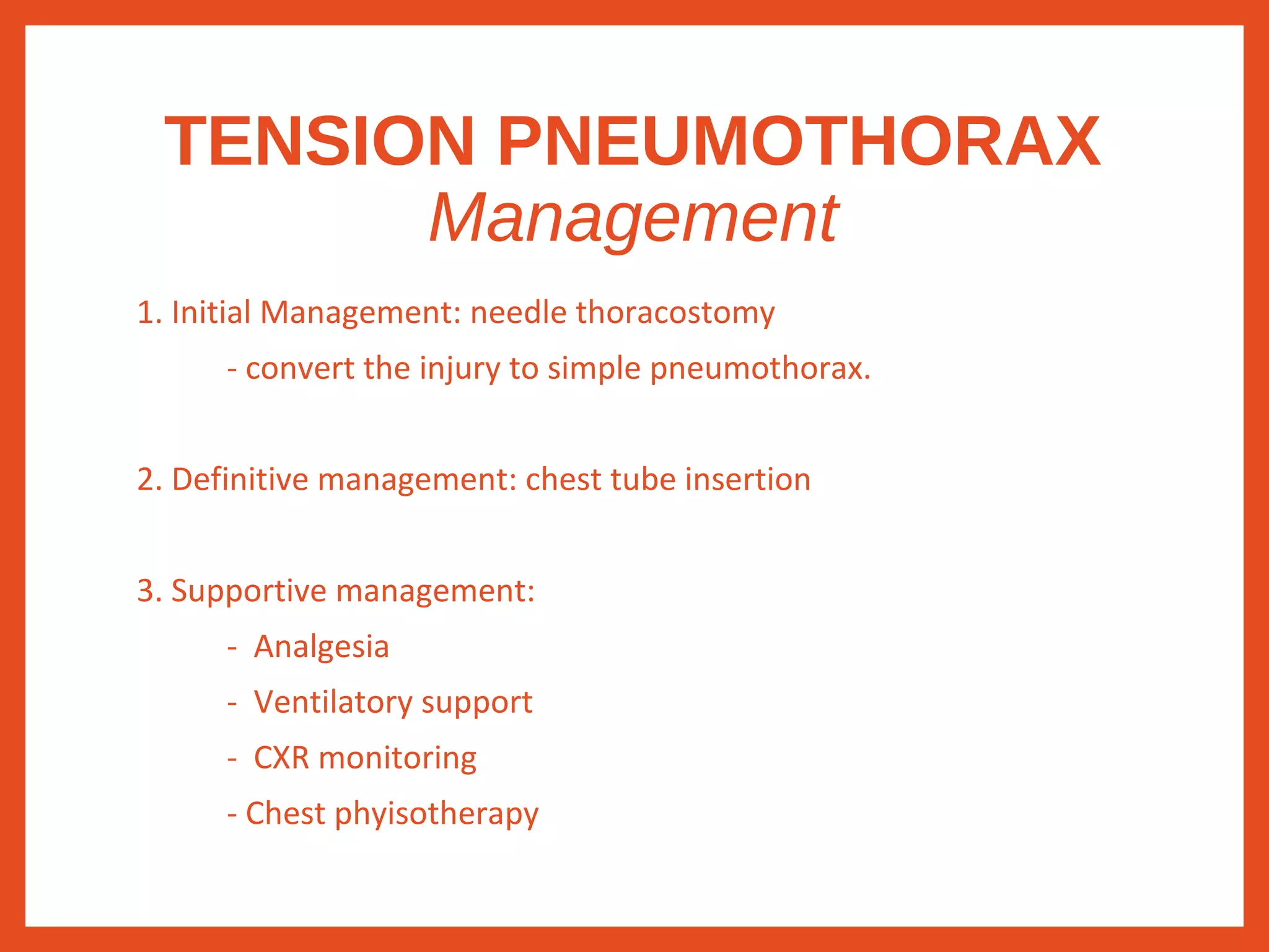


1) Chest injuries account for 20-25% of all trauma deaths and are a leading cause of death worldwide. Life-threatening conditions include tension pneumothorax, open pneumothorax, massive hemothorax, flail chest, and cardiac tamponade. 2) Tension pneumothorax requires immediate needle decompression without waiting for imaging if suspected clinically. Open pneumothorax is managed with an occlusive dressing. 3) Flail chest involves fractures of 3 or more ribs in two places, leading to paradoxical chest wall movement and impaired ventilation. Massive hemothorax involves over 1.5L of blood drained by chest tube or more than 200cc/hour


