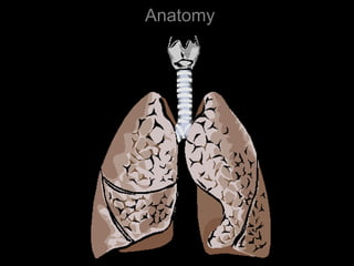
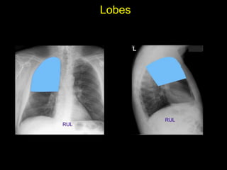
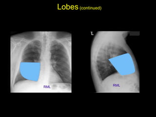
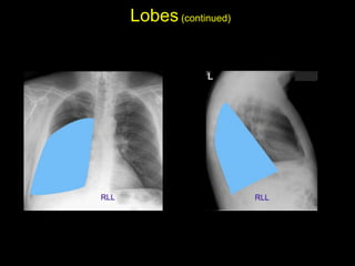
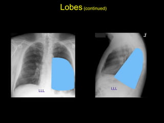
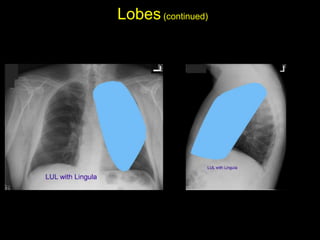
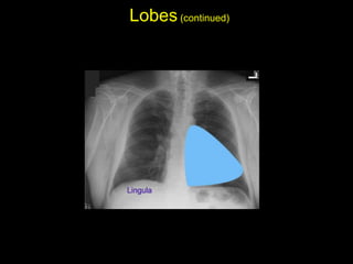
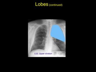
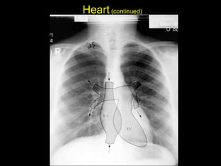
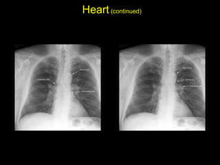
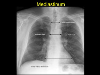
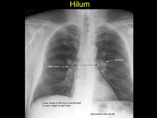
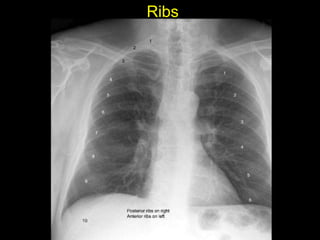
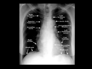
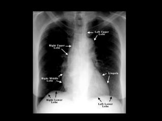
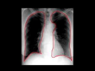
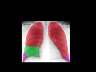
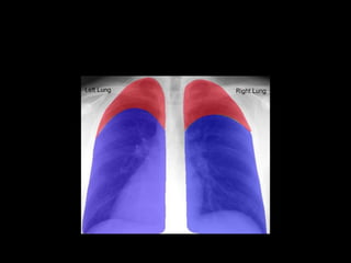
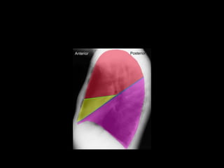
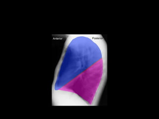
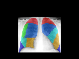
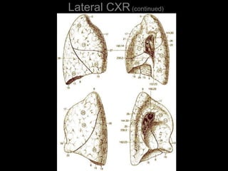
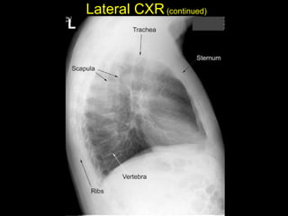
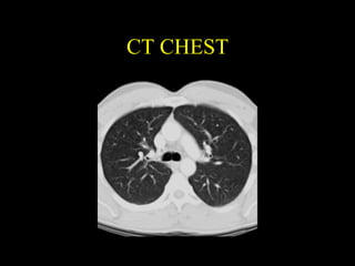
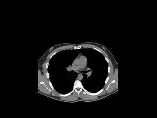
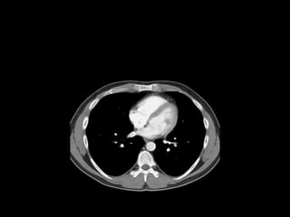
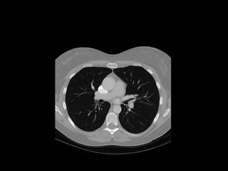
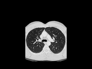
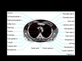
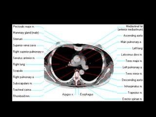
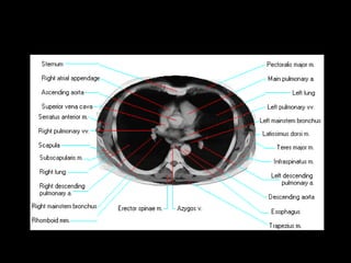
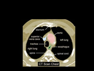
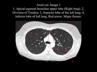
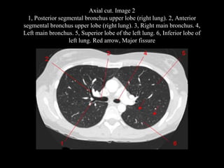
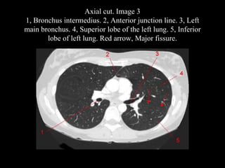
The document discusses the anatomy of the chest x-ray and CT scan by describing the lobes of the lungs and their locations. It also mentions the heart, mediastinum, hilum, and ribs. Several axial, coronal, and sagittal CT images are included with labels pointing out structures like the trachea, bronchi, lobes of the lungs, and fissures. In summary, the document provides an overview of lung and chest anatomy as seen on x-rays and CT scans through text descriptions and labeled medical images.