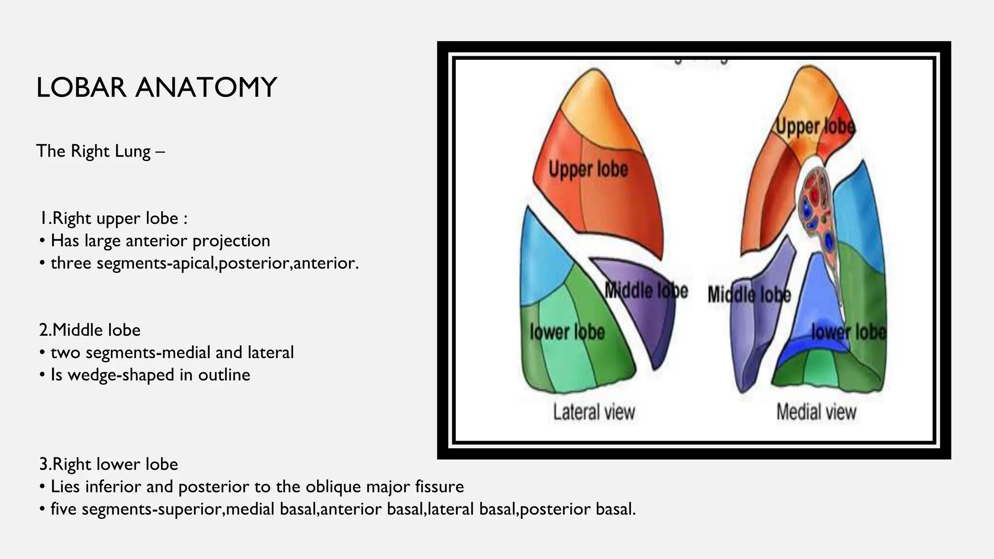
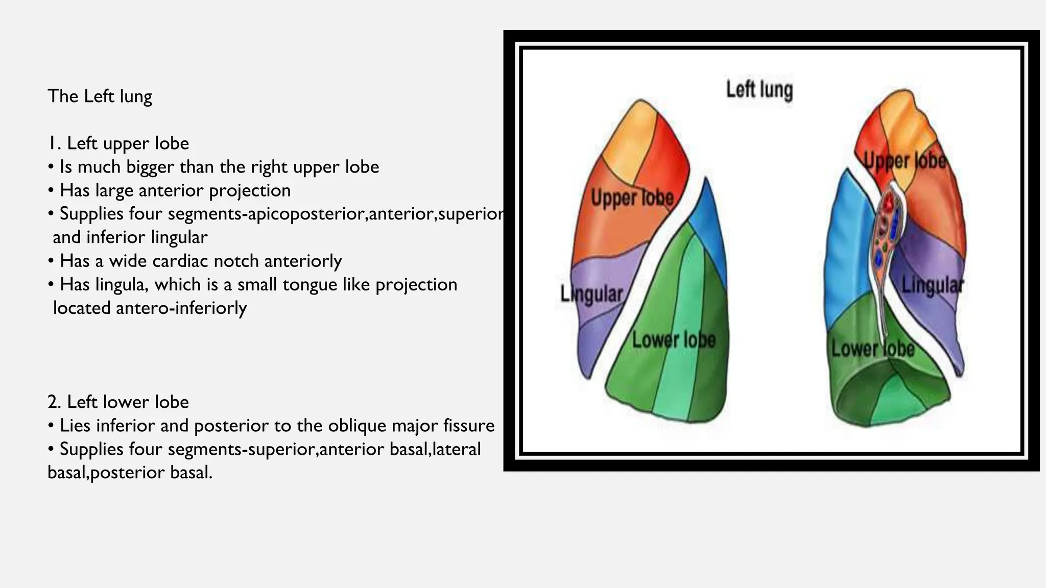
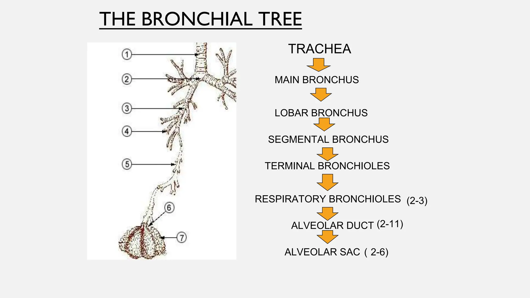
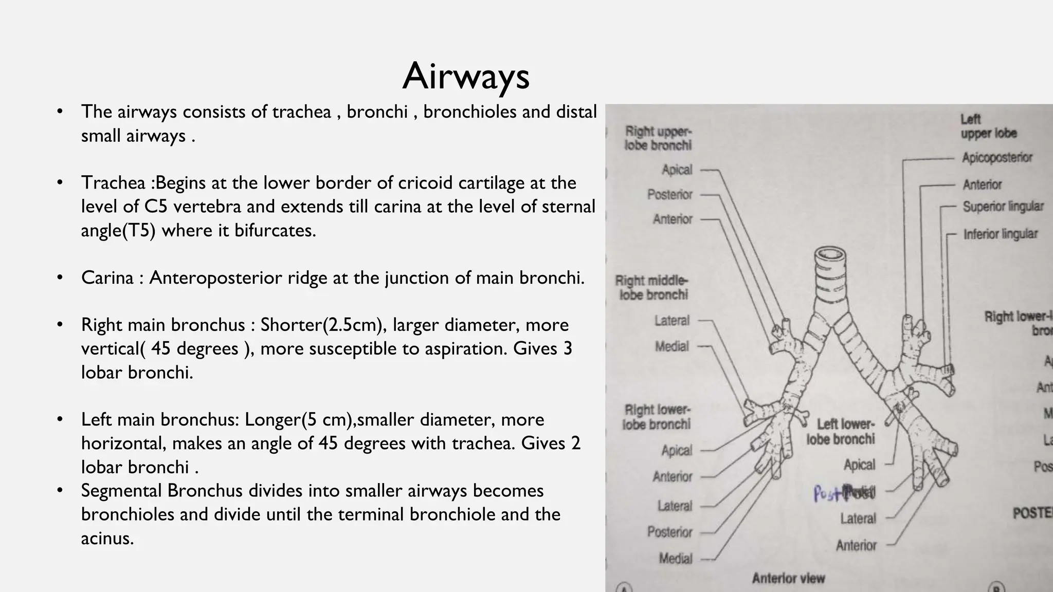
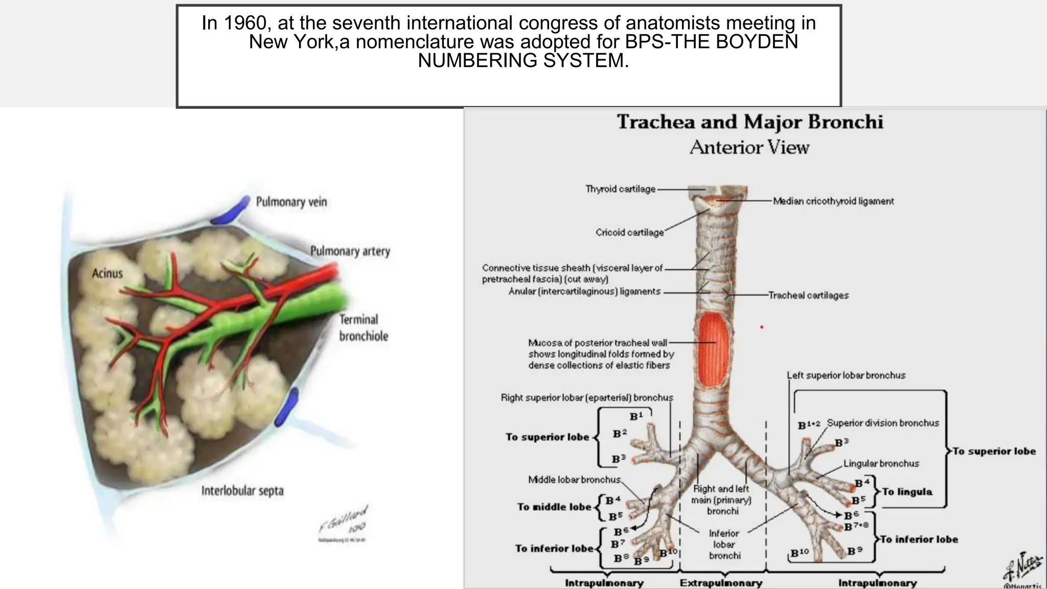
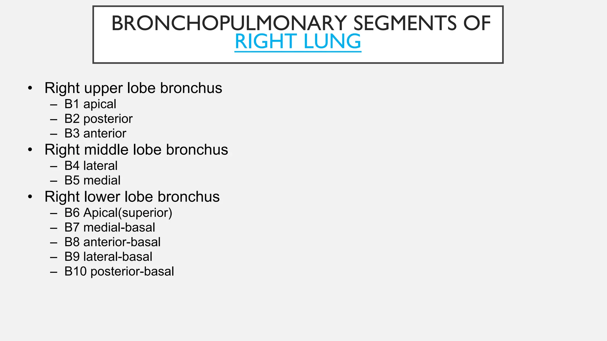
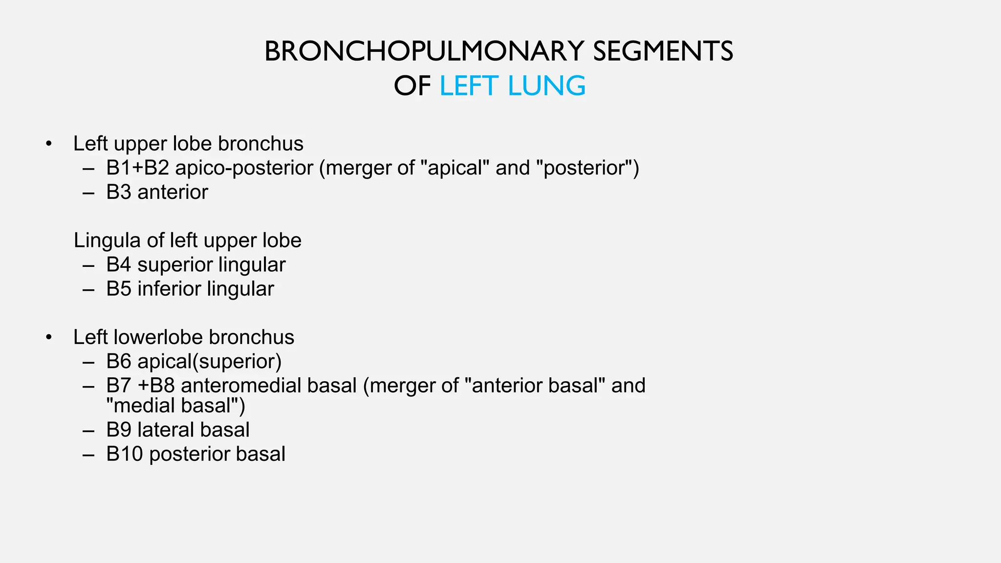
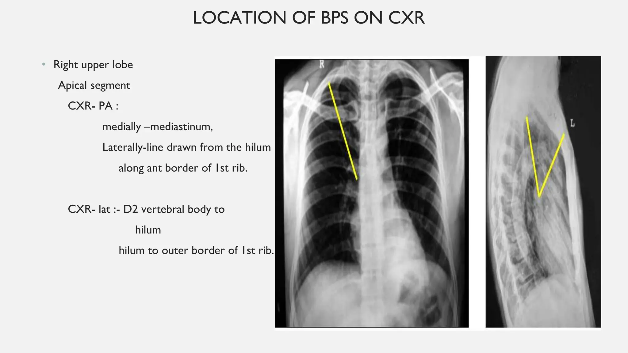
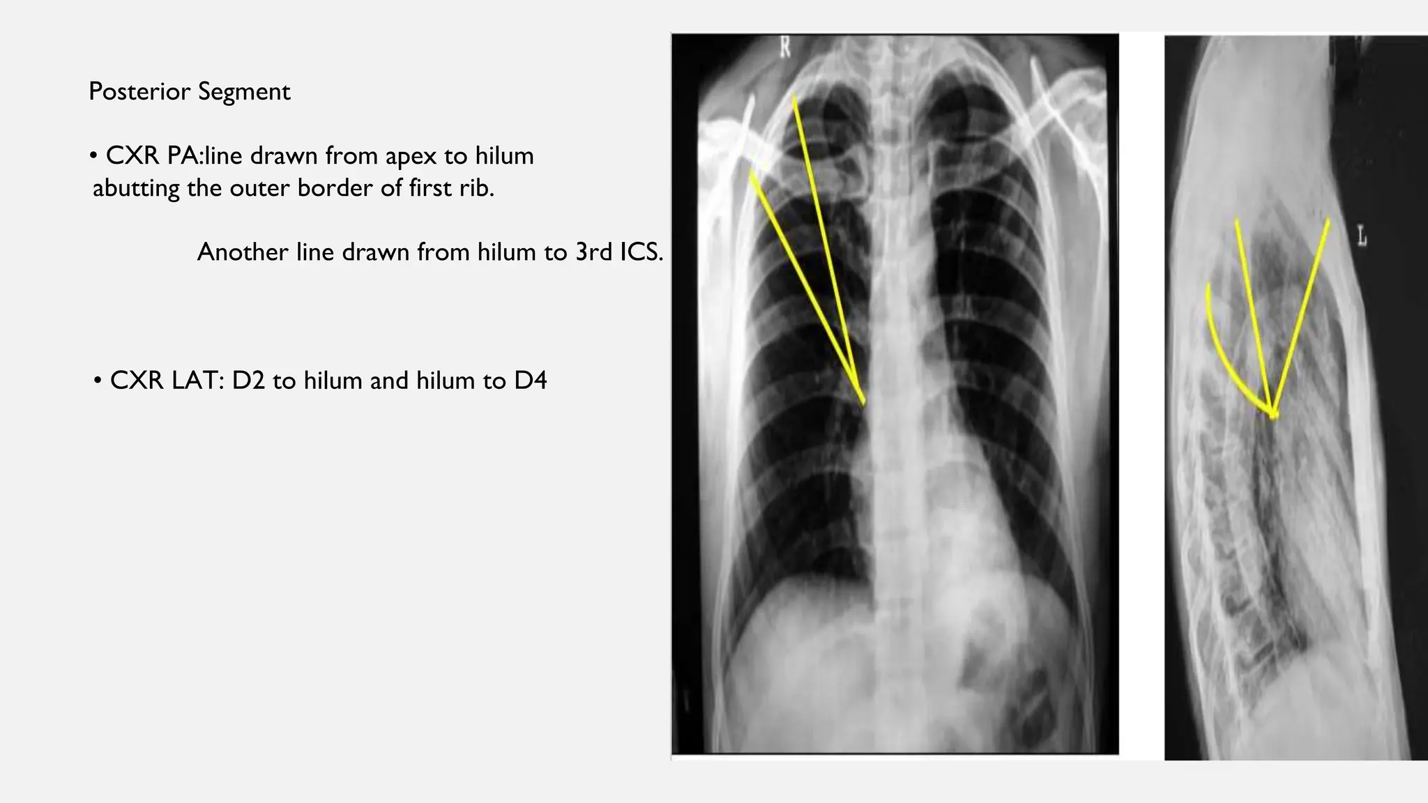
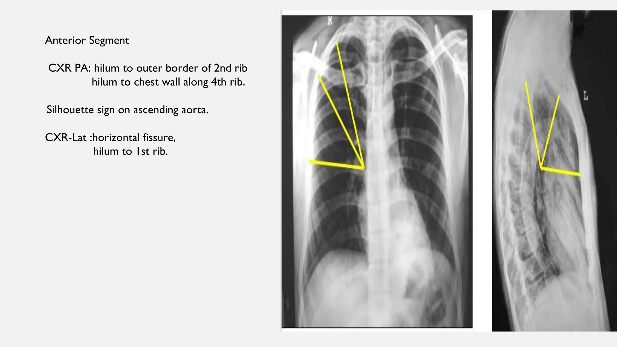
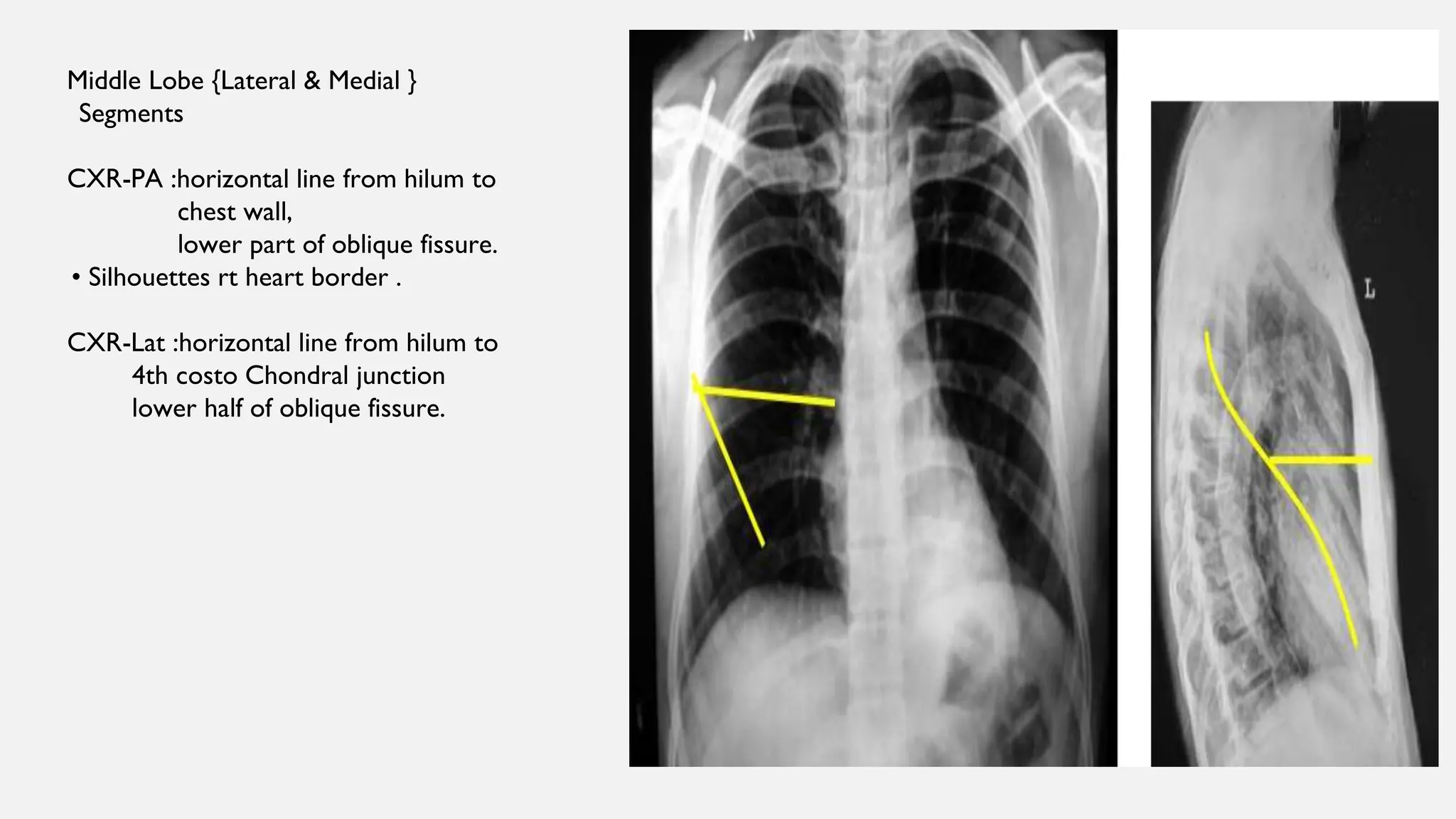
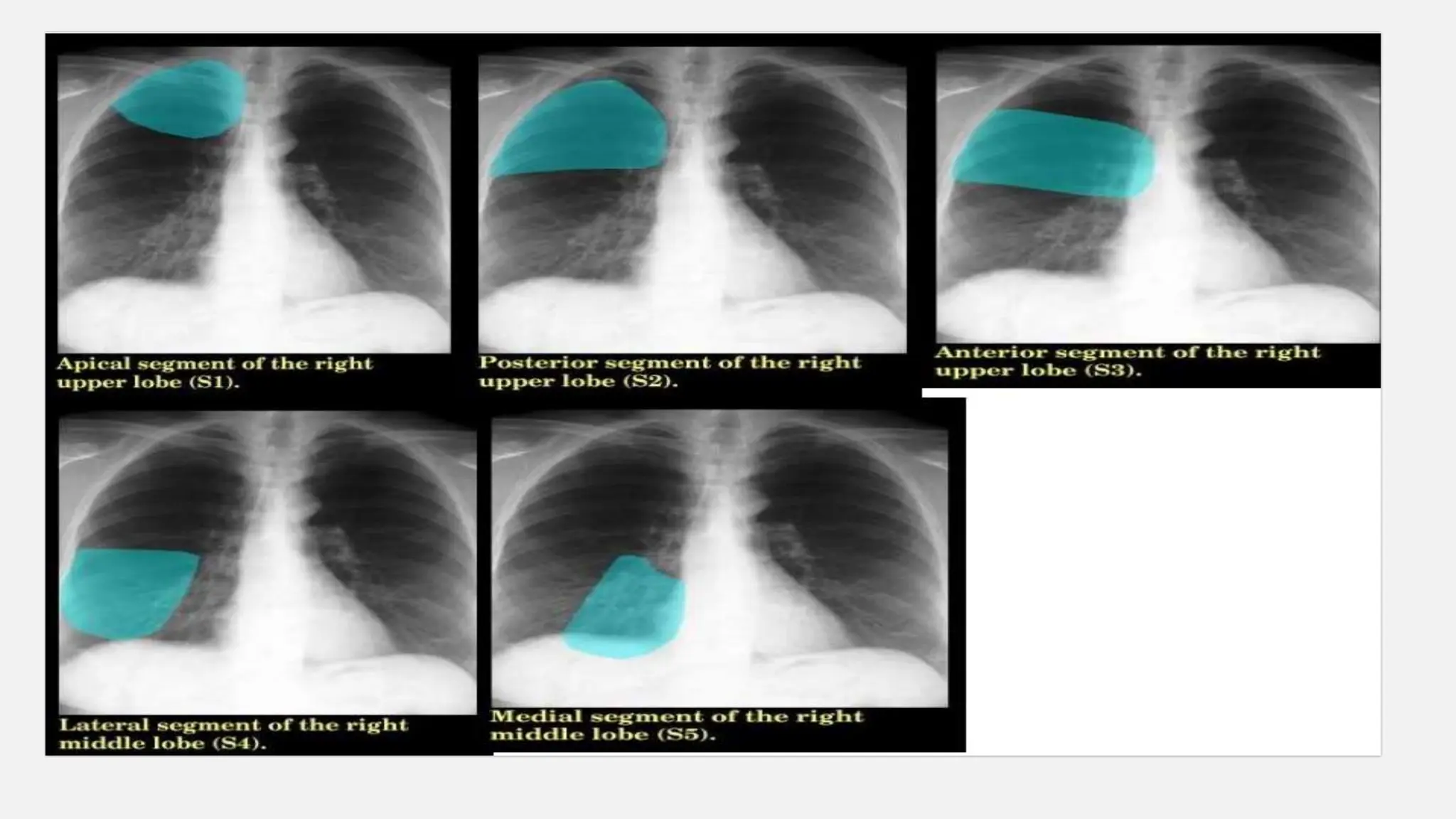
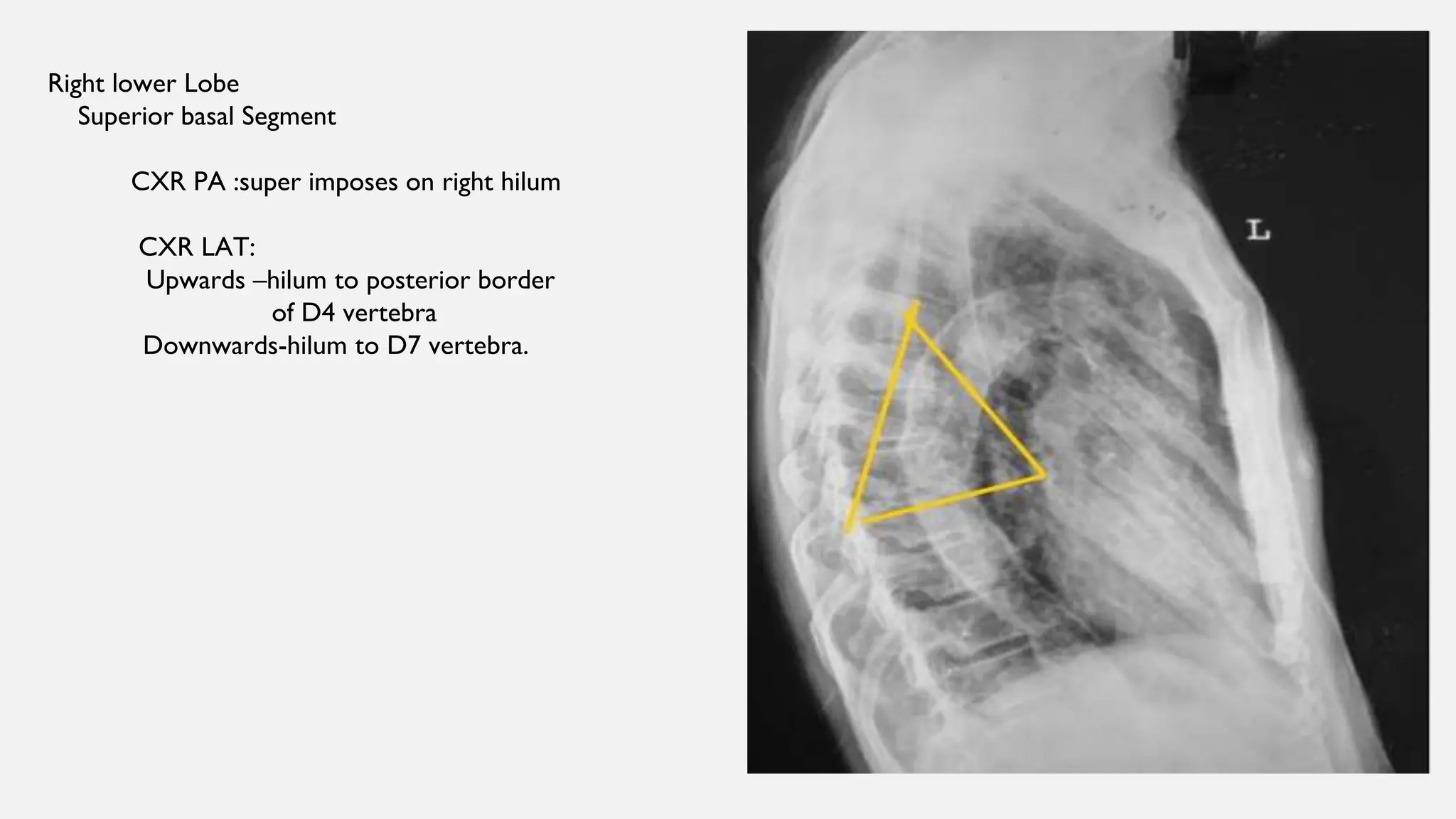
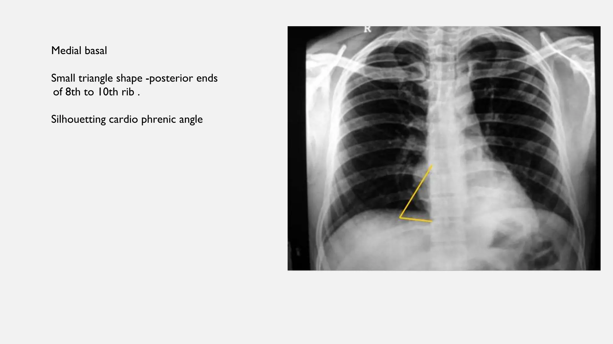
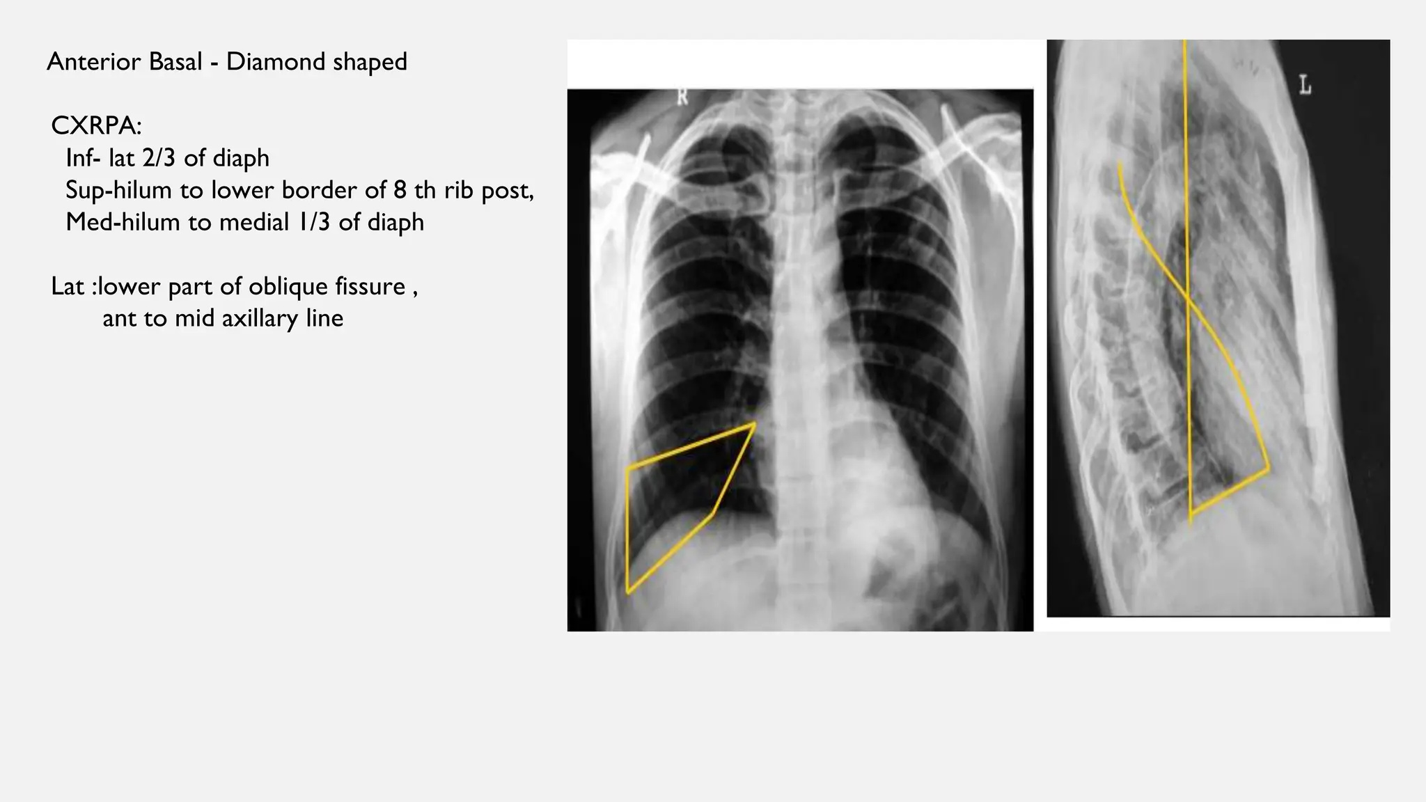
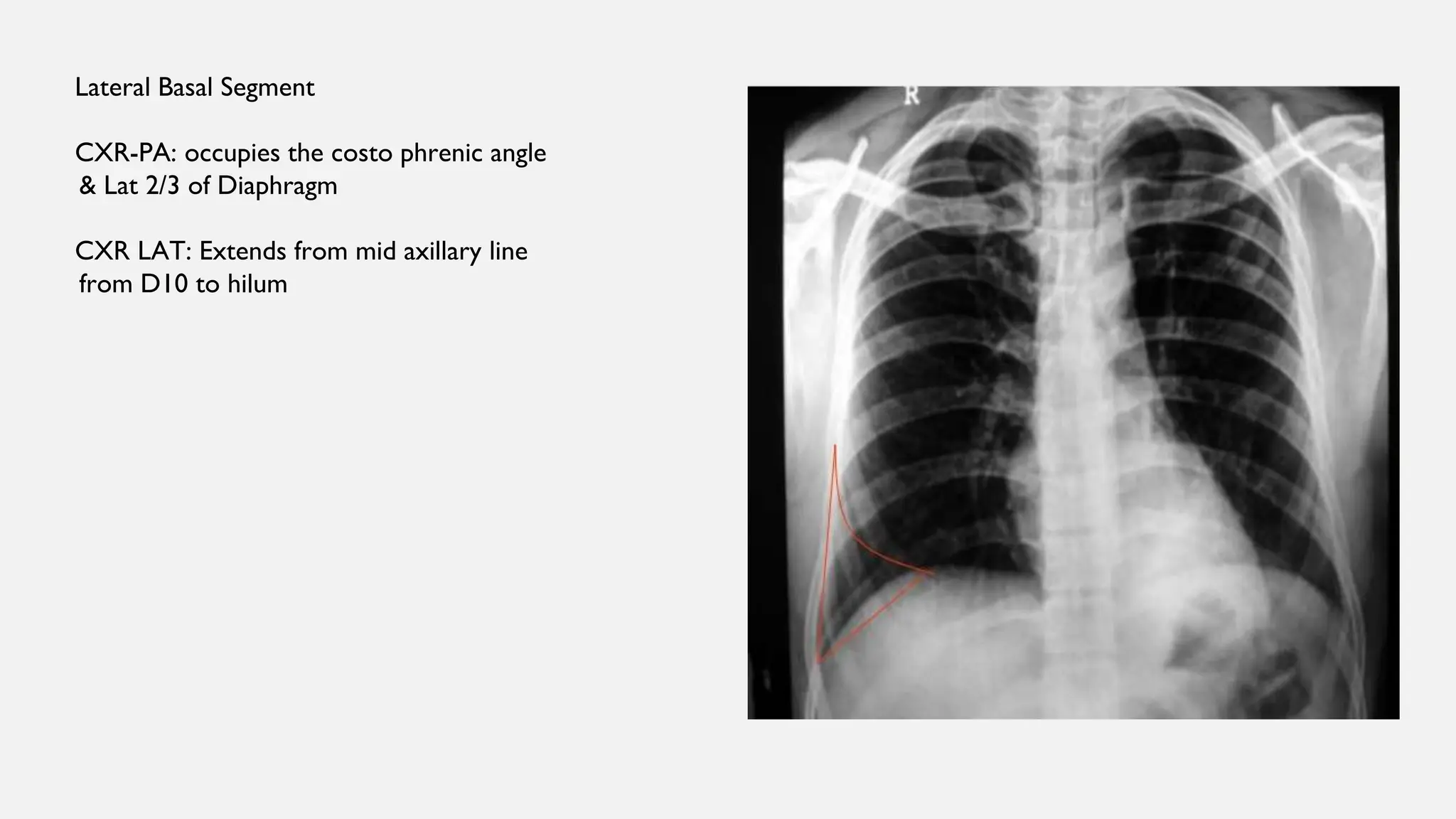
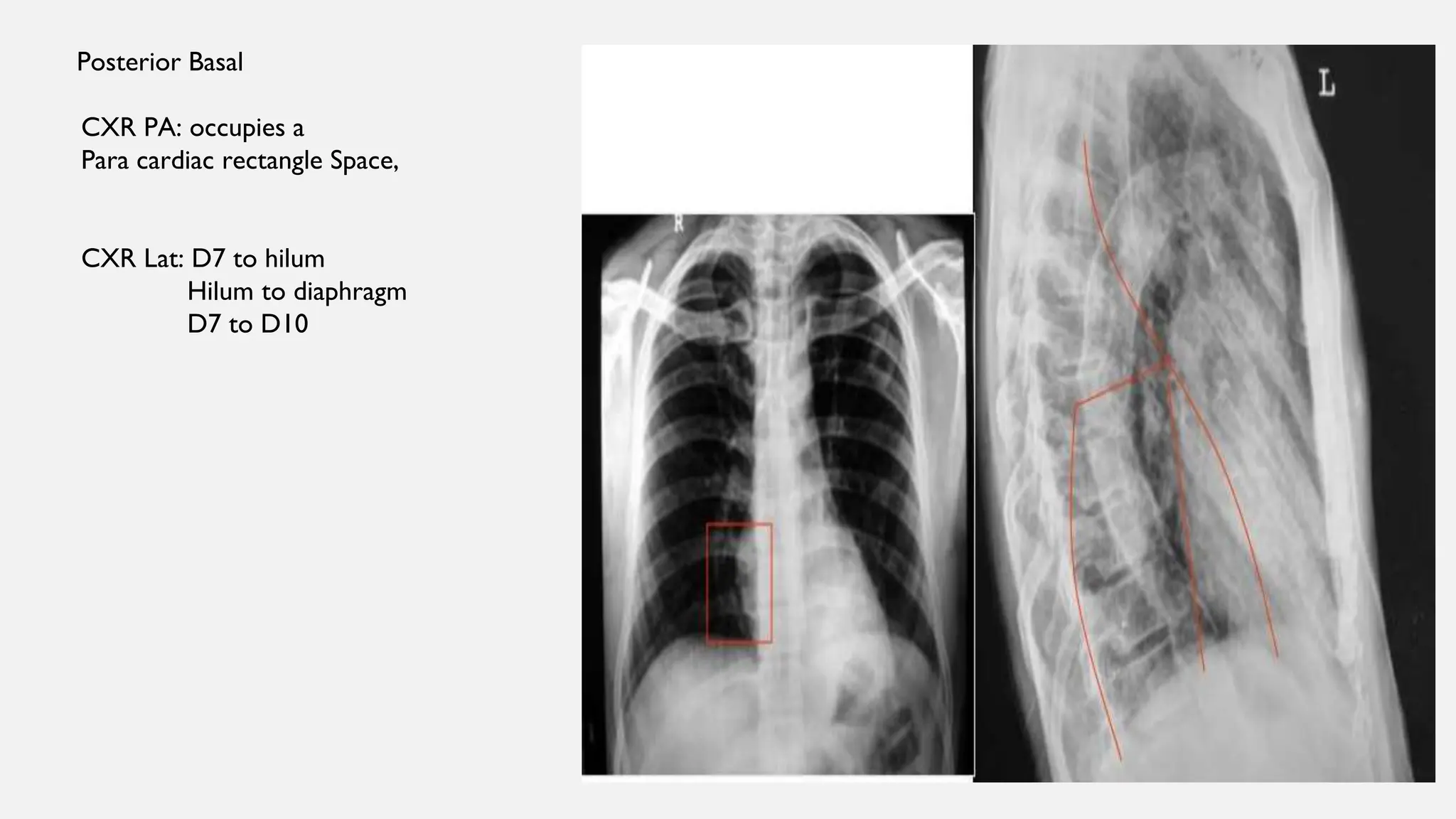
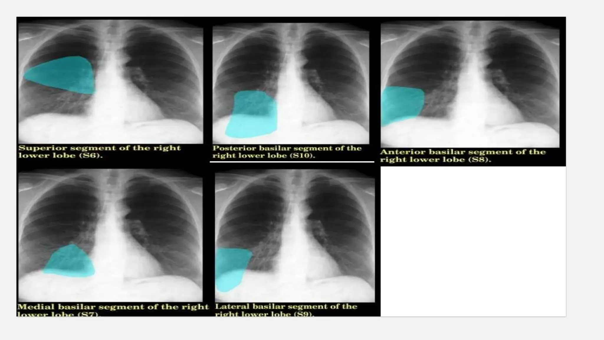
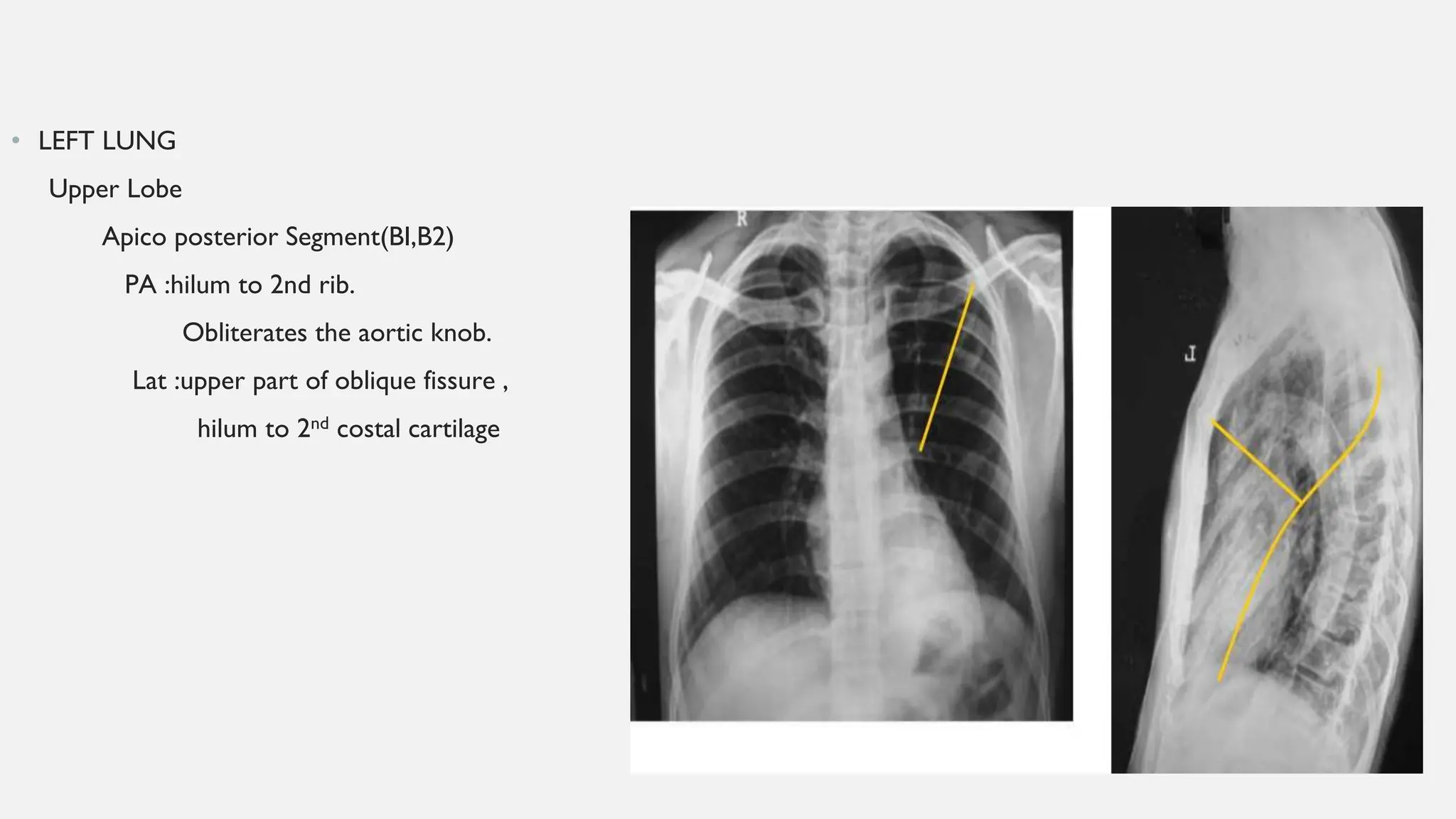
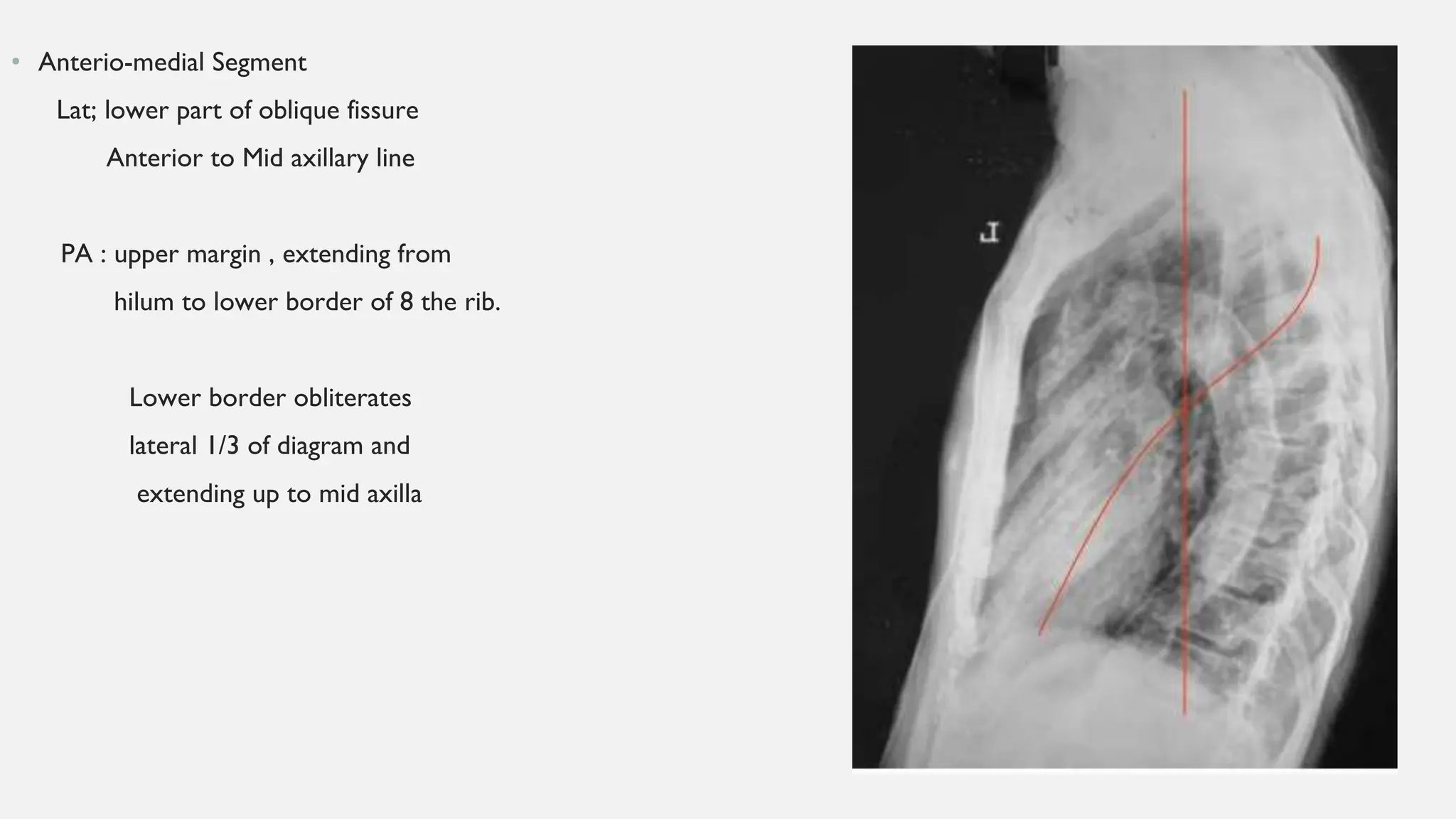
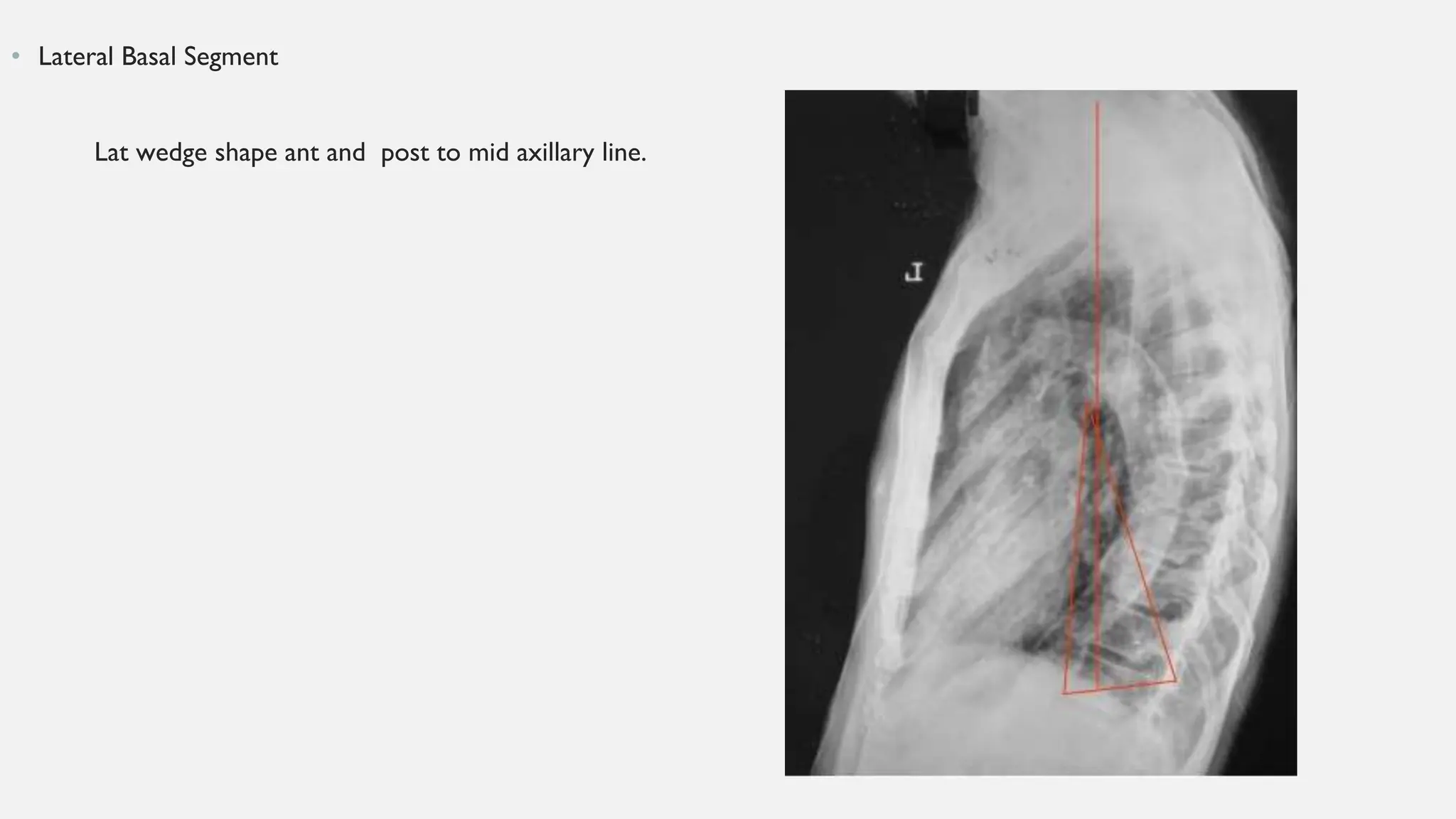
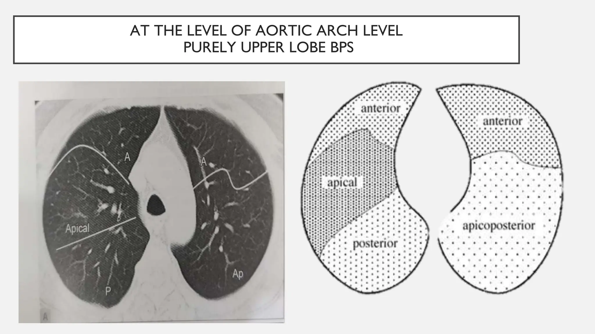
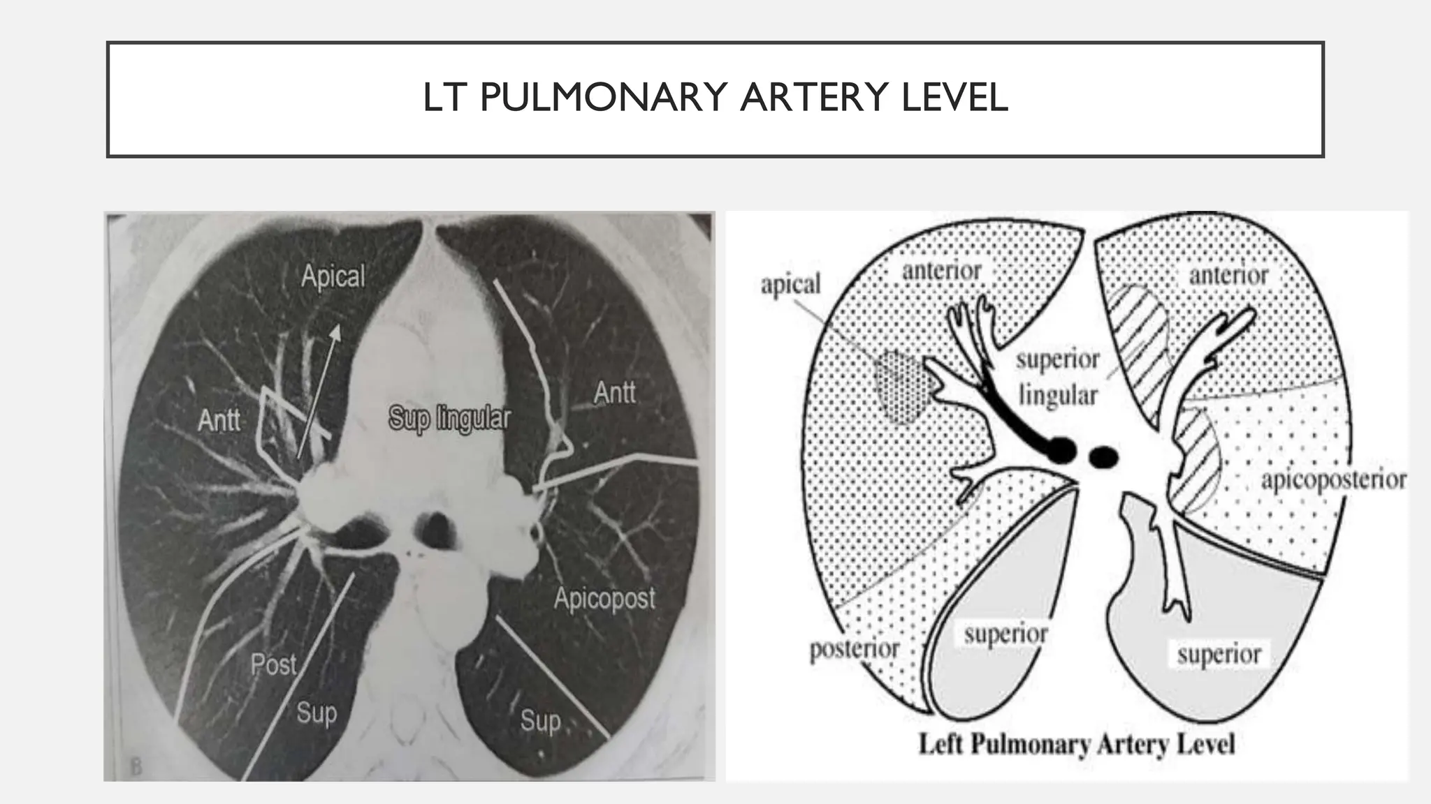
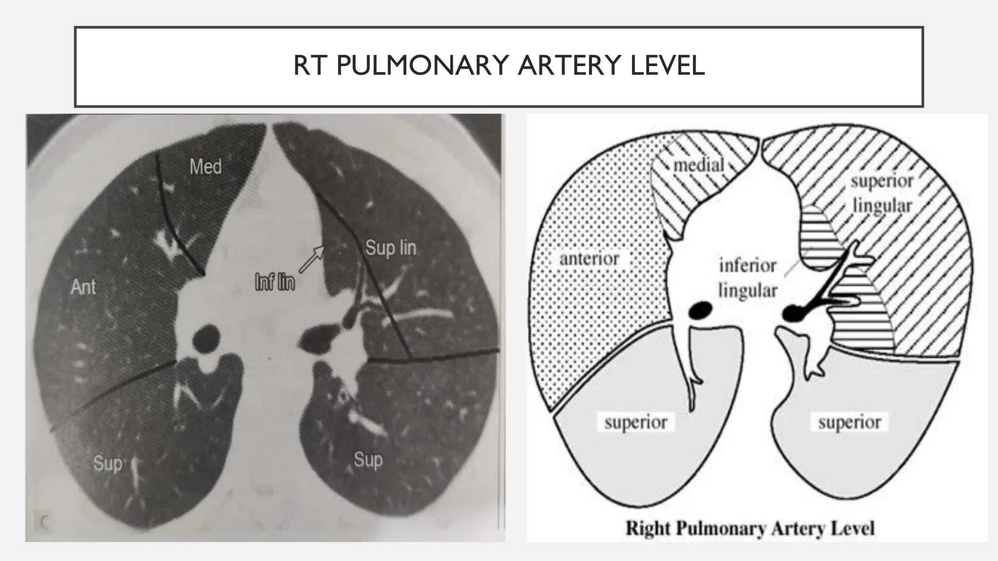
BRONCHOPULMONARY SEGMENTS
DR ANTONY SHAJAN
LOBAR ANATOMY
The Right Lung –
1.Right upper lobe :
• Has large anterior projection
• three segments-apical,posterior,anterior.
2.Middle lobe
• two segments-medial and lateral
• Is wedge-shaped in outline
3.Right lower lobe
• Lies inferior and posterior to the oblique major fissure
• five segments-superior,medial basal,anterior basal,lateral basal,posterior basal.
The Left lung
1. Left upper lobe
• Is much bigger than the right upper lobe
• Has large anterior projection
• Supplies four segments-apicoposterior,anterior,superior
and inferior lingular
• Has a wide cardiac notch anteriorly
• Has lingula, which is a small tongue like projection
located antero-inferiorly
2. Left lower lobe
• Lies inferior and posterior to the oblique major fissure
• Supplies four segments-superior,anterior basal,lateral
basal,posterior basal.
MAIN BRONCHUS
LOBAR BRONCHUS
SEGMENTAL BRONCHUS
TERMINAL BRONCHIOLES
RESPIRATORY BRONCHIOLES
ALVEOLAR DUCT
ALVEOLAR SAC
(2-3)
(2-11)
( 2-6)
TRACHEA
THE BRONCHIAL TREE
Airways
• The airways consists of trachea , bronchi , bronchioles and distal
small airways .
• Trachea :Begins at the lower border of cricoid cartilage at the
level of C5 vertebra and extends till carina at the level of sternal
angle(T5) where it bifurcates.
• Carina : Anteroposterior ridge at the junction of main bronchi.
• Right main bronchus : Shorter(2.5cm), larger diameter, more
vertical( 45 degrees ), more susceptible to aspiration. Gives 3
lobar bronchi.
• Left main bronchus: Longer(5 cm),smaller diameter, more
horizontal, makes an angle of 45 degrees with trachea. Gives 2
lobar bronchi .
• Segmental Bronchus divides into smaller airways becomes
bronchioles and divide until the terminal bronchiole and the
acinus.
• Bronchopulmonary segments are the independent respiratory units of the lung
divided on the basis of the division of the segmental/tertiary bronchi.
Definition:
• Each of the tertiary bronchi serves a specific bronchopulmonary segment. These
segments each have their own artery. Thus, each bronchopulmonary segment is
supplied by a bronchus, and an artery.
• Pulmonary veins do not accompany bronchi or pulmonary artery.They run in the
intersegmental planes.Thus each segment has more than one vein and each vein
drains more than one segment.
• Segments vary considerably in size and shape, but in general , segments are wedge
shaped and radiate from hilum.
• Unlike the lobes these segments are not separated by the pleura.
In 1960, at the seventh international congress of anatomists meeting in
New York,a nomenclature was adopted for BPS-THE BOYDEN
NUMBERING SYSTEM.
BRONCHOPULMONARY SEGMENTS OF
RIGHT LUNG
• Right upper lobe bronchus
– B1 apical
– B2 posterior
– B3 anterior
• Right middle lobe bronchus
– B4 lateral
– B5 medial
• Right lower lobe bronchus
– B6 Apical(superior)
– B7 medial-basal
– B8 anterior-basal
– B9 lateral-basal
– B10 posterior-basal
BRONCHOPULMONARY SEGMENTS
OF LEFT LUNG
• Left upper lobe bronchus
– B1+B2 apico-posterior (merger of "apical" and "posterior")
– B3 anterior
Lingula of left upper lobe
– B4 superior lingular
– B5 inferior lingular
• Left lowerlobe bronchus
– B6 apical(superior)
– B7 +B8 anteromedial basal (merger of "anterior basal" and
"medial basal")
– B9 lateral basal
– B10 posterior basal
LOCATION OF BPS ON CXR
• Right upper lobe
Apical segment
CXR- PA :
medially –mediastinum,
Laterally-line drawn from the hilum
along ant border of 1st rib.
CXR- lat :- D2 vertebral body to
hilum
hilum to outer border of 1st rib.
Posterior Segment
• CXR PA:line drawn from apex to hilum
abutting the outer border of first rib.
Another line drawn from hilum to 3rd ICS.
• CXR LAT: D2 to hilum and hilum to D4
Anterior Segment
CXR PA: hilum to outer border of 2nd rib
hilum to chest wall along 4th rib.
Silhouette sign on ascending aorta.
CXR-Lat :horizontal fissure,
hilum to 1st rib.
Middle Lobe {Lateral & Medial }
Segments
CXR-PA :horizontal line from hilum to
chest wall,
lower part of oblique fissure.
• Silhouettes rt heart border .
CXR-Lat :horizontal line from hilum to
4th costo Chondral junction
lower half of oblique fissure.
Right lower Lobe
Superior basal Segment
CXR PA :super imposes on right hilum
CXR LAT:
Upwards –hilum to posterior border
of D4 vertebra
Downwards-hilum to D7 vertebra.
Medial basal
Small triangle shape -posterior ends
of 8th to 10th rib .
Silhouetting cardio phrenic angle
Anterior Basal - Diamond shaped
CXRPA:
Inf- lat 2/3 of diaph
Sup-hilum to lower border of 8 th rib post,
Med-hilum to medial 1/3 of diaph
Lat :lower part of oblique fissure ,
ant to mid axillary line
Lateral Basal Segment
CXR-PA: occupies the costo phrenic angle
& Lat 2/3 of Diaphragm
CXR LAT: Extends from mid axillary line
from D10 to hilum
Posterior Basal
CXR PA: occupies a
Para cardiac rectangle Space,
CXR Lat: D7 to hilum
Hilum to diaphragm
D7 to D10
• LEFT LUNG
Upper Lobe
Apico posterior Segment(BI,B2)
PA :hilum to 2nd rib.
Obliterates the aortic knob.
Lat :upper part of oblique fissure ,
hilum to 2nd costal cartilage
• Anterior Segment
PA : hilum to 2nd rib
hilum to 4th rib
Lat : hilum to 2nd costal cartilage
hilum to 4th coastal cartilage.
• Lingular Segments
• PA :hilum to 4th rib,
lower part of oblique fissure.
Lat :lower part of oblique fissure ,
hilum to 4th costal cartilage.
silhouettes the left cardiac border
• Left Lower Lobe
Superior Segment
CXR PA : Oval shape 6th rib and 8th rib,
CXR Lat :upper part of oblique fissure ,
D4 to D7,
hilum to D7.
• Anterio-medial Segment
Lat; lower part of oblique fissure
Anterior to Mid axillary line
PA : upper margin , extending from
hilum to lower border of 8 the rib.
Lower border obliterates
lateral 1/3 of diagram and
extending up to mid axilla
• Lateral Basal Segment
Lat wedge shape ant and post to mid axillary line.
•
LOCATION OF BPS ON CT SCAN
• • Most commonly used levels
• 1.At the level of aortic arch
• 2.At the level of Lt pulmonary artery level.
• 3.At the level of Rt pulmonary artery level.
• 4.At the level of cardiac ventricles
AT THE LEVEL OF AORTIC ARCH LEVEL
PURELY UPPER LOBE BPS
LT PULMONARY ARTERY LEVEL
RT PULMONARY ARTERY LEVEL
AT THE LEVEL OF CARDIAC VENTRICLES
CLINICAL IMPORTANCE
• Collapse of multiple lobes or segments in certain combinations almost rules out malignancy
(RUL+RML ,LLL+LUL, RML+ one segment of RLL )
• Exceptions :
• multi centric neoplasm
• primary & metastasis
• Silhoutte sign: is applied to localize parenchymal lesions which can be stated as “an intrathoracic lesion
with similar radiologic density is in contiguity with either heart, aorta, or diaphragm and will obliterate the
border, as the radiographic contrast is lost.”
• Right cardiac border – Right middle lobe .
• Left cardiac border - Lingula
• Aortic knuckle –Apico posterior segment LUL
• Right aortic border –Anterior segment of RUL , middle lobe
• 1.Tuberculosis—apical & posterior segments of upper lobes because of high ventilation perfusion ratios
with elevated alveolar PO2 relative to other zones
• 2.Sequestration of lung-posterior basal segment of lower lobe (left>>right)
• 3.Cancerous lesion-anterior segments of RUL.
• 4.Lung abscess—posterior segment of the upper lobe or in the superior or posterior basal segment of
the lower lobe, especially on the right lung because of more vertical bronchus and also the aspirated
secretions tend to gravitate in these segments in supine position
• 5.Aspiration pneumonia: apical segments of both lower lobes or posterior segment of the RUL.
• Bronchiectasis is more common in left lower lobe because of longer and narrower bronchus leading to
retained secretions.
SUGICAL RESECTION
• PRINCIPLE: A diseased segment, as it is a structural unit can be removed surgically. During surgical
resection, the surgeon works along the segmental veins to isolate a particular segment.
• Indications:
• Aspergilloma
• TB
• Bronchiectasis
• Metastatic disease
• Primary lung cancer
• Contra indications:
• Extrinsic segmental compression
• Presence of endoluminal tumor
• Prerequisites: complete bronchoscopy
• Advantages :
• 1)thorough resection
• 2)better staging
• 3)decreasing local recurrence
THANK YOU…

BRONCHOPULMONARY SEGMENTS RADIOLOGY AND IMAGING

  • 1.
  • 2.
    LOBAR ANATOMY The RightLung – 1.Right upper lobe : • Has large anterior projection • three segments-apical,posterior,anterior. 2.Middle lobe • two segments-medial and lateral • Is wedge-shaped in outline 3.Right lower lobe • Lies inferior and posterior to the oblique major fissure • five segments-superior,medial basal,anterior basal,lateral basal,posterior basal.
  • 3.
    The Left lung 1.Left upper lobe • Is much bigger than the right upper lobe • Has large anterior projection • Supplies four segments-apicoposterior,anterior,superior and inferior lingular • Has a wide cardiac notch anteriorly • Has lingula, which is a small tongue like projection located antero-inferiorly 2. Left lower lobe • Lies inferior and posterior to the oblique major fissure • Supplies four segments-superior,anterior basal,lateral basal,posterior basal.
  • 4.
    MAIN BRONCHUS LOBAR BRONCHUS SEGMENTALBRONCHUS TERMINAL BRONCHIOLES RESPIRATORY BRONCHIOLES ALVEOLAR DUCT ALVEOLAR SAC (2-3) (2-11) ( 2-6) TRACHEA THE BRONCHIAL TREE
  • 5.
    Airways • The airwaysconsists of trachea , bronchi , bronchioles and distal small airways . • Trachea :Begins at the lower border of cricoid cartilage at the level of C5 vertebra and extends till carina at the level of sternal angle(T5) where it bifurcates. • Carina : Anteroposterior ridge at the junction of main bronchi. • Right main bronchus : Shorter(2.5cm), larger diameter, more vertical( 45 degrees ), more susceptible to aspiration. Gives 3 lobar bronchi. • Left main bronchus: Longer(5 cm),smaller diameter, more horizontal, makes an angle of 45 degrees with trachea. Gives 2 lobar bronchi . • Segmental Bronchus divides into smaller airways becomes bronchioles and divide until the terminal bronchiole and the acinus.
  • 6.
    • Bronchopulmonary segmentsare the independent respiratory units of the lung divided on the basis of the division of the segmental/tertiary bronchi. Definition: • Each of the tertiary bronchi serves a specific bronchopulmonary segment. These segments each have their own artery. Thus, each bronchopulmonary segment is supplied by a bronchus, and an artery. • Pulmonary veins do not accompany bronchi or pulmonary artery.They run in the intersegmental planes.Thus each segment has more than one vein and each vein drains more than one segment. • Segments vary considerably in size and shape, but in general , segments are wedge shaped and radiate from hilum. • Unlike the lobes these segments are not separated by the pleura.
  • 7.
    In 1960, atthe seventh international congress of anatomists meeting in New York,a nomenclature was adopted for BPS-THE BOYDEN NUMBERING SYSTEM.
  • 8.
    BRONCHOPULMONARY SEGMENTS OF RIGHTLUNG • Right upper lobe bronchus – B1 apical – B2 posterior – B3 anterior • Right middle lobe bronchus – B4 lateral – B5 medial • Right lower lobe bronchus – B6 Apical(superior) – B7 medial-basal – B8 anterior-basal – B9 lateral-basal – B10 posterior-basal
  • 9.
    BRONCHOPULMONARY SEGMENTS OF LEFTLUNG • Left upper lobe bronchus – B1+B2 apico-posterior (merger of "apical" and "posterior") – B3 anterior Lingula of left upper lobe – B4 superior lingular – B5 inferior lingular • Left lowerlobe bronchus – B6 apical(superior) – B7 +B8 anteromedial basal (merger of "anterior basal" and "medial basal") – B9 lateral basal – B10 posterior basal
  • 10.
    LOCATION OF BPSON CXR • Right upper lobe Apical segment CXR- PA : medially –mediastinum, Laterally-line drawn from the hilum along ant border of 1st rib. CXR- lat :- D2 vertebral body to hilum hilum to outer border of 1st rib.
  • 11.
    Posterior Segment • CXRPA:line drawn from apex to hilum abutting the outer border of first rib. Another line drawn from hilum to 3rd ICS. • CXR LAT: D2 to hilum and hilum to D4
  • 12.
    Anterior Segment CXR PA:hilum to outer border of 2nd rib hilum to chest wall along 4th rib. Silhouette sign on ascending aorta. CXR-Lat :horizontal fissure, hilum to 1st rib.
  • 13.
    Middle Lobe {Lateral& Medial } Segments CXR-PA :horizontal line from hilum to chest wall, lower part of oblique fissure. • Silhouettes rt heart border . CXR-Lat :horizontal line from hilum to 4th costo Chondral junction lower half of oblique fissure.
  • 15.
    Right lower Lobe Superiorbasal Segment CXR PA :super imposes on right hilum CXR LAT: Upwards –hilum to posterior border of D4 vertebra Downwards-hilum to D7 vertebra.
  • 16.
    Medial basal Small triangleshape -posterior ends of 8th to 10th rib . Silhouetting cardio phrenic angle
  • 17.
    Anterior Basal -Diamond shaped CXRPA: Inf- lat 2/3 of diaph Sup-hilum to lower border of 8 th rib post, Med-hilum to medial 1/3 of diaph Lat :lower part of oblique fissure , ant to mid axillary line
  • 18.
    Lateral Basal Segment CXR-PA:occupies the costo phrenic angle & Lat 2/3 of Diaphragm CXR LAT: Extends from mid axillary line from D10 to hilum
  • 19.
    Posterior Basal CXR PA:occupies a Para cardiac rectangle Space, CXR Lat: D7 to hilum Hilum to diaphragm D7 to D10
  • 21.
    • LEFT LUNG UpperLobe Apico posterior Segment(BI,B2) PA :hilum to 2nd rib. Obliterates the aortic knob. Lat :upper part of oblique fissure , hilum to 2nd costal cartilage
  • 22.
    • Anterior Segment PA: hilum to 2nd rib hilum to 4th rib Lat : hilum to 2nd costal cartilage hilum to 4th coastal cartilage.
  • 24.
    • Lingular Segments •PA :hilum to 4th rib, lower part of oblique fissure. Lat :lower part of oblique fissure , hilum to 4th costal cartilage. silhouettes the left cardiac border
  • 26.
    • Left LowerLobe Superior Segment CXR PA : Oval shape 6th rib and 8th rib, CXR Lat :upper part of oblique fissure , D4 to D7, hilum to D7.
  • 27.
    • Anterio-medial Segment Lat;lower part of oblique fissure Anterior to Mid axillary line PA : upper margin , extending from hilum to lower border of 8 the rib. Lower border obliterates lateral 1/3 of diagram and extending up to mid axilla
  • 28.
    • Lateral BasalSegment Lat wedge shape ant and post to mid axillary line.
  • 29.
  • 30.
    LOCATION OF BPSON CT SCAN • • Most commonly used levels • 1.At the level of aortic arch • 2.At the level of Lt pulmonary artery level. • 3.At the level of Rt pulmonary artery level. • 4.At the level of cardiac ventricles
  • 31.
    AT THE LEVELOF AORTIC ARCH LEVEL PURELY UPPER LOBE BPS
  • 32.
  • 33.
  • 34.
    AT THE LEVELOF CARDIAC VENTRICLES
  • 35.
    CLINICAL IMPORTANCE • Collapseof multiple lobes or segments in certain combinations almost rules out malignancy (RUL+RML ,LLL+LUL, RML+ one segment of RLL ) • Exceptions : • multi centric neoplasm • primary & metastasis
  • 36.
    • Silhoutte sign:is applied to localize parenchymal lesions which can be stated as “an intrathoracic lesion with similar radiologic density is in contiguity with either heart, aorta, or diaphragm and will obliterate the border, as the radiographic contrast is lost.” • Right cardiac border – Right middle lobe . • Left cardiac border - Lingula • Aortic knuckle –Apico posterior segment LUL • Right aortic border –Anterior segment of RUL , middle lobe
  • 37.
    • 1.Tuberculosis—apical &posterior segments of upper lobes because of high ventilation perfusion ratios with elevated alveolar PO2 relative to other zones • 2.Sequestration of lung-posterior basal segment of lower lobe (left>>right) • 3.Cancerous lesion-anterior segments of RUL. • 4.Lung abscess—posterior segment of the upper lobe or in the superior or posterior basal segment of the lower lobe, especially on the right lung because of more vertical bronchus and also the aspirated secretions tend to gravitate in these segments in supine position
  • 38.
    • 5.Aspiration pneumonia:apical segments of both lower lobes or posterior segment of the RUL. • Bronchiectasis is more common in left lower lobe because of longer and narrower bronchus leading to retained secretions.
  • 39.
    SUGICAL RESECTION • PRINCIPLE:A diseased segment, as it is a structural unit can be removed surgically. During surgical resection, the surgeon works along the segmental veins to isolate a particular segment. • Indications: • Aspergilloma • TB • Bronchiectasis • Metastatic disease • Primary lung cancer
  • 40.
    • Contra indications: •Extrinsic segmental compression • Presence of endoluminal tumor • Prerequisites: complete bronchoscopy • Advantages : • 1)thorough resection • 2)better staging • 3)decreasing local recurrence
  • 41.