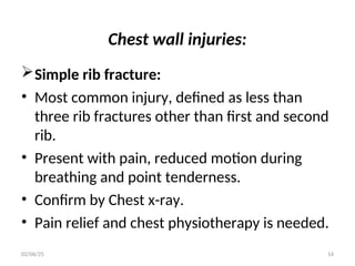
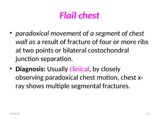
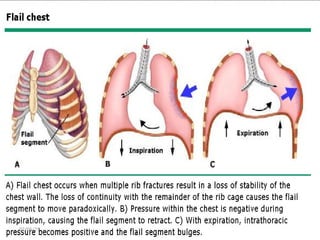
Chest Injuries
02/06/25 1
Introduction
• 25% of all trauma deaths are a result of chest
injuries alone and respiratory problems
contributes significantly in 75% of traumatic
deaths.
• The terrible death toll related to chest injuries
is avoidable by simple measures.
02/06/25 2
Classification
1. Blunt trauma:
• It accounts for 85% of all chest injuries.
• In 70%-80% cases, it results from motor
vehicle accident.
• The rest are due to fall and crush injuries.
• 84% of cases are associated with injury to
other sites.
02/06/25 3
02/06/25 4
2. Penetrating trauma:
• Accounts for 15% of all chest injuries.
• Stab and gunshot wounds are the most
frequent causes.
• It results in hemothorax in more than 80% and
pneumothorax in nearly all cases.
• It should be considered as thoracoabdominal
if penetration is below fourth intercostal
space.
02/06/25 5
02/06/25 6
Pathophyiology
• Inadequate delivery of oxygen to tissue results
from
• 1. Ventilation-perfusion mismatch i.e.
perfusion of non ventilated lung mostly as a
result of lung contusion
02/06/25 7
• 2. Decreased tidal volume due to pain or other
cause
• 3. Hypovolemia from bleeding
• 4. Mechanical obstruction due to tension
pneumothorax and cardiac tamponade
02/06/25 8
Initial Assessment and Management
• Ensuring adequate airway: Keep cervical spine
neutral.
• The oropharyngeal airway should be cleared
from debris and secretion.
• Cricothyroidothomy till intubation or
tracheostomy is made if indicated.
02/06/25 9
Ensuring adequate ventilation:
• Give 100% oxygen.
• Tightly dress any sucking wound and look for
signs of tension pneumothorax (distended neck
veins, shift of the trachea, hyper resonance with
decreased air entry), cardiac tamponade
(hypotension, distended neck vein and distant
heart sounds), massive hemothorax and flail
chest all of which can compromise ventilation
despite patent airway and adequate
oxygenation.
02/06/25 10
Control extreme hemorrhage and restore
circulation:
• Insert wide bore cannula for fluid and blood
transfusion.
• N.B: If one suspects tension pneumothorax,
massive hemothorax or cardiac tamponade,
the management should be dealt as part of
resuscitation and patients should not be sent
for confirmatory investigations.
02/06/25 11
• Besides, in case of suspected cardiac
tamponade, simple insertion of a needle
through xiphoid angle pointing towards the
left shoulder tip can help enter the
pericardium and aspirate accumulated blood.
• This could salvage the patient’s life to reach a
proper centre.
02/06/25 12
Generally injury to the chest can involve:
• A. The chest wall
• B. Lung
• C. Mediastinal structures
• D. The diaphragm
02/06/25 13
Chest wall injuries:
Simple rib fracture:
• Most common injury, defined as less than
three rib fractures other than first and second
rib.
• Present with pain, reduced motion during
breathing and point tenderness.
• Confirm by Chest x-ray.
• Pain relief and chest physiotherapy is needed.
02/06/25 14
Flail chest
• paradoxical movement of a segment of chest
wall as a result of fracture of four or more ribs
at two points or bilateral costochondral
junction separation.
• Diagnosis: Usually clinical, by closely
observing paradoxical chest motion, chest x-
ray shows multiple segmental fractures.
02/06/25 15
16
02/06/25
Treatment:
• Aggressive chest physiotherapy, effective
analgesia, close ICU observation if available,
Oxygen supplement to keep saturation ≥ 90%.
• Administer fluid only to restore hemodynamic
stability.
02/06/25 17
Fracture of first, second rib and the sternum:
• These are considered to be major injuries
since a considerable force, which usually
causes associated injury to underlying
structures like vessels or nerves, is required.
• It is otherwise managed as simple rib fracture.
02/06/25 18
Injury to mediastinal structure:
• Injury to trachea, bronchus, major vessel and
heart are fortunately rare. But if they occur,
they are usually fatal and patient often does
not reach health facility.
02/06/25 19
Lung contusion:
• This presents with bloody sputum upon
coughing.
• Diagnosis: Chest x-ray (parenchymal opacity
immediately after injury and increasing in the
next 24-48 hours).
• Treatment: Pulmonary physiotherapy and
prevention of fluid load.
02/06/25 20
Diaphragmatic rupture:
• Mostly occurs on the left side and diagnosis
needs high index of suspicion.
• Symptoms and signs are usually due to
herniation of intra abdominal organ like
stomach or colon in to the chest.
02/06/25 21
• Diagnosis: Insert nasogastric tube. Auscultate
the chest simultaneously while insufflating air
in to the tube.
• Chest x-ray may reveal tube, loop of bowel or
fluid level in the thorax
• Treatment: Immediate repair
02/06/25 22
Pneumothorax
• Pneumothorax is a presence of air in pleural
cavity.
Type:
• Open: This is associated with chest wall
wound which communicate with the external
environment.
02/06/25 23
• Tension: This is a surgical emergency
associated with development of pressure
which compromise breathing as well as
circulation.
• Simple: This is collection which is not
associated with compromised breathing and
no breach of chest wall
02/06/25 24
02/06/25 25
Cause
• Blunt and penetrating injuries
Mechanism
• Fractured rib penetrating the lung
• Deceleration and crush disrupting the alveoli
• Sucking effect of negative intrapleural pressure
• N.B: In most cases of traumatic pneumothorax,
there will be associated bleeding which may not
be apparent.
02/06/25 26
Clinical Features
• Presume pneumothorax in any chest injury
until proved otherwise since pain and splinting
make physical examination difficult.
• Look for decreased chest expansion, tracheal
shift, hyper resonant percussion note and
decreased air entry.
• If patient’s condition is stable, confirm by
erect chest x-ray.
02/06/25 27
Treatment
• The principal objective is to remove trapped
air through tube thoracostomy (chest tube).
• In case of tension pneumothorax, insertion of
needle at second intercostal space over the
mid clavicular line of the same side relives the
tension until chest tube insertion.
02/06/25 28
02/06/25 29
Hemothorax
• Hemothorax is collection of blood in the
pleural cavity. Bleeding usually occurs from
intercostal or internal mammary arteries.
• Bleeding from parenchymal injury is nearly
always self-limiting.
• Massive Hemothorax is a bleeding of more
than 1500ml in to pleural cavity and rarely
occurs in blunt trauma.
02/06/25 30
Clinical Feature
• There is a history of trauma to chest. Signs of
fluid collection in the pleural cavity (decreased
air entry, dull percussion note) are found on P/E.
• Chest x-ray: Erect chest film reveals costophrenic
angle obliteration if more than 500 ml blood
exists.
02/06/25 31
• Lateral decubitus film can demonstrate as
small as 250 ml of blood.
• Ultrasonography can reveal a small amount of
fluid in the pleural recess.
Treatment: Chest tube insertion if sign of
collection is visible on erect chest x-ray.
02/06/25 32
02/06/25 33
Abdominal trauma
• Abdominal injuries are present in 7-10% of
trauma patients. These injuries, if
unrecognized, can cause preventable deaths.
• Blunt trauma: most frequent injuries are
spleen (45%), liver (40%), and retroperitoneal
haematoma (15%).
02/06/25 34
• Blunt trauma may cause:
• compression or crushing causing rupture of
solid or hollow organs;
• deceleration injury due to differential
movement of fixed and nonfixed parts of
organs causing tearing or avulsion from
their vascular supply, e.g. liver tear and
vena caval rupture.
02/06/25 35
• Blunt abdominal trauma is very common in
road traffic accidents where:
• there have been fatalities;
• any casualty has been ejected from the
vehicle;
• the closing speed is greater than 50mph.
02/06/25 36
• Penetrating trauma: these may be:
• stab wounds and low velocity gunshot
wounds: cause damage by laceration or
cutting. Stab wounds commonly involve the
liver (40%), small bowel (30%), diaphragm
(20%), colon (15%);
• high velocity gunshot wounds transfer more
kinetic energy and also cause further injury
by cavitation effect, tumble, and
fragmentation. Commonly involve the small
bowel (50%), colon (40%), liver (30%), and
02/06/25 37
02/06/25 38
02/06/25 39
Management primary survey
• Any patient persistently hypotensive despite
resuscitation, for whom no obvious cause of
blood loss has been identified by the primary
survey, can be assumed to have
intraabdominal bleeding.
• If the patient is stable an emergency
abdominal CT scan is indicated.
• If the patient remains critically unstable an
emergency laparotomy is usually indicated.
02/06/25 40
Management secondary survey of the abdomen
• History
• Obtain from patient, other passengers, observers,
police, and emergency medical personnel.
• Mechanism of injury: seat belt usage, steering
wheel deformation, speed, damage to vehicle,
ejection of victim, etc. in automobile collision;
velocity, calibre, presumed path of bullet, distance
from weapon, etc. in penetrating injuries.
• Prehospital condition and treatment of patient.
02/06/25 41
Physical examination
• Inspect anterior abdomen, which includes
lower thorax, perineum, and log roll to inspect
posterior abdomen. Look for abrasions,
contusions, lacerations, penetrating wounds,
distension, evisceration of viscera.
02/06/25 42
• Palpate abdomen for tenderness, involuntary
muscle guarding, rebound tenderness.
• Auscultate for presence/absence of bowel
sounds.
• Percuss to elicit subtle rebound tenderness.
• Assess pelvic stability.
• Penile, perineum, rectal, vaginal examinations,
and examination of gluteal regions
02/06/25 43
Investigations
• Blood and urine sampling ,
• Plain radiography
• Focused abdominal sonography for trauma
(FAST)
• It consists of imaging of the four Ps:
Morrison's pouch, pouch of Douglas (or
pelvic), perisplenic, and pericardium.
02/06/25 44
• It is used to identify the peritoneal cavity as a
source of significant haemorrhage.
• It is also used as a screening test for patients
without major risk factors for abdominal
injury.
02/06/25 45
• Computerized tomography
• The investigation of choice in
haemodynamically stable patients in whom
there is no apparent indication for an
emergency laparotomy.
• It provides detailed information relative to
specific organ injury and its extent and may
guide/inform conservative management.
02/06/25 46
• Indications for resuscitative laparotomy
• Blunt abdominal trauma: unresponsive
hypotension despite adequate resuscitation
and no other cause for bleeding found.
02/06/25 47
Indications for urgent laparotomy
• Blunt trauma with positive DPL or free blood
on ultrasound and an unstable circulatory
status.
• Blunt trauma with CT features of solid organ
injury not suitable for conservative
management.
02/06/25 48
• Clinical features of peritonitis.
• Any knife injury associated with visible viscera,
clinical features of peritonitis, haemodynamic
instability, or developing fever/signs of sepsis.
• Any gunshot wound.
02/06/25 49

Chapter 3 a, chest and abdominal trauma.ppt

  • 1.
  • 2.
    Introduction • 25% ofall trauma deaths are a result of chest injuries alone and respiratory problems contributes significantly in 75% of traumatic deaths. • The terrible death toll related to chest injuries is avoidable by simple measures. 02/06/25 2
  • 3.
    Classification 1. Blunt trauma: •It accounts for 85% of all chest injuries. • In 70%-80% cases, it results from motor vehicle accident. • The rest are due to fall and crush injuries. • 84% of cases are associated with injury to other sites. 02/06/25 3
  • 4.
  • 5.
    2. Penetrating trauma: •Accounts for 15% of all chest injuries. • Stab and gunshot wounds are the most frequent causes. • It results in hemothorax in more than 80% and pneumothorax in nearly all cases. • It should be considered as thoracoabdominal if penetration is below fourth intercostal space. 02/06/25 5
  • 6.
  • 7.
    Pathophyiology • Inadequate deliveryof oxygen to tissue results from • 1. Ventilation-perfusion mismatch i.e. perfusion of non ventilated lung mostly as a result of lung contusion 02/06/25 7
  • 8.
    • 2. Decreasedtidal volume due to pain or other cause • 3. Hypovolemia from bleeding • 4. Mechanical obstruction due to tension pneumothorax and cardiac tamponade 02/06/25 8
  • 9.
    Initial Assessment andManagement • Ensuring adequate airway: Keep cervical spine neutral. • The oropharyngeal airway should be cleared from debris and secretion. • Cricothyroidothomy till intubation or tracheostomy is made if indicated. 02/06/25 9
  • 10.
    Ensuring adequate ventilation: •Give 100% oxygen. • Tightly dress any sucking wound and look for signs of tension pneumothorax (distended neck veins, shift of the trachea, hyper resonance with decreased air entry), cardiac tamponade (hypotension, distended neck vein and distant heart sounds), massive hemothorax and flail chest all of which can compromise ventilation despite patent airway and adequate oxygenation. 02/06/25 10
  • 11.
    Control extreme hemorrhageand restore circulation: • Insert wide bore cannula for fluid and blood transfusion. • N.B: If one suspects tension pneumothorax, massive hemothorax or cardiac tamponade, the management should be dealt as part of resuscitation and patients should not be sent for confirmatory investigations. 02/06/25 11
  • 12.
    • Besides, incase of suspected cardiac tamponade, simple insertion of a needle through xiphoid angle pointing towards the left shoulder tip can help enter the pericardium and aspirate accumulated blood. • This could salvage the patient’s life to reach a proper centre. 02/06/25 12
  • 13.
    Generally injury tothe chest can involve: • A. The chest wall • B. Lung • C. Mediastinal structures • D. The diaphragm 02/06/25 13
  • 14.
    Chest wall injuries: Simplerib fracture: • Most common injury, defined as less than three rib fractures other than first and second rib. • Present with pain, reduced motion during breathing and point tenderness. • Confirm by Chest x-ray. • Pain relief and chest physiotherapy is needed. 02/06/25 14
  • 15.
    Flail chest • paradoxicalmovement of a segment of chest wall as a result of fracture of four or more ribs at two points or bilateral costochondral junction separation. • Diagnosis: Usually clinical, by closely observing paradoxical chest motion, chest x- ray shows multiple segmental fractures. 02/06/25 15
  • 16.
  • 17.
    Treatment: • Aggressive chestphysiotherapy, effective analgesia, close ICU observation if available, Oxygen supplement to keep saturation ≥ 90%. • Administer fluid only to restore hemodynamic stability. 02/06/25 17
  • 18.
    Fracture of first,second rib and the sternum: • These are considered to be major injuries since a considerable force, which usually causes associated injury to underlying structures like vessels or nerves, is required. • It is otherwise managed as simple rib fracture. 02/06/25 18
  • 19.
    Injury to mediastinalstructure: • Injury to trachea, bronchus, major vessel and heart are fortunately rare. But if they occur, they are usually fatal and patient often does not reach health facility. 02/06/25 19
  • 20.
    Lung contusion: • Thispresents with bloody sputum upon coughing. • Diagnosis: Chest x-ray (parenchymal opacity immediately after injury and increasing in the next 24-48 hours). • Treatment: Pulmonary physiotherapy and prevention of fluid load. 02/06/25 20
  • 21.
    Diaphragmatic rupture: • Mostlyoccurs on the left side and diagnosis needs high index of suspicion. • Symptoms and signs are usually due to herniation of intra abdominal organ like stomach or colon in to the chest. 02/06/25 21
  • 22.
    • Diagnosis: Insertnasogastric tube. Auscultate the chest simultaneously while insufflating air in to the tube. • Chest x-ray may reveal tube, loop of bowel or fluid level in the thorax • Treatment: Immediate repair 02/06/25 22
  • 23.
    Pneumothorax • Pneumothorax isa presence of air in pleural cavity. Type: • Open: This is associated with chest wall wound which communicate with the external environment. 02/06/25 23
  • 24.
    • Tension: Thisis a surgical emergency associated with development of pressure which compromise breathing as well as circulation. • Simple: This is collection which is not associated with compromised breathing and no breach of chest wall 02/06/25 24
  • 25.
  • 26.
    Cause • Blunt andpenetrating injuries Mechanism • Fractured rib penetrating the lung • Deceleration and crush disrupting the alveoli • Sucking effect of negative intrapleural pressure • N.B: In most cases of traumatic pneumothorax, there will be associated bleeding which may not be apparent. 02/06/25 26
  • 27.
    Clinical Features • Presumepneumothorax in any chest injury until proved otherwise since pain and splinting make physical examination difficult. • Look for decreased chest expansion, tracheal shift, hyper resonant percussion note and decreased air entry. • If patient’s condition is stable, confirm by erect chest x-ray. 02/06/25 27
  • 28.
    Treatment • The principalobjective is to remove trapped air through tube thoracostomy (chest tube). • In case of tension pneumothorax, insertion of needle at second intercostal space over the mid clavicular line of the same side relives the tension until chest tube insertion. 02/06/25 28
  • 29.
  • 30.
    Hemothorax • Hemothorax iscollection of blood in the pleural cavity. Bleeding usually occurs from intercostal or internal mammary arteries. • Bleeding from parenchymal injury is nearly always self-limiting. • Massive Hemothorax is a bleeding of more than 1500ml in to pleural cavity and rarely occurs in blunt trauma. 02/06/25 30
  • 31.
    Clinical Feature • Thereis a history of trauma to chest. Signs of fluid collection in the pleural cavity (decreased air entry, dull percussion note) are found on P/E. • Chest x-ray: Erect chest film reveals costophrenic angle obliteration if more than 500 ml blood exists. 02/06/25 31
  • 32.
    • Lateral decubitusfilm can demonstrate as small as 250 ml of blood. • Ultrasonography can reveal a small amount of fluid in the pleural recess. Treatment: Chest tube insertion if sign of collection is visible on erect chest x-ray. 02/06/25 32
  • 33.
  • 34.
    Abdominal trauma • Abdominalinjuries are present in 7-10% of trauma patients. These injuries, if unrecognized, can cause preventable deaths. • Blunt trauma: most frequent injuries are spleen (45%), liver (40%), and retroperitoneal haematoma (15%). 02/06/25 34
  • 35.
    • Blunt traumamay cause: • compression or crushing causing rupture of solid or hollow organs; • deceleration injury due to differential movement of fixed and nonfixed parts of organs causing tearing or avulsion from their vascular supply, e.g. liver tear and vena caval rupture. 02/06/25 35
  • 36.
    • Blunt abdominaltrauma is very common in road traffic accidents where: • there have been fatalities; • any casualty has been ejected from the vehicle; • the closing speed is greater than 50mph. 02/06/25 36
  • 37.
    • Penetrating trauma:these may be: • stab wounds and low velocity gunshot wounds: cause damage by laceration or cutting. Stab wounds commonly involve the liver (40%), small bowel (30%), diaphragm (20%), colon (15%); • high velocity gunshot wounds transfer more kinetic energy and also cause further injury by cavitation effect, tumble, and fragmentation. Commonly involve the small bowel (50%), colon (40%), liver (30%), and 02/06/25 37
  • 38.
  • 39.
  • 40.
    Management primary survey •Any patient persistently hypotensive despite resuscitation, for whom no obvious cause of blood loss has been identified by the primary survey, can be assumed to have intraabdominal bleeding. • If the patient is stable an emergency abdominal CT scan is indicated. • If the patient remains critically unstable an emergency laparotomy is usually indicated. 02/06/25 40
  • 41.
    Management secondary surveyof the abdomen • History • Obtain from patient, other passengers, observers, police, and emergency medical personnel. • Mechanism of injury: seat belt usage, steering wheel deformation, speed, damage to vehicle, ejection of victim, etc. in automobile collision; velocity, calibre, presumed path of bullet, distance from weapon, etc. in penetrating injuries. • Prehospital condition and treatment of patient. 02/06/25 41
  • 42.
    Physical examination • Inspectanterior abdomen, which includes lower thorax, perineum, and log roll to inspect posterior abdomen. Look for abrasions, contusions, lacerations, penetrating wounds, distension, evisceration of viscera. 02/06/25 42
  • 43.
    • Palpate abdomenfor tenderness, involuntary muscle guarding, rebound tenderness. • Auscultate for presence/absence of bowel sounds. • Percuss to elicit subtle rebound tenderness. • Assess pelvic stability. • Penile, perineum, rectal, vaginal examinations, and examination of gluteal regions 02/06/25 43
  • 44.
    Investigations • Blood andurine sampling , • Plain radiography • Focused abdominal sonography for trauma (FAST) • It consists of imaging of the four Ps: Morrison's pouch, pouch of Douglas (or pelvic), perisplenic, and pericardium. 02/06/25 44
  • 45.
    • It isused to identify the peritoneal cavity as a source of significant haemorrhage. • It is also used as a screening test for patients without major risk factors for abdominal injury. 02/06/25 45
  • 46.
    • Computerized tomography •The investigation of choice in haemodynamically stable patients in whom there is no apparent indication for an emergency laparotomy. • It provides detailed information relative to specific organ injury and its extent and may guide/inform conservative management. 02/06/25 46
  • 47.
    • Indications forresuscitative laparotomy • Blunt abdominal trauma: unresponsive hypotension despite adequate resuscitation and no other cause for bleeding found. 02/06/25 47
  • 48.
    Indications for urgentlaparotomy • Blunt trauma with positive DPL or free blood on ultrasound and an unstable circulatory status. • Blunt trauma with CT features of solid organ injury not suitable for conservative management. 02/06/25 48
  • 49.
    • Clinical featuresof peritonitis. • Any knife injury associated with visible viscera, clinical features of peritonitis, haemodynamic instability, or developing fever/signs of sepsis. • Any gunshot wound. 02/06/25 49