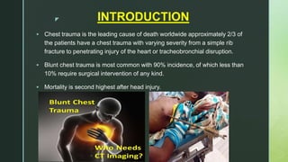
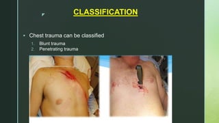
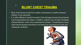
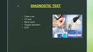
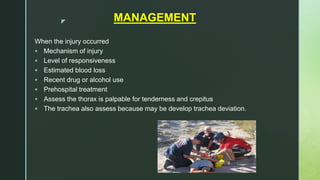
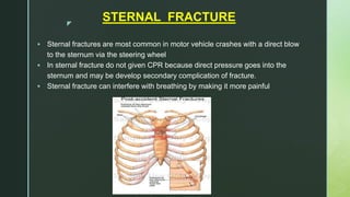
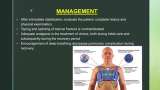
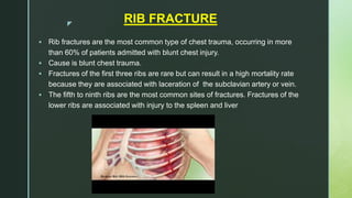
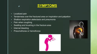
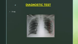
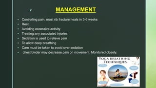
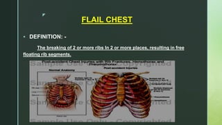
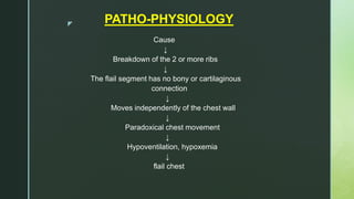
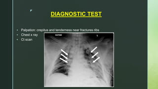
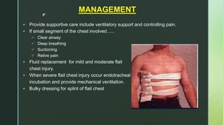
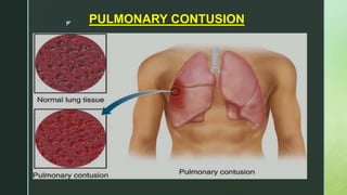
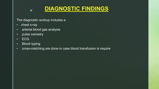
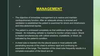
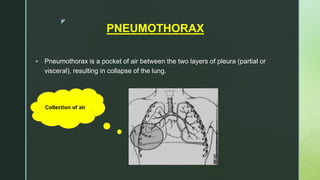
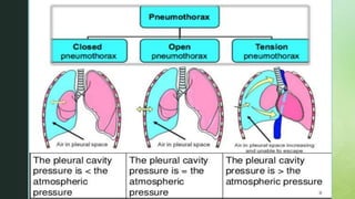
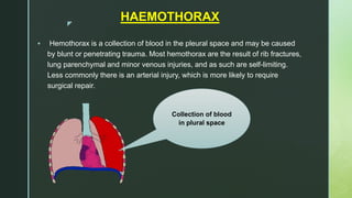
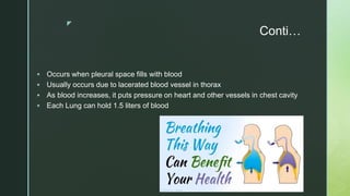
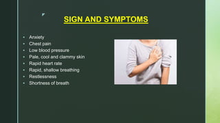
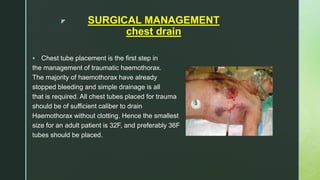
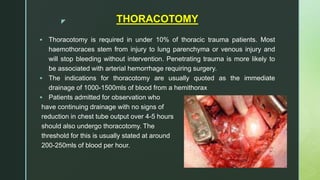
z
zPRESENTED BY:- DIPIKA PATEL
I.K.D.R.C COLLEGE OF
NURSING
F. Y. M.SC NURSING
CHEST TRAUMA
z
OUTLINE
• Introduction of chest trauma
• Blunt trauma
• Rib and sternal fracture
• Fail chest
• Penetrating trauma
• Haemothorax
• Emergency management
• Surgical management
• Nursing management
z INTRODUCTION
 Chest trauma is the leading cause of death worldwide approximately 2/3 of
the patients have a chest trauma with varying severity from a simple rib
fracture to penetrating injury of the heart or tracheobronchial disruption.
 Blunt chest trauma is most common with 90% incidence, of which less than
10% require surgical intervention of any kind.
 Mortality is second highest after head injury.
z CLASSIFICATION
 Chest trauma can be classified
1. Blunt trauma
2. Penetrating trauma
z
Other type of chest injury:-
 Rib fractures
 Flail chest
 Pneumothorax
 Hemothorax
 Sternal fractures
 Pulmonary contusion
z
BLUNT CHEST TRAUMA
 Blunt chest trauma results from sudden compression or positive pressure
inflicted to the chest wall.
 It is often difficult to identify the extent of the damage because the symptoms
may be generalized and vague. In addition, patients may not seek immediate
medical attention, which may complicate the problem. Complications in blunt
chest trauma develop secondary to rib fracture as consequence of pain and
inadequate ventilation.
z
CAUSE
fall Vehicle
accident
Cycle
accident
z
PATHO-PHYSIOLOGY
Cause (vehicle accident)
↓
Develop hypoxemia due to disruption of airway, lung collapse, rib
Fracture
↓
Hypovolemia from massive blood/fluid loss, cardiac contusion
↓
Cardiac failure due to increase intrathoracic pressure
↓
Acute renal failure
↓
Death
z DIAGNOSTIC TEST
 Chest x-ray
 CT scan
 Blood count
 Oxygen saturation
 ECG
z MANAGEMENT
When the injury occurred
 Mechanism of injury
 Level of responsiveness
 Estimated blood loss
 Recent drug or alcohol use
 Prehospital treatment
 Assess the thorax is palpable for tenderness and crepitus
 The trachea also assess because may be develop trachea deviation.
z
STERNAL FRACTURE
 Sternal fractures are most common in motor vehicle crashes with a direct blow
to the sternum via the steering wheel
 In sternal fracture do not given CPR because direct pressure goes into the
sternum and may be develop secondary complication of fracture.
 Sternal fracture can interfere with breathing by making it more painful
z MANAGEMENT
 After immediate stabilization, evaluate the patient, complete history and
physical examination.
 Taping and splinting of sternal fracture is contraindicated
 Adequate analgesia is the treatment of choice, both during initial care and
subsequently during the recovery period
 Encouragement of deep breathing decreases pulmonary complication during
recovery.
z
RIB FRACTURE
 Rib fractures are the most common type of chest trauma, occurring in more
than 60% of patients admitted with blunt chest injury.
 Cause is blunt chest trauma.
 Fractures of the first three ribs are rare but can result in a high mortality rate
because they are associated with laceration of the subclavian artery or vein.
 The fifth to ninth ribs are the most common sites of fractures. Fractures of the
lower ribs are associated with injury to the spleen and liver
z SYMPTOMS
 Localized pain
 Tenderness over the fractured area on inspiration and palpation
 Shallow respiration atelectasis and pneumonia
 Pain when coughing
 Swelling and brushing in the fracture area
 Internal bleeding
 Pneumothorax or hemothorax
z DIAGNOSTIC TEST
 X ray
z MANAGEMENT
 Controlling pain, most rib fracture heals in 3-6 weeks
 Rest
 Avoiding excessive activity
 Treating any associated injuries
 Sedation is used to relieve pain
 To allow deep breathing
 Care must be taken to avoid over sedation
 chest binder may decrease pain on movement. Monitored closely.
z
FLAIL CHEST
 DEFINITION: -
The breaking of 2 or more ribs In 2 or more places, resulting in free
floating rib segments.
z PATHO-PHYSIOLOGY
Cause
↓
Breakdown of the 2 or more ribs
↓
The flail segment has no bony or cartilaginous
connection
↓
Moves independently of the chest wall
↓
Paradoxical chest movement
↓
Hypoventilation, hypoxemia
↓
flail chest
z PARADOXICAL MOVEMENT
 By chart
z SIGN AND SYMPOTOMS
 Shortness of breath
 Paradoxical movement
 Bruising/swelling
 Crepitus (grinding of bone ends on palpation)
 Tachycardia
 Hypotension
 Hypoxemia
z
DIAGNOSTIC TEST
 Palpation: crepitus and tenderness near fractures ribs
 Chest x ray
 Ct scan
z
MANAGEMENT
 Provide supportive care include ventilatory support and controlling pain.
 If small segment of the chest involved…..
 Clear airway
 Deep breathing
 Suctioning
 Relive pain
 Fluid replacement for mild and moderate flail
chest injury.
 When severe flail chest injury occur endotracheal
incubation and provide mechanical ventilation.
 Bulky dressing for splint of flail chest
z PULMONARY CONTUSION
z INTRODUCTION
 A pulmonary contusion, also known as lung contusion, is a bruise of
the lung, caused by chest trauma.
 As a result of damage to capillaries, blood and other fluids accumulate in the
lung tissue.
 The excess fluid interferes with gas exchange, potentially leading to
inadequate oxygen levels (hypoxia).
 Unlike pulmonary laceration, another type of lung injury, pulmonary contusion
does not involve a cut or tear of the lung tissue.
z SIGN AND SYMPTOMS
 Decrease breath sound
 Tachypnea
 Tachycardia
 Chest pain
 Hypoxemia
 Crackles
 Frank bleeding
 Constant coughing
z
MANAGEMENT
 Maintaining the airway,
 providing adequate oxygenation
 controlling pain.
 In mild pulmonary contusion intravenous fluids and oral intake is important to
mobilize secretions.
 postural drainage, physiotherapy including coughing, and endotracheal suctioning
are used to remove the secretions.
 antimicrobial therapy is administered.
 Supplemental oxygen is usually given by mask or cannula for 24 to 36 hours.
 The patient with moderate pulmonary contusion may require; intubation and
mechanical ventilation with PEEP may also be necessary to maintain the pressure.
 Diuretics
 The patient with severe contusion may develop respiratory failure and may require
aggressive treatment with endotracheal intubation and ventilatory support,
diuretics, and. fluid restriction. Colloids and crystalloid solutions may be used to
treat hypovolemia.
z
PENETRATING TRAUMA
 Gunshot and stab wounds are the most common types of penetrating chest
trauma. They are classified according to their velocity.

Stab wounds are
generally considered of
low velocity because
the weapon destroys a
small area around
the wound. Knives and
switchblades cause
most stab wounds.
z
z DIAGNOSTIC FINDINGS
The diagnostic workup includes a
 chest x-ray
 arterial blood gas analysis
 pulse oximetry
 ECG
 Blood typing
 cross-matching are done in case blood transfusion is require
z
MANAGEMENT
 The objective of immediate management is to restore and maintain
cardiopulmonary function. After an adequate airway is ensured and
ventilation is established the patient is examined for shock and intrathoracic
and intra-abdominal injuries.
 The patient is undressed completely so that additional injuries will not be
missed. An indwelling catheter is inserted to monitor urinary output. Shock
is treated simultaneously with colloid solutions, crystalloids, or blood, as
indicated by the patient’s condition
 A chest tube is inserted into the pleural space in most patients with
penetrating wounds of the chest to achieve rapid and continuing re-
expansion of the lungs. The insertion of the chest tube frequently results in a
complete evacuation of the blood and air.
z
PNEUMOTHORAX
 Pneumothorax is a pocket of air between the two layers of pleura (partial or
visceral), resulting in collapse of the lung.
Collection of air
z
z
HAEMOTHORAX
 Hemothorax is a collection of blood in the pleural space and may be caused
by blunt or penetrating trauma. Most hemothorax are the result of rib fractures,
lung parenchymal and minor venous injuries, and as such are self-limiting.
Less commonly there is an arterial injury, which is more likely to require
surgical repair.
Collection of blood
in plural space
z
Conti…
 Occurs when pleural space fills with blood
 Usually occurs due to lacerated blood vessel in thorax
 As blood increases, it puts pressure on heart and other vessels in chest cavity
 Each Lung can hold 1.5 liters of blood
z
SIGN AND SYMPTOMS
 Anxiety
 Chest pain
 Low blood pressure
 Pale, cool and clammy skin
 Rapid heart rate
 Rapid, shallow breathing
 Restlessness
 Shortness of breath
z
DIAGNOSTIC TEST
 Chest x-ray
 CT scan
 Pleural fluid analysis (often very bloody or blood-tinged)
 Thoracentesis (drainage of pleural fluid through a needle or catheter)
z MANAGEMENT
 The goal of treatment is to get the person stable, stop the bleeding, and
remove the blood and air in the pleural space.
 A chest tube is inserted through the chest wall between the ribs to drain the
blood and air.
 It is left in place and attached to suction for several days to re-expand the
lung.
 If a chest tube alone does not control the bleeding, surgery (thoracotomy)
may be needed to stop the bleeding.
 The cause of the hemothorax should be also treated. The underlying lung
may have collapsed. This can lead to breathing difficulty. In people who have
had an injury, chest tube drainage may be all that is needed. Surgery may not
be necessary.
z SURGICAL MANAGEMENT
chest drain
 Chest tube placement is the first step in
the management of traumatic haemothorax.
The majority of haemothorax have already
stopped bleeding and simple drainage is all
that is required. All chest tubes placed for trauma
should be of sufficient caliber to drain
Haemothorax without clotting. Hence the smallest
size for an adult patient is 32F, and preferably 36F
tubes should be placed.
z THORACOTOMY
 Thoracotomy is required in under 10% of thoracic trauma patients. Most
haemothoraces stem from injury to lung parenchyma or venous injury and
will stop bleeding without intervention. Penetrating trauma is more likely to
be associated with arterial hemorrhage requiring surgery.
 The indications for thoracotomy are usually quoted as the immediate
drainage of 1000-1500mls of blood from a hemithorax
 Patients admitted for observation who
have continuing drainage with no signs of
reduction in chest tube output over 4-5 hours
should also undergo thoracotomy. The
threshold for this is usually stated at around
200-250mls of blood per hour.
z
z
EMERGENCY MANAGEMENT AND
GENARAL MANAGEMENT
 By leaflet
z
NURSING MANAGEMENT
1. Ineffective breathing pattern related to decreased lung expansion as
evidence by dyspnea.
2. Increase risk of hypoxia and respiratory failure related to injury.
3. Increased risk of hypovolemia and shock related to hemorrhage and
impaired cardiac function
4. Fear related breathing difficulty as evidence by facial expression.
5. Decrease cardiac output related to mediastinal shift.
6. Acute pain related to trauma, altered pressure in the chest cavity
7. Risk for infection related to traumatic injury and chest tube insertion
8. Activity intolerance related to activity restriction
9. Deficient knowledge related to complexity of treatment as evidence by
inappropriate behaviour’s
z
ASSIGNMENT
 Write down nursing intervention of nursing diagnosis of chest
trauma
 submitted on 17 / 02/ 2018
z
z

Chest trauma

  • 1.
  • 2.
    zPRESENTED BY:- DIPIKAPATEL I.K.D.R.C COLLEGE OF NURSING F. Y. M.SC NURSING CHEST TRAUMA
  • 3.
    z OUTLINE • Introduction ofchest trauma • Blunt trauma • Rib and sternal fracture • Fail chest • Penetrating trauma • Haemothorax • Emergency management • Surgical management • Nursing management
  • 4.
    z INTRODUCTION  Chesttrauma is the leading cause of death worldwide approximately 2/3 of the patients have a chest trauma with varying severity from a simple rib fracture to penetrating injury of the heart or tracheobronchial disruption.  Blunt chest trauma is most common with 90% incidence, of which less than 10% require surgical intervention of any kind.  Mortality is second highest after head injury.
  • 5.
    z CLASSIFICATION  Chesttrauma can be classified 1. Blunt trauma 2. Penetrating trauma
  • 6.
    z Other type ofchest injury:-  Rib fractures  Flail chest  Pneumothorax  Hemothorax  Sternal fractures  Pulmonary contusion
  • 7.
    z BLUNT CHEST TRAUMA Blunt chest trauma results from sudden compression or positive pressure inflicted to the chest wall.  It is often difficult to identify the extent of the damage because the symptoms may be generalized and vague. In addition, patients may not seek immediate medical attention, which may complicate the problem. Complications in blunt chest trauma develop secondary to rib fracture as consequence of pain and inadequate ventilation.
  • 8.
  • 9.
    z PATHO-PHYSIOLOGY Cause (vehicle accident) ↓ Develophypoxemia due to disruption of airway, lung collapse, rib Fracture ↓ Hypovolemia from massive blood/fluid loss, cardiac contusion ↓ Cardiac failure due to increase intrathoracic pressure ↓ Acute renal failure ↓ Death
  • 10.
    z DIAGNOSTIC TEST Chest x-ray  CT scan  Blood count  Oxygen saturation  ECG
  • 11.
    z MANAGEMENT When theinjury occurred  Mechanism of injury  Level of responsiveness  Estimated blood loss  Recent drug or alcohol use  Prehospital treatment  Assess the thorax is palpable for tenderness and crepitus  The trachea also assess because may be develop trachea deviation.
  • 12.
    z STERNAL FRACTURE  Sternalfractures are most common in motor vehicle crashes with a direct blow to the sternum via the steering wheel  In sternal fracture do not given CPR because direct pressure goes into the sternum and may be develop secondary complication of fracture.  Sternal fracture can interfere with breathing by making it more painful
  • 13.
    z MANAGEMENT  Afterimmediate stabilization, evaluate the patient, complete history and physical examination.  Taping and splinting of sternal fracture is contraindicated  Adequate analgesia is the treatment of choice, both during initial care and subsequently during the recovery period  Encouragement of deep breathing decreases pulmonary complication during recovery.
  • 14.
    z RIB FRACTURE  Ribfractures are the most common type of chest trauma, occurring in more than 60% of patients admitted with blunt chest injury.  Cause is blunt chest trauma.  Fractures of the first three ribs are rare but can result in a high mortality rate because they are associated with laceration of the subclavian artery or vein.  The fifth to ninth ribs are the most common sites of fractures. Fractures of the lower ribs are associated with injury to the spleen and liver
  • 15.
    z SYMPTOMS  Localizedpain  Tenderness over the fractured area on inspiration and palpation  Shallow respiration atelectasis and pneumonia  Pain when coughing  Swelling and brushing in the fracture area  Internal bleeding  Pneumothorax or hemothorax
  • 16.
  • 17.
    z MANAGEMENT  Controllingpain, most rib fracture heals in 3-6 weeks  Rest  Avoiding excessive activity  Treating any associated injuries  Sedation is used to relieve pain  To allow deep breathing  Care must be taken to avoid over sedation  chest binder may decrease pain on movement. Monitored closely.
  • 18.
    z FLAIL CHEST  DEFINITION:- The breaking of 2 or more ribs In 2 or more places, resulting in free floating rib segments.
  • 19.
    z PATHO-PHYSIOLOGY Cause ↓ Breakdown ofthe 2 or more ribs ↓ The flail segment has no bony or cartilaginous connection ↓ Moves independently of the chest wall ↓ Paradoxical chest movement ↓ Hypoventilation, hypoxemia ↓ flail chest
  • 20.
  • 21.
    z SIGN ANDSYMPOTOMS  Shortness of breath  Paradoxical movement  Bruising/swelling  Crepitus (grinding of bone ends on palpation)  Tachycardia  Hypotension  Hypoxemia
  • 22.
    z DIAGNOSTIC TEST  Palpation:crepitus and tenderness near fractures ribs  Chest x ray  Ct scan
  • 23.
    z MANAGEMENT  Provide supportivecare include ventilatory support and controlling pain.  If small segment of the chest involved…..  Clear airway  Deep breathing  Suctioning  Relive pain  Fluid replacement for mild and moderate flail chest injury.  When severe flail chest injury occur endotracheal incubation and provide mechanical ventilation.  Bulky dressing for splint of flail chest
  • 24.
  • 25.
    z INTRODUCTION  Apulmonary contusion, also known as lung contusion, is a bruise of the lung, caused by chest trauma.  As a result of damage to capillaries, blood and other fluids accumulate in the lung tissue.  The excess fluid interferes with gas exchange, potentially leading to inadequate oxygen levels (hypoxia).  Unlike pulmonary laceration, another type of lung injury, pulmonary contusion does not involve a cut or tear of the lung tissue.
  • 26.
    z SIGN ANDSYMPTOMS  Decrease breath sound  Tachypnea  Tachycardia  Chest pain  Hypoxemia  Crackles  Frank bleeding  Constant coughing
  • 27.
    z MANAGEMENT  Maintaining theairway,  providing adequate oxygenation  controlling pain.  In mild pulmonary contusion intravenous fluids and oral intake is important to mobilize secretions.  postural drainage, physiotherapy including coughing, and endotracheal suctioning are used to remove the secretions.  antimicrobial therapy is administered.  Supplemental oxygen is usually given by mask or cannula for 24 to 36 hours.  The patient with moderate pulmonary contusion may require; intubation and mechanical ventilation with PEEP may also be necessary to maintain the pressure.  Diuretics  The patient with severe contusion may develop respiratory failure and may require aggressive treatment with endotracheal intubation and ventilatory support, diuretics, and. fluid restriction. Colloids and crystalloid solutions may be used to treat hypovolemia.
  • 28.
    z PENETRATING TRAUMA  Gunshotand stab wounds are the most common types of penetrating chest trauma. They are classified according to their velocity.  Stab wounds are generally considered of low velocity because the weapon destroys a small area around the wound. Knives and switchblades cause most stab wounds.
  • 29.
  • 30.
    z DIAGNOSTIC FINDINGS Thediagnostic workup includes a  chest x-ray  arterial blood gas analysis  pulse oximetry  ECG  Blood typing  cross-matching are done in case blood transfusion is require
  • 31.
    z MANAGEMENT  The objectiveof immediate management is to restore and maintain cardiopulmonary function. After an adequate airway is ensured and ventilation is established the patient is examined for shock and intrathoracic and intra-abdominal injuries.  The patient is undressed completely so that additional injuries will not be missed. An indwelling catheter is inserted to monitor urinary output. Shock is treated simultaneously with colloid solutions, crystalloids, or blood, as indicated by the patient’s condition  A chest tube is inserted into the pleural space in most patients with penetrating wounds of the chest to achieve rapid and continuing re- expansion of the lungs. The insertion of the chest tube frequently results in a complete evacuation of the blood and air.
  • 32.
    z PNEUMOTHORAX  Pneumothorax isa pocket of air between the two layers of pleura (partial or visceral), resulting in collapse of the lung. Collection of air
  • 33.
  • 34.
    z HAEMOTHORAX  Hemothorax isa collection of blood in the pleural space and may be caused by blunt or penetrating trauma. Most hemothorax are the result of rib fractures, lung parenchymal and minor venous injuries, and as such are self-limiting. Less commonly there is an arterial injury, which is more likely to require surgical repair. Collection of blood in plural space
  • 35.
    z Conti…  Occurs whenpleural space fills with blood  Usually occurs due to lacerated blood vessel in thorax  As blood increases, it puts pressure on heart and other vessels in chest cavity  Each Lung can hold 1.5 liters of blood
  • 36.
    z SIGN AND SYMPTOMS Anxiety  Chest pain  Low blood pressure  Pale, cool and clammy skin  Rapid heart rate  Rapid, shallow breathing  Restlessness  Shortness of breath
  • 37.
    z DIAGNOSTIC TEST  Chestx-ray  CT scan  Pleural fluid analysis (often very bloody or blood-tinged)  Thoracentesis (drainage of pleural fluid through a needle or catheter)
  • 38.
    z MANAGEMENT  Thegoal of treatment is to get the person stable, stop the bleeding, and remove the blood and air in the pleural space.  A chest tube is inserted through the chest wall between the ribs to drain the blood and air.  It is left in place and attached to suction for several days to re-expand the lung.  If a chest tube alone does not control the bleeding, surgery (thoracotomy) may be needed to stop the bleeding.  The cause of the hemothorax should be also treated. The underlying lung may have collapsed. This can lead to breathing difficulty. In people who have had an injury, chest tube drainage may be all that is needed. Surgery may not be necessary.
  • 39.
    z SURGICAL MANAGEMENT chestdrain  Chest tube placement is the first step in the management of traumatic haemothorax. The majority of haemothorax have already stopped bleeding and simple drainage is all that is required. All chest tubes placed for trauma should be of sufficient caliber to drain Haemothorax without clotting. Hence the smallest size for an adult patient is 32F, and preferably 36F tubes should be placed.
  • 40.
    z THORACOTOMY  Thoracotomyis required in under 10% of thoracic trauma patients. Most haemothoraces stem from injury to lung parenchyma or venous injury and will stop bleeding without intervention. Penetrating trauma is more likely to be associated with arterial hemorrhage requiring surgery.  The indications for thoracotomy are usually quoted as the immediate drainage of 1000-1500mls of blood from a hemithorax  Patients admitted for observation who have continuing drainage with no signs of reduction in chest tube output over 4-5 hours should also undergo thoracotomy. The threshold for this is usually stated at around 200-250mls of blood per hour.
  • 41.
  • 42.
    z EMERGENCY MANAGEMENT AND GENARALMANAGEMENT  By leaflet
  • 43.
    z NURSING MANAGEMENT 1. Ineffectivebreathing pattern related to decreased lung expansion as evidence by dyspnea. 2. Increase risk of hypoxia and respiratory failure related to injury. 3. Increased risk of hypovolemia and shock related to hemorrhage and impaired cardiac function 4. Fear related breathing difficulty as evidence by facial expression. 5. Decrease cardiac output related to mediastinal shift. 6. Acute pain related to trauma, altered pressure in the chest cavity 7. Risk for infection related to traumatic injury and chest tube insertion 8. Activity intolerance related to activity restriction 9. Deficient knowledge related to complexity of treatment as evidence by inappropriate behaviour’s
  • 44.
    z ASSIGNMENT  Write downnursing intervention of nursing diagnosis of chest trauma  submitted on 17 / 02/ 2018
  • 45.
  • 46.