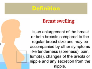
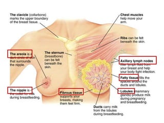
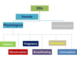
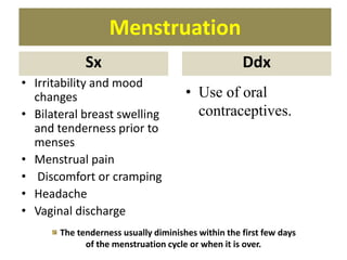
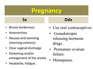
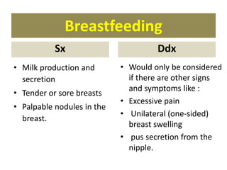
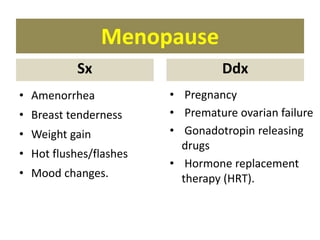
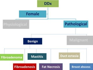
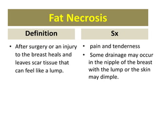
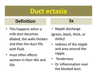
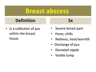
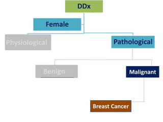
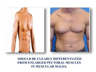
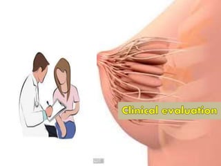
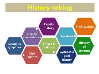
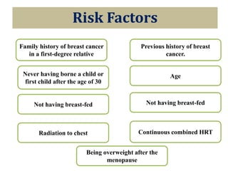
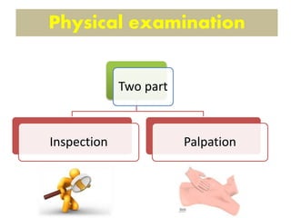
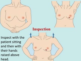
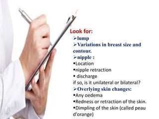
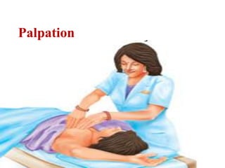
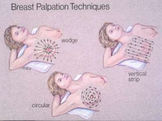
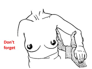
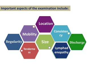
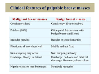
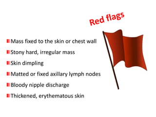
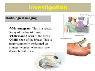
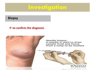
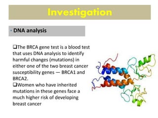
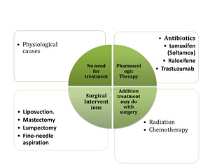
Maryam AL-Qahtani
Learning objectives
Definition of breast swelling
Short anatomy of breast
Differential Diagnosis of breast
swelling
Clinical Evaluations
History
Physical Examinations
Investigations
Management
is an enlargement of the breast
or both breasts compared to the
regular breast size and may be
accompanied by other symptoms
like tenderness (soreness), pain,
lump(s), changes of the areola or
nipple and any secretion from the
nipple.
Definition
Breast swelling
DDx
Physiological Pathological
Female
Pathological
Puberty
Menstruation
Pregnancy
Breastfeeding Contraceptives
Menopause
Puberty
Sx
• Increasing breast size
• Androgenic hair (pubic
hair and/or hair of the
armpits)
• Onset of menstruation
(menarche)
• Fat accumulation on the
thighs, buttocks and/or
lower abdomen.
Ddx
• Use of oral
contraceptives.
No reason to worry unless the pain becomes
unbearable.
Menstruation
Sx
• Irritability and mood
changes
• Bilateral breast swelling
and tenderness prior to
menses
• Menstrual pain
• Discomfort or cramping
• Headache
• Vaginal discharge
Ddx
• Use of oral
contraceptives.
The tenderness usually diminishes within the first few days
of the menstruation cycle or when it is over.
Pregnancy
Sx
• Breast tenderness
• Amenorrhea
• Nausea and vomiting
(morning sickness)
• Clear vaginal discharge
• Darkening and/or
enlargement of the areola
• Headache, Fatigue.
Ddx
• Use oral contraceptives
• Gonadotropin
releasing hormone
drugs .
• Premature ovarian
failure.
• Menopause.
Breastfeeding
Sx
• Milk production and
secretion
• Tender or sore breasts
• Palpable nodules in the
breast.
Ddx
• Would only be considered
if there are other signs
and symptoms like :
• Excessive pain
• Unilateral (one-sided)
breast swelling
• pus secretion from the
nipple.
Menopause
Sx
• Amenorrhea
• Breast tenderness
• Weight gain
• Hot flushes/flashes
• Mood changes.
Ddx
• Pregnancy
• Premature ovarian failure
• Gonadotropin releasing
drugs
• Hormone replacement
therapy (HRT).
Contraceptives
• Women who use oral contraceptive pills can
experience problems with their breasts because of
the hormonal imbalance that pills can cause.
• This is usually not a reason for concern, but if it
happens with
unusual bleeding, bloating, headache and uneasiness,
the woman should seek immediate medical care.
DDx
Physiological Pathological
Female
Physiological
Fibroadenoma
Fibroadenosis
Mastitis
Fat Necrosis Breast abscess
Duct ectasia
Benign MalignantMalignant
Fibroadenoma
Definition
• are the most common
breast lesions
• commonly found in
women under the age
of 30.
Sx
• Painless
Fibroadenosis
Definition
• Fibrosis is firmness in the
connective tissues,
and cysts are fluid-filled
sacs.
• These changes often are
affected by hormones.
Sx
• Areas of lumpiness,
thickening
• Tenderness
• Nipple discharge
• Pain
Mastitis
Definition
• Bacterial infection and
can cause inflammation
around the nipple
• Often occurs when
breastfeeding.
Sx
• Breast pain
• Fever
• Tenderness/soreness
• Redness
• Heat or warmth of
affected area.
Fat Necrosis
Definition
• After surgery or an injury
to the breast heals and
leaves scar tissue that
can feel like a lump.
Sx
• pain and tenderness
• Some drainage may occur
in the nipple of the breast
with the lump or the skin
may dimple.
Duct ectasia
• This happens when a
milk duct becomes
dilated, the walls thicken
and then the duct fills
with fluid.
• most often affects
women in their 40s and
50s
Sx
• Nipple discharge
(green, black, thick, or
sticky)
• redness of the nipple
and area around the
nipple
• Tenderness
• Or inflammation near
the blocked duct.
Definition
Breast abscess
Definition
• Is a collection of pus
within the breast
tissue.
Sx
• Severe breast pain
• Fever, chills
• Redness, heat/warmth
• Discharge of pus
• Deviated nipple
• Visible lump
DDx
Physiological Pathological
Female
Physiological
Benign MalignantBenign
Breast Cancer
Breast cancer
Most common type of
breast cancer :
Inflammatory breast cancer
Symptoms and signs:
painless lump in the breast.
Changes in the size or shape of a breast.
Dimpling or thickening of some of the skin
on a part of a breast.
The nipple becoming inverted (turning in).
Rarely, a discharge occurring from a nipple
redness and warmth
The most commonly used tool that doctors use to describe
tumor stage is the TNM system. TNM is an abbreviation for
tumor (T), node (N), and metastasis (M).
Gynecomastia
Risk factorsDefinition
•Is swelling of the
breast tissue in boys or
men, caused by an
imbalance of the
hormones estrogen
and testosterone.
•Older age
•obese
•Use of anabolic steroids or
androgens to enhance
athletic performance
•Certain health conditions,
including liver and kidney
disease, thyroid disease,
hormonally active tumors,
and Klinefelter syndrome
SHOULD BE CLEARLY DIFFERENTIATED
FROM ENLARGED PECTORAL MUSCLES
IN MUSCULAR MALES.
Break Time
Risk
Factors
Onset &
Pattern
Duration
Family
history
History
of
trauma
Family
history
Associated
symptoms
Gynaecolo
gical
history
Medications
Medical
comorbidities
History taking
Risk Factors
Previous history of breast
cancer.
Family history of breast cancer
in a first-degree relative
AgeNever having borne a child or
first child after the age of 30
Not having breast-fedNot having breast-fed
Continuous combined HRTRadiation to chest
Being overweight after the
menopause
Physical examination
Risk
Factors
Two part
Inspection Palpation
Inspect with the
patient sitting
and then with
their hands
raised above
head.
Inspection
Look for:
lump
Variations in breast size and
contour.
nipple :
Location
nipple retraction
 discharge
if so, is it unilateral or bilateral?
Overlying skin changes:
Any oedema
Redness or retraction of the skin.
Dimpling of the skin (called peau
d'orange)
Palpation
Don't
forget
Tenderne
ss
Size
Consisten
cy
Family
history
Discharge
Location
Regularity
Lymphad
enopathy
Mobility
Important aspects of the examination include:
Clinical features of palpable breast masses
Malignant breast masses Benign breast masses
Consistency: hard Consistency: firm or rubbery
Painless (90%) Often painful (consistent with
benign breast conditions)
Irregular margins Regular or smooth margins
Fixation to skin or chest wall Mobile and not fixed
Skin dimpling may occur Skin dimpling unlikely
Discharge: bloody, unilateral Discharge: no blood and bilateral
discharge. Green or yellow colour
Nipple retraction may be present No nipple retraction
Mass fixed to the skin or chest wall
Stony hard, irregular mass
Skin dimpling
Matted or fixed axillary lymph nodes
Bloody nipple discharge
Thickened, erythematous skin
Investigation
Mammogram. This is a special
X-ray of the breast tissue.
Ultrasound scan of the breast.
MRI scan of the breast. This is
more commonly performed on
younger women, who may have
denser breast tissue.
Radiological imaging
Investigation
-to confirm the diagnosis
• Biopsy
Investigation
The BRCA gene test is a blood test
that uses DNA analysis to identify
harmful changes (mutations) in
either one of the two breast cancer
susceptibility genes — BRCA1 and
BRCA2.
Women who have inherited
mutations in these genes face a
much higher risk of developing
breast cancer
• DNA analysis
Investigation
Human epidermal growth factor
receptor 2 (HER2).
This tumor marker is a specialized
protein on breast cancer cells that
helps control cancer growth and
spread
• Blood Marker Tests
Investigation
 an ultrasound scan of
the liver
chest X-ray,
a bone scan or other
types of scan.
• Assessing the extent and spread
• Treatment options for breast swelling vary
depending on the underlying etiology.
Treatment
• Radiation
• Chemotherapy
• Liposuction.
• Mastectomy
• Lumpectomy
• Fine-needle
aspiration
• Antibiotics
• tamoxifen
(Soltamox)
• Raloxifene
• Trastuzumab
• Physiological
causes
No need
for
treatment
Pharmacol
ogic
Therapy
Addition
treatment
may do
with
surgery
Surgical
Intervent
ions
Break Time
• 61-year-old woman who presents to her internist alarmed
by a lump in her right breast that she discovered while
showering. She report painless, swelling, and skin changes.
She has not experienced fever, weight loss, headache,
nausea, vomiting, dizziness, or bone pain.
• PMH (past medical history): SM is moderately overweight,
but otherwise in good health. experienced menopause at
age 55. She took hormone replacement therapy from age
54 until age 59.
• FH (family history): history of malignancy in first-degree
relatives.
CASE SCENARIO
Most of causes are self-limited and benign.
Physiological : premenstrual syndrome is
a common cause of breast swelling.
Pathological : fibroadenoma is the most
common breast lesion.
The initial approach to the adolescent
patient with a breast mass includes a careful
history and physical examination.
Imaging may be necessary to differentiate
References
Uptodate ; Overview of breast masses in
children and adolescents.
Healthline; breast-swelling.
Patient education website ; Breast cancer.
The Merck Manual, Professional Edition.
National Cancer Institute; Breast Cancer.
American Cancer Society ;Breast Cancer.
Medlineplus; Premenstrual breast
changes.
Med-health; Swollen Breast.
Breast swelling

Breast swelling

  • 1.
  • 2.
    Learning objectives Definition ofbreast swelling Short anatomy of breast Differential Diagnosis of breast swelling Clinical Evaluations History Physical Examinations Investigations Management
  • 4.
    is an enlargementof the breast or both breasts compared to the regular breast size and may be accompanied by other symptoms like tenderness (soreness), pain, lump(s), changes of the areola or nipple and any secretion from the nipple. Definition Breast swelling
  • 8.
  • 9.
    Puberty Sx • Increasing breastsize • Androgenic hair (pubic hair and/or hair of the armpits) • Onset of menstruation (menarche) • Fat accumulation on the thighs, buttocks and/or lower abdomen. Ddx • Use of oral contraceptives. No reason to worry unless the pain becomes unbearable.
  • 10.
    Menstruation Sx • Irritability andmood changes • Bilateral breast swelling and tenderness prior to menses • Menstrual pain • Discomfort or cramping • Headache • Vaginal discharge Ddx • Use of oral contraceptives. The tenderness usually diminishes within the first few days of the menstruation cycle or when it is over.
  • 11.
    Pregnancy Sx • Breast tenderness •Amenorrhea • Nausea and vomiting (morning sickness) • Clear vaginal discharge • Darkening and/or enlargement of the areola • Headache, Fatigue. Ddx • Use oral contraceptives • Gonadotropin releasing hormone drugs . • Premature ovarian failure. • Menopause.
  • 12.
    Breastfeeding Sx • Milk productionand secretion • Tender or sore breasts • Palpable nodules in the breast. Ddx • Would only be considered if there are other signs and symptoms like : • Excessive pain • Unilateral (one-sided) breast swelling • pus secretion from the nipple.
  • 13.
    Menopause Sx • Amenorrhea • Breasttenderness • Weight gain • Hot flushes/flashes • Mood changes. Ddx • Pregnancy • Premature ovarian failure • Gonadotropin releasing drugs • Hormone replacement therapy (HRT).
  • 14.
    Contraceptives • Women whouse oral contraceptive pills can experience problems with their breasts because of the hormonal imbalance that pills can cause. • This is usually not a reason for concern, but if it happens with unusual bleeding, bloating, headache and uneasiness, the woman should seek immediate medical care.
  • 15.
  • 16.
    Fibroadenoma Definition • are themost common breast lesions • commonly found in women under the age of 30. Sx • Painless
  • 17.
    Fibroadenosis Definition • Fibrosis isfirmness in the connective tissues, and cysts are fluid-filled sacs. • These changes often are affected by hormones. Sx • Areas of lumpiness, thickening • Tenderness • Nipple discharge • Pain
  • 18.
    Mastitis Definition • Bacterial infectionand can cause inflammation around the nipple • Often occurs when breastfeeding. Sx • Breast pain • Fever • Tenderness/soreness • Redness • Heat or warmth of affected area.
  • 19.
    Fat Necrosis Definition • Aftersurgery or an injury to the breast heals and leaves scar tissue that can feel like a lump. Sx • pain and tenderness • Some drainage may occur in the nipple of the breast with the lump or the skin may dimple.
  • 20.
    Duct ectasia • Thishappens when a milk duct becomes dilated, the walls thicken and then the duct fills with fluid. • most often affects women in their 40s and 50s Sx • Nipple discharge (green, black, thick, or sticky) • redness of the nipple and area around the nipple • Tenderness • Or inflammation near the blocked duct. Definition
  • 21.
    Breast abscess Definition • Isa collection of pus within the breast tissue. Sx • Severe breast pain • Fever, chills • Redness, heat/warmth • Discharge of pus • Deviated nipple • Visible lump
  • 22.
  • 23.
    Breast cancer Most commontype of breast cancer : Inflammatory breast cancer
  • 24.
    Symptoms and signs: painlesslump in the breast. Changes in the size or shape of a breast. Dimpling or thickening of some of the skin on a part of a breast. The nipple becoming inverted (turning in). Rarely, a discharge occurring from a nipple redness and warmth
  • 25.
    The most commonlyused tool that doctors use to describe tumor stage is the TNM system. TNM is an abbreviation for tumor (T), node (N), and metastasis (M).
  • 27.
    Gynecomastia Risk factorsDefinition •Is swellingof the breast tissue in boys or men, caused by an imbalance of the hormones estrogen and testosterone. •Older age •obese •Use of anabolic steroids or androgens to enhance athletic performance •Certain health conditions, including liver and kidney disease, thyroid disease, hormonally active tumors, and Klinefelter syndrome
  • 28.
    SHOULD BE CLEARLYDIFFERENTIATED FROM ENLARGED PECTORAL MUSCLES IN MUSCULAR MALES.
  • 29.
  • 31.
  • 32.
    Risk Factors Previous historyof breast cancer. Family history of breast cancer in a first-degree relative AgeNever having borne a child or first child after the age of 30 Not having breast-fedNot having breast-fed Continuous combined HRTRadiation to chest Being overweight after the menopause
  • 33.
  • 34.
    Inspect with the patientsitting and then with their hands raised above head. Inspection
  • 35.
    Look for: lump Variations inbreast size and contour. nipple : Location nipple retraction  discharge if so, is it unilateral or bilateral? Overlying skin changes: Any oedema Redness or retraction of the skin. Dimpling of the skin (called peau d'orange)
  • 36.
  • 38.
  • 39.
  • 40.
    Clinical features ofpalpable breast masses Malignant breast masses Benign breast masses Consistency: hard Consistency: firm or rubbery Painless (90%) Often painful (consistent with benign breast conditions) Irregular margins Regular or smooth margins Fixation to skin or chest wall Mobile and not fixed Skin dimpling may occur Skin dimpling unlikely Discharge: bloody, unilateral Discharge: no blood and bilateral discharge. Green or yellow colour Nipple retraction may be present No nipple retraction
  • 41.
    Mass fixed tothe skin or chest wall Stony hard, irregular mass Skin dimpling Matted or fixed axillary lymph nodes Bloody nipple discharge Thickened, erythematous skin
  • 43.
    Investigation Mammogram. This isa special X-ray of the breast tissue. Ultrasound scan of the breast. MRI scan of the breast. This is more commonly performed on younger women, who may have denser breast tissue. Radiological imaging
  • 44.
  • 45.
    Investigation The BRCA genetest is a blood test that uses DNA analysis to identify harmful changes (mutations) in either one of the two breast cancer susceptibility genes — BRCA1 and BRCA2. Women who have inherited mutations in these genes face a much higher risk of developing breast cancer • DNA analysis
  • 46.
    Investigation Human epidermal growthfactor receptor 2 (HER2). This tumor marker is a specialized protein on breast cancer cells that helps control cancer growth and spread • Blood Marker Tests
  • 47.
    Investigation  an ultrasoundscan of the liver chest X-ray, a bone scan or other types of scan. • Assessing the extent and spread
  • 49.
    • Treatment optionsfor breast swelling vary depending on the underlying etiology. Treatment
  • 50.
    • Radiation • Chemotherapy •Liposuction. • Mastectomy • Lumpectomy • Fine-needle aspiration • Antibiotics • tamoxifen (Soltamox) • Raloxifene • Trastuzumab • Physiological causes No need for treatment Pharmacol ogic Therapy Addition treatment may do with surgery Surgical Intervent ions
  • 51.
  • 52.
    • 61-year-old womanwho presents to her internist alarmed by a lump in her right breast that she discovered while showering. She report painless, swelling, and skin changes. She has not experienced fever, weight loss, headache, nausea, vomiting, dizziness, or bone pain. • PMH (past medical history): SM is moderately overweight, but otherwise in good health. experienced menopause at age 55. She took hormone replacement therapy from age 54 until age 59. • FH (family history): history of malignancy in first-degree relatives. CASE SCENARIO
  • 53.
    Most of causesare self-limited and benign. Physiological : premenstrual syndrome is a common cause of breast swelling. Pathological : fibroadenoma is the most common breast lesion. The initial approach to the adolescent patient with a breast mass includes a careful history and physical examination. Imaging may be necessary to differentiate
  • 54.
    References Uptodate ; Overviewof breast masses in children and adolescents. Healthline; breast-swelling. Patient education website ; Breast cancer. The Merck Manual, Professional Edition. National Cancer Institute; Breast Cancer. American Cancer Society ;Breast Cancer. Medlineplus; Premenstrual breast changes. Med-health; Swollen Breast.

Editor's Notes

  • #6  The structure of the male breast is nearly identical to that of the female breast, except that the male breast tissue lacks the specialized lobules, as there is no physiologic need for milk production by the male breast.
  • #9 Puberty. Puberty is often the first time women have problems with their growing breasts. Many girls feel temporary pain because of the hormonal changes in their bodies. There is no reason to worry unless the pain becomes unbearable. Menstruation. Most women experience changes in their breasts during the menstruation cycle. The breasts often become tender and swollen before their periods begin. This is caused by the increase of the hormones called estrogen and progesterone during this time of the month. The tenderness usually diminishes within the first few days of the menstruation cycle or when it is over. Pregnancy. Swollen breasts are one of the first signs a woman notices when she becomes pregnant. If pregnancy is a possibility, the woman should talk to her doctor as soon as possible. Again, the tenderness is caused by the hormones from pregnancy. Breastfeeding. If a woman chooses to breastfeed, she could experience swelling as a result of milk production and secretion. Sometimes, the baby sucking the breasts can cause temporary swelling. There is no reason for concern. Menopause. These changes are caused by hormones and are not considered a serious issue unless it is accompanied by excessive pain or any fluid secretion. If that occurs, contact a doctor as soon as possible. Contraceptives. Women who use oral contraceptive pills can experience problems with their breasts because of the hormonal imbalance that pills can cause. This is usually not a reason for concern, but if it happens with unusual bleeding, bloating, headache and uneasiness, the woman should seek immediate medical care.
  • #30 English television personality, talent judge and television producer most famous for his role as the blunt-talking judge on singing shows American Idol and X Factor. سايمون كاول
  • #32 gynaecological history : Pregnancy and Menstruation history History of present illness should include how long the mass has been present and whether it comes and goes or is painful. Previous occurrence of a mass and the outcome of its evaluation should be queried. Review of systems should determine whether nipple discharge is present and, if present, whether it is clear, milky, or bloody. Symptoms of advanced cancer (eg, weight loss, malaise, bone pain) should be sought. Past medical history should include risk factors for breast cancer, including previous diagnosis of breast cancer, history of radiation therapy to the chest area before age 30 (eg, for Hodgkin lymphoma). Family history should note breast cancer in a 1st-degree relative (mother, sister, daughter) and, if family history is positive, whether the relative carried one of the 2 known breast cancer genes, BRCA1 or BRCA2 .
  • #33 Previous history of breast cancer. Family history of breast cancer in a first-degree relative. A number of genetic mutations are implicated. The BRCA1, BRCA2 and TP53 mutations carry very high risk but only around 5% of women diagnosed with breast cancer carry a relevant genetic mutation on their chromosomes. Between 6% and 19% of women will have a family history but this may be due to chance, shared environmental or lifestyle risk factors, or increased genetic susceptibility. Risk increases with age. In Europe, ≤5% of cases present before age the age of 35, ≤25% before the age of 50.[4] Cancer Research UK statistics in 2009-2011 showed in the UK 80% of cancers were diagnosed in women over the age of 50, and 24% over the age of 75.[1]  Never having borne a child or first child after the age of 30. Not having breast-fed (breast-feeding is protective). Early menarche and late menopause. Continuous combined HRT increases risk.[5]  Radiation to chest (even quite small doses). Being overweight after the menopause. High alcohol intake - may increase risk in a dose-related manner
  • #37 Technique for palpation of the breast[13]  There is no proven "best method" to examine the breast. Different people have different techniques and the following description is by no means the only approach. Ask the patient to lie supine with their hands above their head. Examine from the clavicle medially to the mid-sternum, laterally to the mid-axillary line and to the inferior portion of the breast. Remember the axillary tail of breast tissue. Examine the axilla for palpable lymphadenopathy
  • #38 Technique for palpation of the breast[13]  There is no proven "best method" to examine the breast. Different people have different techniques and the following description is by no means the only approach. Ask the patient to lie supine with their hands above their head. Examine from the clavicle medially to the mid-sternum, laterally to the mid-axillary line and to the inferior portion of the breast. Remember the axillary tail of breast tissue. Examine the axilla for palpable lymphadenopathy
  • #40 Examination — Important aspects of the examination include [7,15]: ●Location of the mass – Fibrocystic change and fibroadenoma are usually located in the upper outer breast quadrants. The well-circumscribed nodules of intraductal papilloma may be located under the areola or in the ducts at the breast periphery. Mammary duct ectasia and cysts of Montgomery are subareolar. ●Consistency of the mass (cystic versus solid) – Mammary duct ectasia and cysts of Montgomery are cystic, whereas fibroadenoma, phyllodes tumors, fat necrosis, and malignant breast tumors are usually solid. ●Size of the mass – Fibroadenomas are usually smaller than phyllodes tumors (average of 2 to 3 cm versus 7 cm) [9,27]. The size of the mass can be monitored through the menstrual cycle. ●Mobility of the mass – Fibroadenomas are usually mobile, whereas malignant breast tumors are usually (but not always) fixed to the underlying tissue. ●Tenderness – Tenderness may be present before the onset of menses in adolescents with fibrocystic change and fibroadenoma. Tenderness also may occur in patients with infection or trauma. ●Overlying skin changes – Overlying skin changes may occur in large fibroadenomas, phyllodes tumors (the skin is shiny and stretched from rapid growth), and in breast cancer (peau d'orange, retraction). ●Nipple discharge – Nipple discharge may occur in fibrocystic disease (nonbloody green or brown), cysts of Montgomery (clear to brown), intraductal papilloma (bloody), mammary duct ectasia (multicolored, sticky), phyllodes tumor (bloody), infection (purulent) and breast cancer (bloody). ●Appearance of the nipple – The nipple may appear to be blue or to have a blue mass under it in patients with mammary duct ectasia. Nipple retraction may occur in patients with breast cancer. ●Lymphadenopathy – Lymphadenopathy may be present in patients with breast infection or cancer. ●Hepatosplenomegaly – Hepatosplenomegaly may be an indication of metastatic cancer.
  • #41 Interpretation of findings Painful, tender, rubbery masses in women who have a history of similar findings and who are of reproductive age suggest fibrocystic changes. Red flag findings suggest cancer. However, the characteristics of benign and malignant lesions, including presence or absence of risk factors, overlap considerably. For this reason and because failure to recognize cancer has serious consequences, most patients require testing to more conclusively exclude breast cancer.
  • #46 The BRCA gene test is a blood test that uses DNA analysis to identify harmful changes (mutations) in either one of the two breast cancer susceptibility genes — BRCA1 and BRCA2. Women who have inherited mutations in these genes face a much higher risk of developing breast cancer
  • #52 أنجلينا جولي