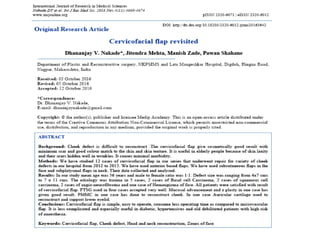
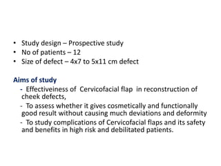
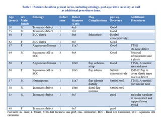
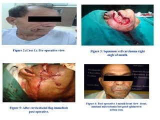
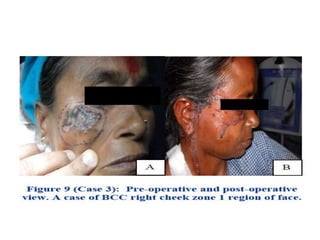
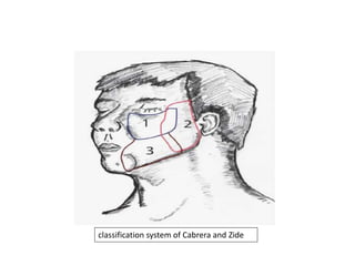
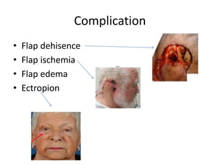
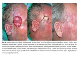
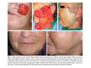
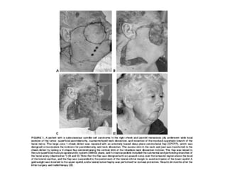
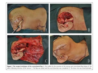
CERVICOFACIAL FALP : REVISTED
Dr Kundan
Department of Surgical oncology
Mahavir Cancer Sansthan
• Study design – Prospective study
• No of patients – 12
• Size of defect – 4x7 to 5x11 cm defect
Aims of study
- Effectiveiness of Cervicofacial flap in reconstruction of
cheek defects,
- To assess whether it gives cosmetically and functionally
good result without causing much deviations and deformity
- To study complications of Cervicofacial flaps and its safety
and benefits in high risk and debilitated patients.
• Large cutaneous defects of the cheek and
external ear present a reconstructive challenge.
• While free tissue transfer has been increasingly
used for such defects, many patients with these
malignancies are poor candidates for lengthy
reconstructive surgery.
• The use of a cervicofacial or cervicothoracic
advancement flap in head and neck surgery was
first described by Esser in 1918 and the technique
has since evolved considerably
classification system of Cabrera and Zide
• Zone 1 is bounded by the inferior orbital margin superiorly, the nasolabial
crease medially and the gingival sulcus inferiorly. The lateral border extends up
the medial border of the sideburn and joins the lateral canthus along an
imaginary line.
• Zone - 2 is bounded by the lateral border of the sideburn descending inferiorly
to the angle of the mandible following it medially, then curving superomedially
over the malar prominence, and finally extending superolaterally toward the
lateral canthus. Zone 2 refers to preauricular defects.
• Zone - 3 is bordered laterally by the midpoint of the mandible, the lower lip
medially, gingival sulcus superiorly, and extends out to its lateral border that
overlaps with zone 2. Zone 3 refers to buccomandibular defects and defines an
area in the lower cheek region.
classification system of Cabrera and Zide
• Reconstruction of cheek lesions will vary according to the
size and location of the defect.
• Small lesions less than 3 cm in diameter may be excised
and closed primarily.
• Lesions larger than this dimension if closed primarily would
cause distortion of the surrounding facial features impairing
cosmesis and perhaps function.
• Cheek defects from 3 to 5 cm in diameter may be closed
satisfactorily with local flaps from within the cheek unit. For
• cheek defects larger than 5 cm, however, one must
generally consider using regional flaps.
• Blood supply of subcutaneous
Cervicofacial flap is random pattern.
• multiple perforators to the skin which
arise from deep facial artery, transeverse
facial artery, and superficial temporal
artery supply to flap by forming vascular
channels.
• Anteriorly based flap derives its blood
supply from submental artery and
perforators of facial artery
• Cervicofacial flap carries dissection below
superficial musculoaponeurotic system in
the face and deep to platysma in the
neck.
• On occasion, a skin graft for the donor site
may be used.
Advantages
• Operative time is short.
• It causes minimum deviations in relations to
important structures around cheek.
• reduce surgical risk in high risk patients like
old age, diabetic patients, un-controlled
hypertension
• It can provide excellent skin colour and
texture match.
Complication
• Flap dehisence
• Flap ischemia
• Flap edema
• Ectropion
References
1. Mustarde JC. Repair and reconstruction in the orbital region. A practical guide. Consultant plastic surgeon, Royal
infirmary, Glasgow. ES. Livingstone Ltd. Edinbirgh and London. 1969;174-5,184-5,189.
2. International Journal of Research in Medical Sciences | November 2016 | Vol 4 | Issue 11 Page 4674
3. Kaplon I, Goldwyn RM. The Versatility of the laterally based Cervico facial flap for cheek repaires Plast Reconst Surg.
1978;61;390-3.
4. Liu FY, Xu ZF. The Versatile applications of cervicofacial and Cervicothoracic rotation flaps in head and Neck surgery.
World J Surg Oncol. 2011;9:135.
5. Coock TA, Israel JM. Cervical rotation flap for midface resurfacing. Arch Otolaryngology head Neck surg. 1991;117:77-82.
6. Bokhari WA, Wang SJ. Modified approach to the cervicofacial rotation flap in the head and neck reconstruction. The
Open Otorhinolaryngology J. 2011;5:18-24.
7. Tann ST, Mac Kinnan. Deep plane cervicofacial flap a useful versatile technique in head and neck surgery. Head and Neck.
2006;28:46-55.
8. Becker FF, Langford FPJ. Deep plane Cervicofacial flap for reconstruction of large defects. Arch Otolaryngology Head and
neck sur. 1996:122(9):997-9.
9. Crow ML, Crow FJ. Resurfacing large cheek defects with rotation flaps from neck. Plast Reconstr Surg. 1976;2;196-200.
10. Austen WG, Parrett BM. The subcutaneous Cervicofacial flap revisited. Ann Plast surg. 2009;62:149-53.
11. Hakim SG, Jacobsen HC. Including the Platysma muscle in a Cervicofacial skin rotation flap to enhance blood supply for
reconstruction of vast orbital and cheek defects, anatomic considerations and surgical techniques. Int J Oral Maxillofac
Surg. 2009;38:1316-9.
12. Mitz V, Peyronie M. The superficial musculo-aponeurotic system (SMAS) in the parotid and cheek area. Plast Reconstr
Surg. 1976;58(1):80-8.
13. Juri J, Juri C. Cheek reconstruction with advancement rotation flaps. Clin Plast Surg. 1981;2: 223-6.
14. Juri J, Juri C. Advancement and rotation of a large Cervicofacial flap for cheek repairs. Plast Reconstr Surg. 1979;5:692-6.
15. Stark RB, Kaplan JM. Rotation of flaps neck to cheek. Plast Reconstr Surg. 1972;3:230-3.

Cervicofacial flap : revisted

  • 1.
    CERVICOFACIAL FALP :REVISTED Dr Kundan Department of Surgical oncology Mahavir Cancer Sansthan
  • 3.
    • Study design– Prospective study • No of patients – 12 • Size of defect – 4x7 to 5x11 cm defect Aims of study - Effectiveiness of Cervicofacial flap in reconstruction of cheek defects, - To assess whether it gives cosmetically and functionally good result without causing much deviations and deformity - To study complications of Cervicofacial flaps and its safety and benefits in high risk and debilitated patients.
  • 7.
    • Large cutaneousdefects of the cheek and external ear present a reconstructive challenge. • While free tissue transfer has been increasingly used for such defects, many patients with these malignancies are poor candidates for lengthy reconstructive surgery. • The use of a cervicofacial or cervicothoracic advancement flap in head and neck surgery was first described by Esser in 1918 and the technique has since evolved considerably
  • 8.
    classification system ofCabrera and Zide • Zone 1 is bounded by the inferior orbital margin superiorly, the nasolabial crease medially and the gingival sulcus inferiorly. The lateral border extends up the medial border of the sideburn and joins the lateral canthus along an imaginary line. • Zone - 2 is bounded by the lateral border of the sideburn descending inferiorly to the angle of the mandible following it medially, then curving superomedially over the malar prominence, and finally extending superolaterally toward the lateral canthus. Zone 2 refers to preauricular defects. • Zone - 3 is bordered laterally by the midpoint of the mandible, the lower lip medially, gingival sulcus superiorly, and extends out to its lateral border that overlaps with zone 2. Zone 3 refers to buccomandibular defects and defines an area in the lower cheek region.
  • 9.
    classification system ofCabrera and Zide
  • 10.
    • Reconstruction ofcheek lesions will vary according to the size and location of the defect. • Small lesions less than 3 cm in diameter may be excised and closed primarily. • Lesions larger than this dimension if closed primarily would cause distortion of the surrounding facial features impairing cosmesis and perhaps function. • Cheek defects from 3 to 5 cm in diameter may be closed satisfactorily with local flaps from within the cheek unit. For • cheek defects larger than 5 cm, however, one must generally consider using regional flaps.
  • 11.
    • Blood supplyof subcutaneous Cervicofacial flap is random pattern. • multiple perforators to the skin which arise from deep facial artery, transeverse facial artery, and superficial temporal artery supply to flap by forming vascular channels. • Anteriorly based flap derives its blood supply from submental artery and perforators of facial artery • Cervicofacial flap carries dissection below superficial musculoaponeurotic system in the face and deep to platysma in the neck. • On occasion, a skin graft for the donor site may be used.
  • 12.
    Advantages • Operative timeis short. • It causes minimum deviations in relations to important structures around cheek. • reduce surgical risk in high risk patients like old age, diabetic patients, un-controlled hypertension • It can provide excellent skin colour and texture match.
  • 13.
    Complication • Flap dehisence •Flap ischemia • Flap edema • Ectropion
  • 31.
    References 1. Mustarde JC.Repair and reconstruction in the orbital region. A practical guide. Consultant plastic surgeon, Royal infirmary, Glasgow. ES. Livingstone Ltd. Edinbirgh and London. 1969;174-5,184-5,189. 2. International Journal of Research in Medical Sciences | November 2016 | Vol 4 | Issue 11 Page 4674 3. Kaplon I, Goldwyn RM. The Versatility of the laterally based Cervico facial flap for cheek repaires Plast Reconst Surg. 1978;61;390-3. 4. Liu FY, Xu ZF. The Versatile applications of cervicofacial and Cervicothoracic rotation flaps in head and Neck surgery. World J Surg Oncol. 2011;9:135. 5. Coock TA, Israel JM. Cervical rotation flap for midface resurfacing. Arch Otolaryngology head Neck surg. 1991;117:77-82. 6. Bokhari WA, Wang SJ. Modified approach to the cervicofacial rotation flap in the head and neck reconstruction. The Open Otorhinolaryngology J. 2011;5:18-24. 7. Tann ST, Mac Kinnan. Deep plane cervicofacial flap a useful versatile technique in head and neck surgery. Head and Neck. 2006;28:46-55. 8. Becker FF, Langford FPJ. Deep plane Cervicofacial flap for reconstruction of large defects. Arch Otolaryngology Head and neck sur. 1996:122(9):997-9. 9. Crow ML, Crow FJ. Resurfacing large cheek defects with rotation flaps from neck. Plast Reconstr Surg. 1976;2;196-200. 10. Austen WG, Parrett BM. The subcutaneous Cervicofacial flap revisited. Ann Plast surg. 2009;62:149-53. 11. Hakim SG, Jacobsen HC. Including the Platysma muscle in a Cervicofacial skin rotation flap to enhance blood supply for reconstruction of vast orbital and cheek defects, anatomic considerations and surgical techniques. Int J Oral Maxillofac Surg. 2009;38:1316-9. 12. Mitz V, Peyronie M. The superficial musculo-aponeurotic system (SMAS) in the parotid and cheek area. Plast Reconstr Surg. 1976;58(1):80-8. 13. Juri J, Juri C. Cheek reconstruction with advancement rotation flaps. Clin Plast Surg. 1981;2: 223-6. 14. Juri J, Juri C. Advancement and rotation of a large Cervicofacial flap for cheek repairs. Plast Reconstr Surg. 1979;5:692-6. 15. Stark RB, Kaplan JM. Rotation of flaps neck to cheek. Plast Reconstr Surg. 1972;3:230-3.