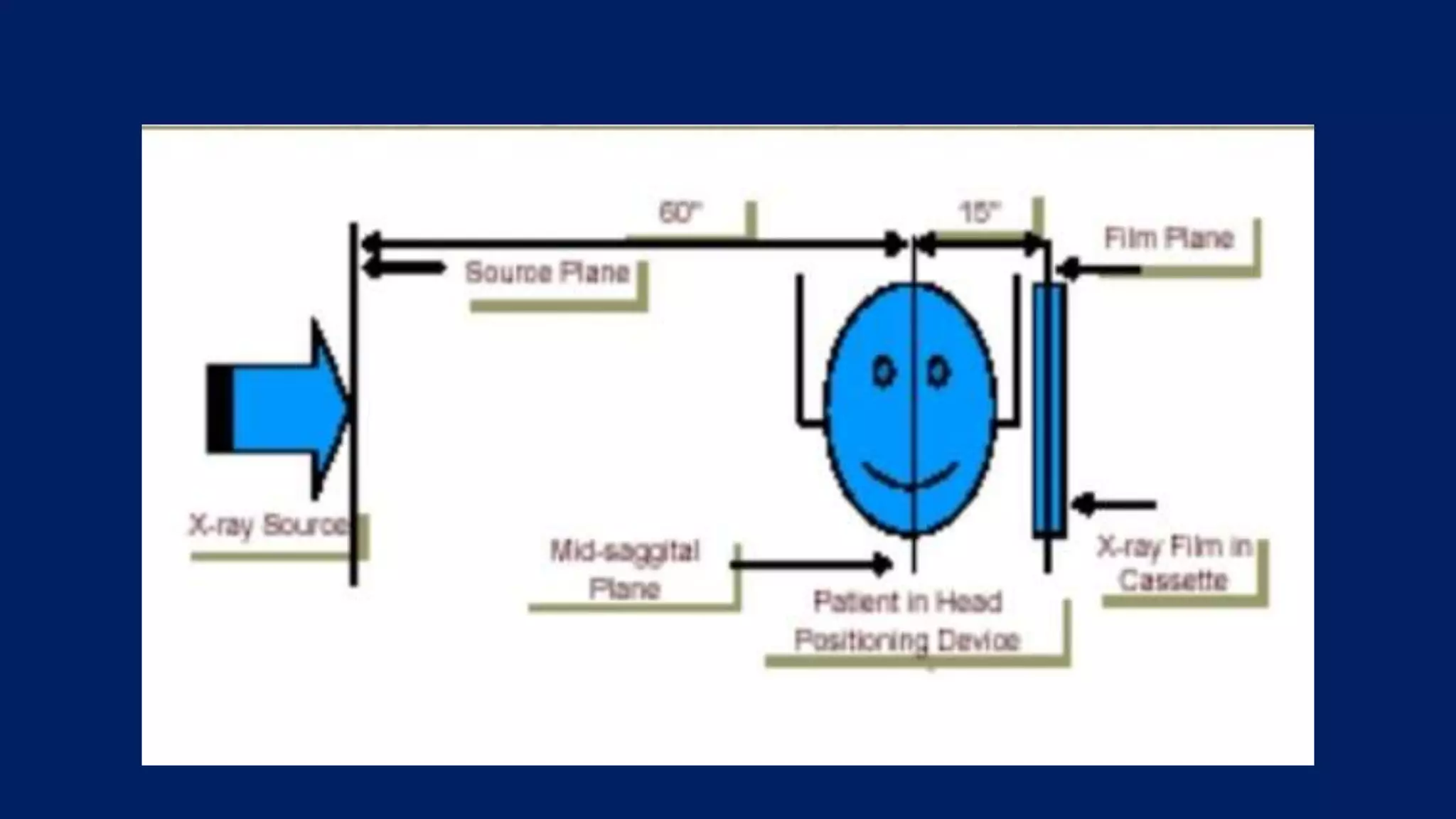
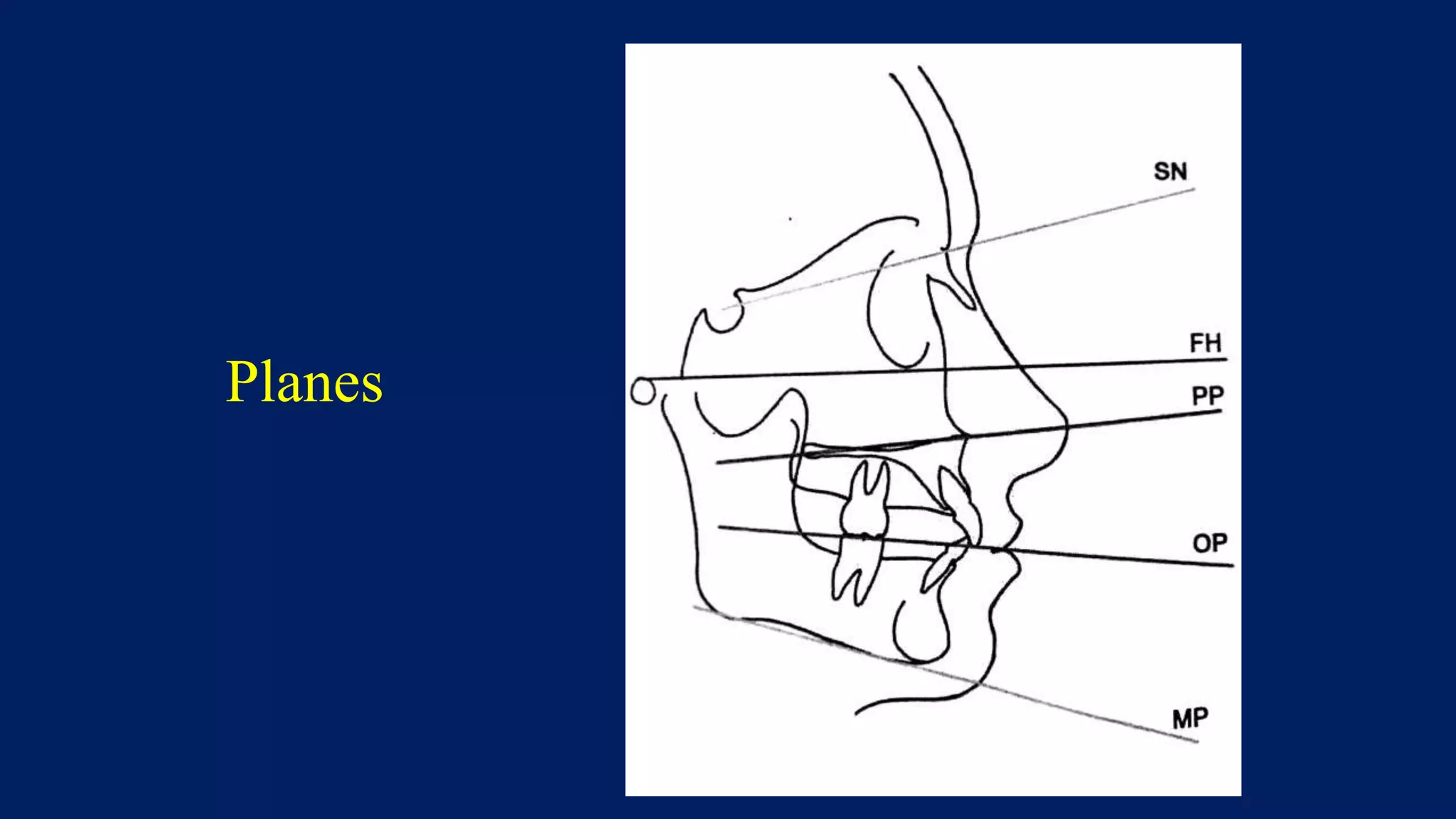
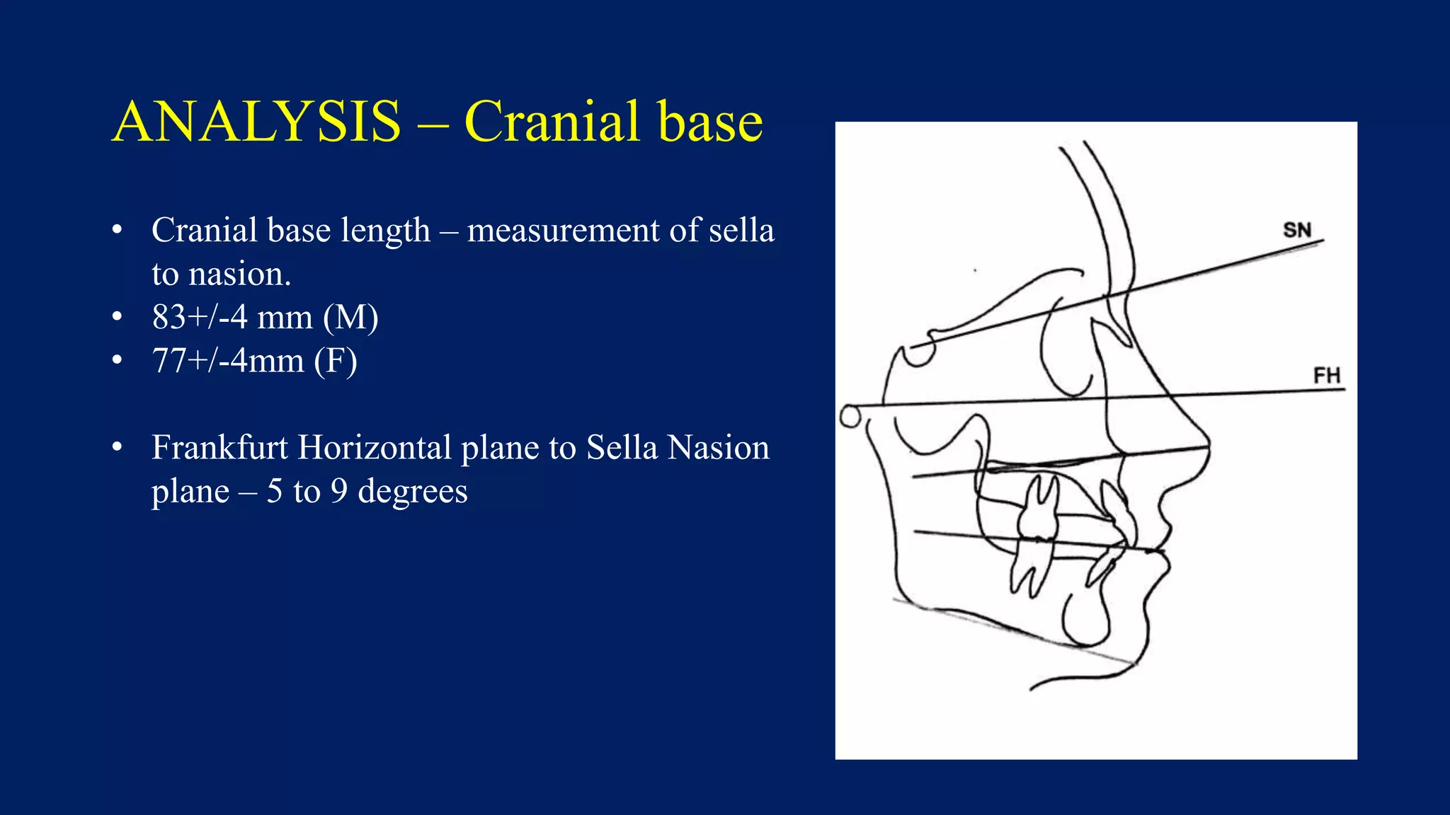
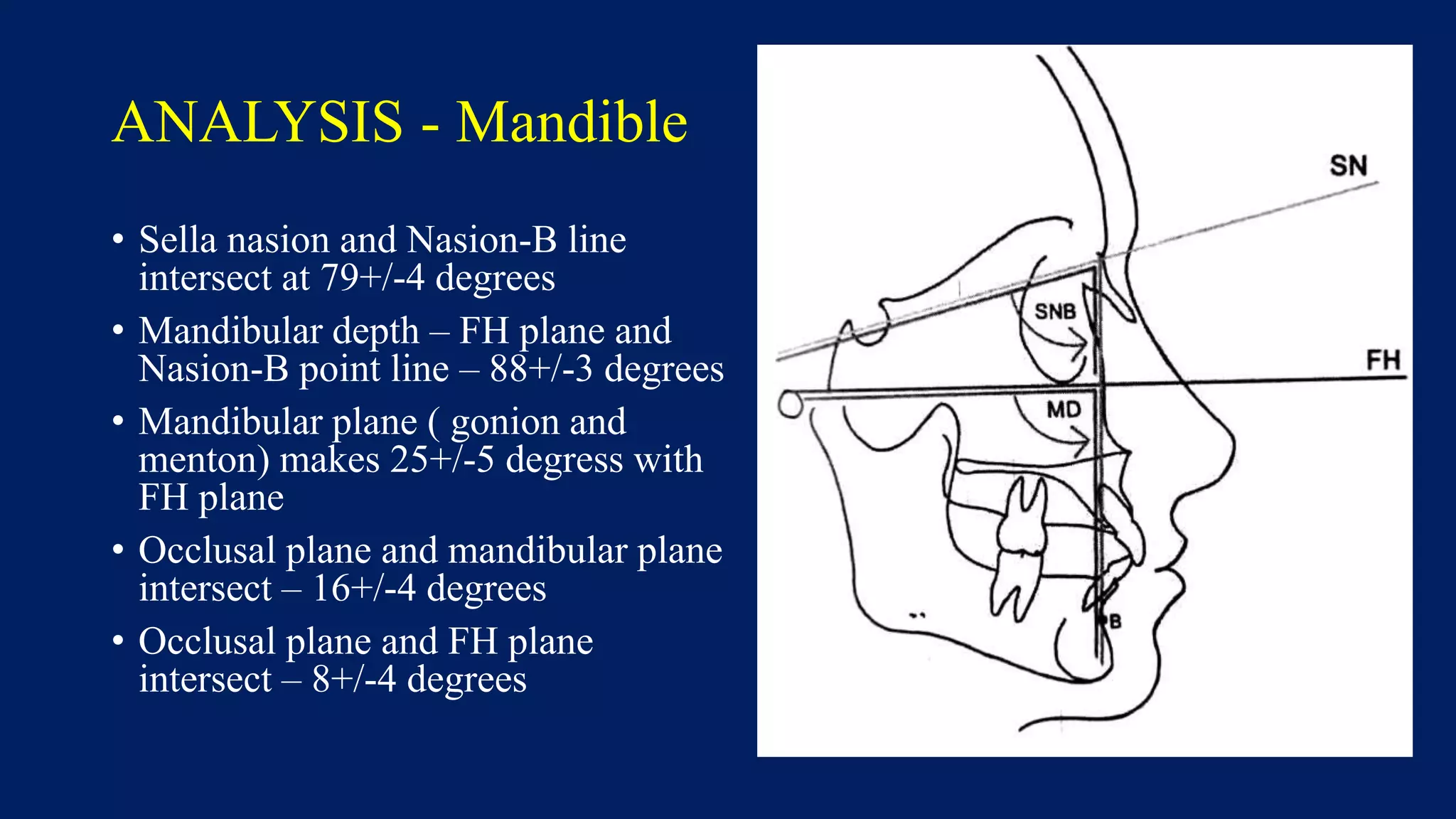
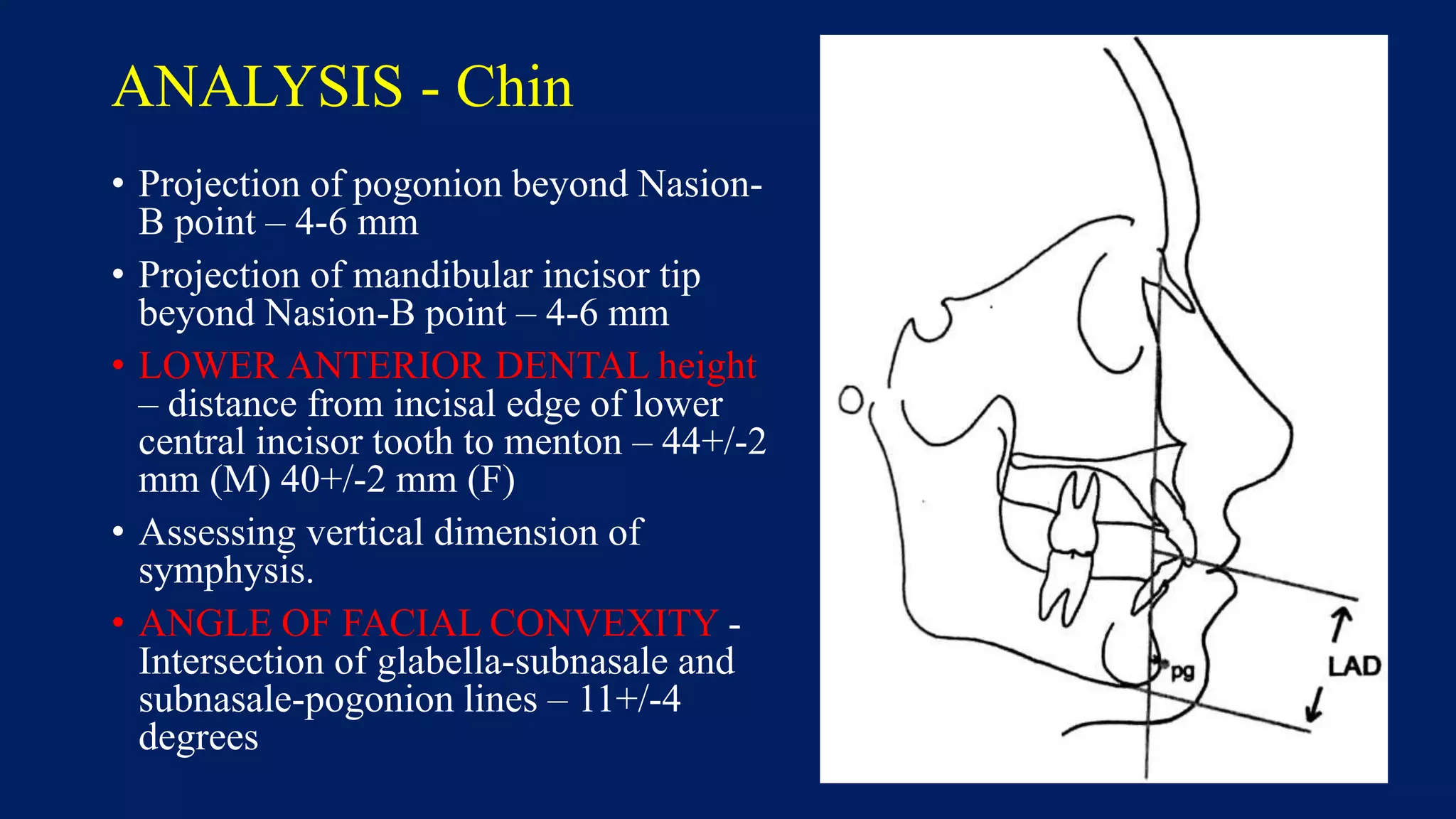
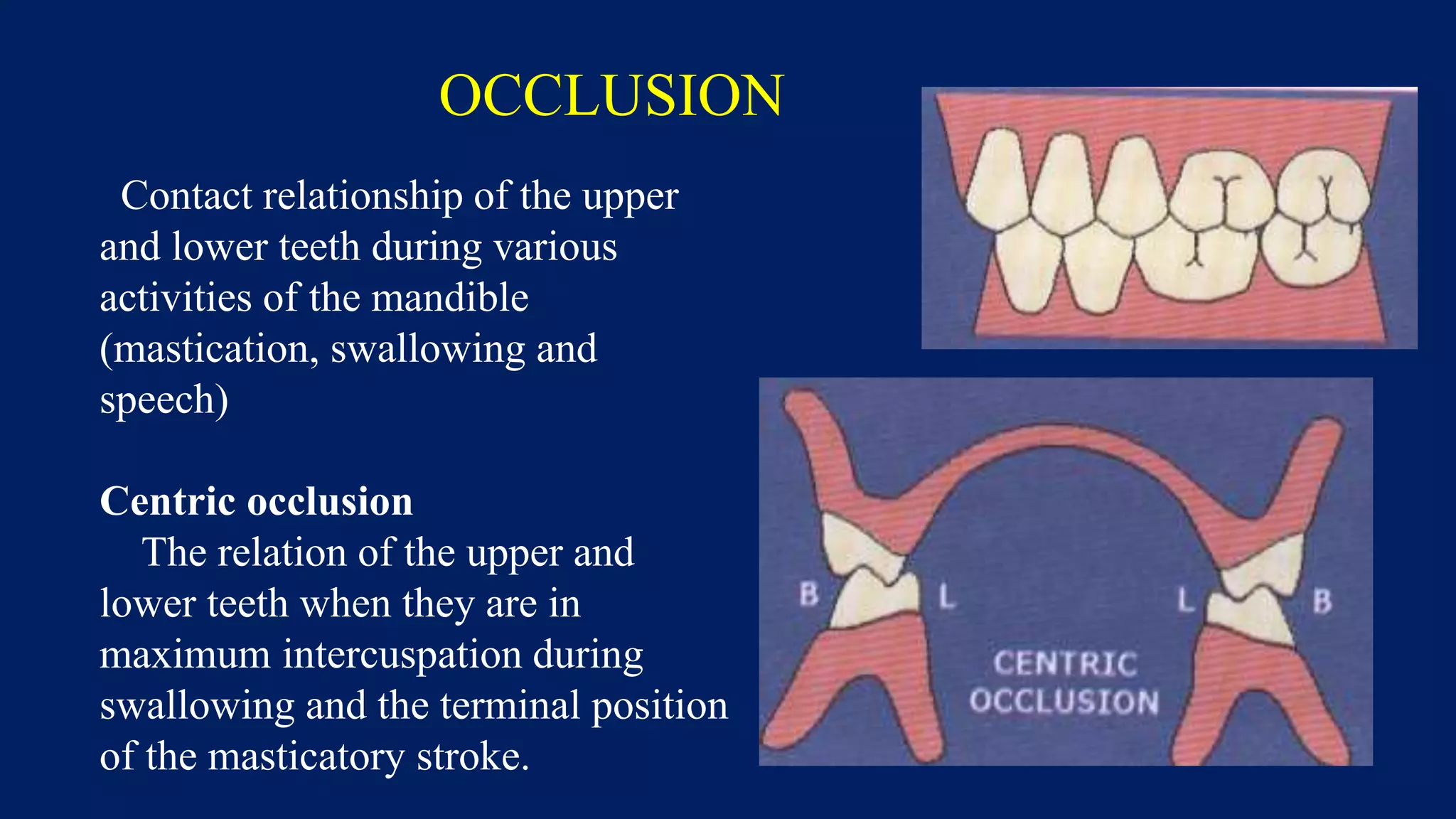
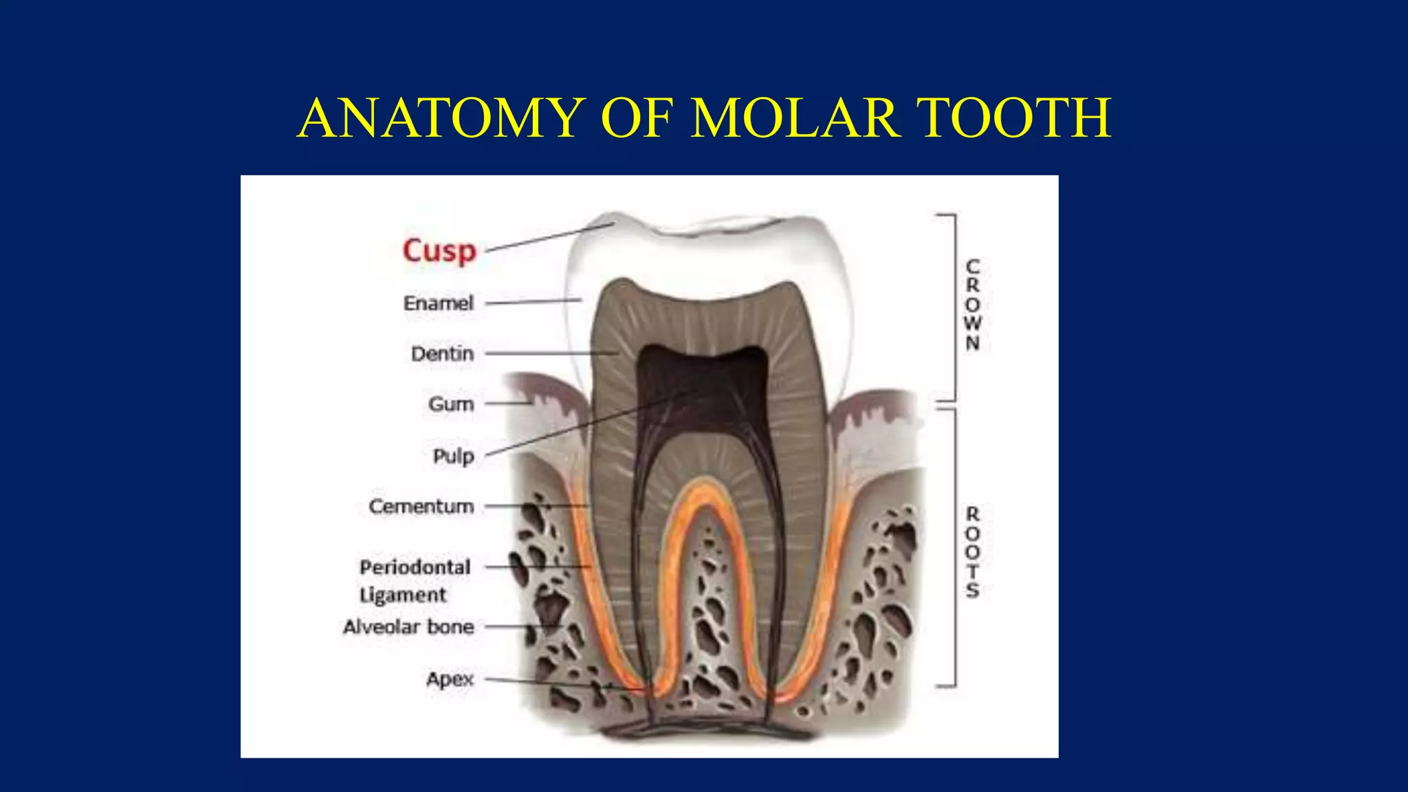
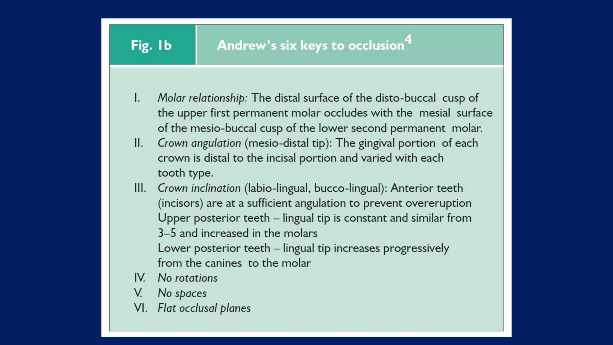
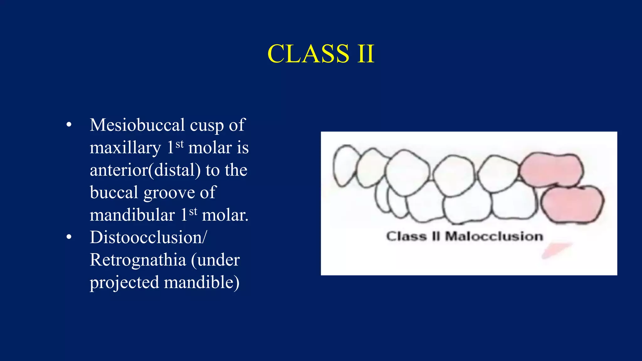
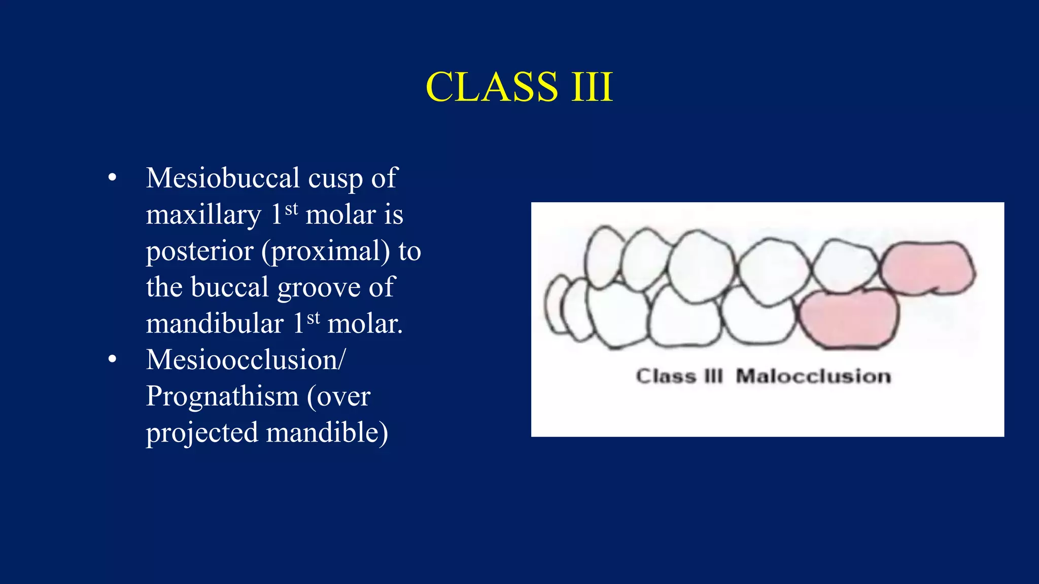
CEPHALOMETRY &
OCCLUSION
Dr. S.Satish Kumar
CEPHALOMETRY
DEFINITION
• Cephalometry – Measurement of the head and face by use of bone
and soft tissue points derived from a specific reproducible radiograph
called a cephalogram.
USES
• Assess bony relationships of face and relationships of jaws and teeth.
• Used for planning orthodontic and surgical treatment.
• Studying growth and development of head and face.
TECHNIQUE
• Standardized lateral head radiograph is done by keeping x-
ray beam, subject and film distances constant.
• Subject head held in position by CEPHALOSTAT.
• Lateral cephalometric head radiograph produced –
CEPHALOGRAM.
• Analysis done by x-ray view box and acetate film for
tracing.
CEPHALOSTAT
CEPHALOGRAM
TRACING A CEPHALOGRAM
• Tape acetate paper to cephalogram, with
rough surface outside.
• Marking
• Frontal bone
• Nasal bone
• Sella turcica – extend anteriorly to frontal
• Orbital rim – backwards J shaped
• Anterior nasal spine and maxillary alveolus
• Incissor and 1st molar
• Mandible with coronoid and condyle
• Soft tissue profile
Landmarks
Planes
ANALYSIS – Cranial base
• Cranial base length – measurement of sella
to nasion.
• 83+/-4 mm (M)
• 77+/-4mm (F)
• Frankfurt Horizontal plane to Sella Nasion
plane – 5 to 9 degrees
ANALYSIS - Orbits
• Porion to orbitale – 74.5+/-5 mm
(M) 70.5+/-4.5 mm (F)
• Line from superior orbital rim to
inferior orbital rim intersects FH
plane at 72+/-9 degrees (M) and
75.8+/-7.6 degrees (F)
ANALYSIS - Maxilla
• Assess anterior-posterior position
of maxilla relative to cranial
base.
• SNA angle
• 82+/-4 degrees
• Maxillary depth – FH plane and
Nasion-A point plane angle
• 90+/-3 degrees
ANALYSIS – Dental relationships
• Occlusal plane of maxilla – FH plane –
angle of 8+/-4 degrees
• Long axis of maxillary central incisor
angle with nasion-A point – 22+/-2
degrees
• Labial surface of maxillary central
incisor projects 4+/-2 mm beyond NA
plane
• Long axis of mandibular incisor angle
with nasion-B point – 20+/-2 degrees.
• Labial surface of mandibular central
incisor projects 4+/-2 mm beyond NB
plane
• Long axes of maxillary and mandible
incissors make angle of 130+/-10
degrees
ANALYSIS - Mandible
• Sella nasion and Nasion-B line
intersect at 79+/-4 degrees
• Mandibular depth – FH plane and
Nasion-B point line – 88+/-3 degrees
• Mandibular plane ( gonion and
menton) makes 25+/-5 degress with
FH plane
• Occlusal plane and mandibular plane
intersect – 16+/-4 degrees
• Occlusal plane and FH plane
intersect – 8+/-4 degrees
ANALYSIS - Mandible
• Angle of mandibular ramus and body
– GONIAL angle.
• 125+/-5 degrees
• Height of ramus – superior head of
condyle to gonion – 68+/-2 mm (M)
60+/-2 mm (F)
• Length of body – Gonion to
Pogonion – 83+/-3 mm (M) 78+/-3
mm (F)
• Ramus height : Body length = 1:1.22
(M) 1:1.27 (F)
ANALYSIS - Chin
• Projection of pogonion beyond Nasion-
B point – 4-6 mm
• Projection of mandibular incisor tip
beyond Nasion-B point – 4-6 mm
• LOWER ANTERIOR DENTAL height
– distance from incisal edge of lower
central incisor tooth to menton – 44+/-2
mm (M) 40+/-2 mm (F)
• Assessing vertical dimension of
symphysis.
• ANGLE OF FACIAL CONVEXITY -
Intersection of glabella-subnasale and
subnasale-pogonion lines – 11+/-4
degrees
FACIAL HEIGHTS
• Upper facial height – nasion to ant nasal spine
• 52-57 mm
• Lower facial height – ant nasal spine to menton
• 63-68 mm
• Ratio – 1:1.2
CEPHALOMETRIC
WORK SHEET
Routine analysis
Planes
• SN plane
• FH plane
• Palatal plane
• Occlusal plane
• Mandibular plane
Measurements
• SNA angle – Maxillary ant-pos
position
• SNB angle – Mandibular ant-pos
position
• Maxillary depth angle
• Mandibular depth angle
• Angle of facial convexity – chin
projection in soft tissue profile
• Max and mand incisors angulation
– jaw position and lip support
OCCLUSION
OCCLUSION
Contact relationship of the upper
and lower teeth during various
activities of the mandible
(mastication, swallowing and
speech)
Centric occlusion
The relation of the upper and
lower teeth when they are in
maximum intercuspation during
swallowing and the terminal position
of the masticatory stroke.
HISTORY
• The founder of contemporary
orthodontics, Edward Angle,
attempted to treat his patients’
occlusion with reference to the
occlusion of a skull displayed on a
shelf in his clinic.
• This skull was affectionately termed
‘Old Glory’.
ANATOMY OF MOLAR TOOTH
NORMAL OCCLUSION - ANGLE
• Mesiobuccal cusp of
maxillary 1st molar
occludes into the buccal
groove of the mandibular
1st molar
OVERBITE & OVERJET
• Overbite
Amount of vertical overlap
maxillary central incisors have
over mandibular central incisors.
• Overjet
Amount of anterior projection
the maxillary central incisors have
beyond the mandibular incisors.
• Normal value – 1-3 mm
• Lack of overbite – Open bite
OPEN BITE
MALOCCLUSION – ANGLE CLASSIFICATION
CLASS I
• Mesiobuccal cusp of
maxillary first molar
occluding into the buccal
groove of the mandibular
1st molar
• Neutroocclusion with
other problems
CLASS II
• Mesiobuccal cusp of
maxillary 1st molar is
anterior(distal) to the
buccal groove of
mandibular 1st molar.
• Distoocclusion/
Retrognathia (under
projected mandible)
CLASS III
• Mesiobuccal cusp of
maxillary 1st molar is
posterior (proximal) to
the buccal groove of
mandibular 1st molar.
• Mesioocclusion/
Prognathism (over
projected mandible)
THANK YOU

Cephalometry and occlusion of teeth

  • 1.
  • 2.
  • 3.
    DEFINITION • Cephalometry –Measurement of the head and face by use of bone and soft tissue points derived from a specific reproducible radiograph called a cephalogram. USES • Assess bony relationships of face and relationships of jaws and teeth. • Used for planning orthodontic and surgical treatment. • Studying growth and development of head and face.
  • 4.
    TECHNIQUE • Standardized lateralhead radiograph is done by keeping x- ray beam, subject and film distances constant. • Subject head held in position by CEPHALOSTAT. • Lateral cephalometric head radiograph produced – CEPHALOGRAM. • Analysis done by x-ray view box and acetate film for tracing.
  • 5.
  • 7.
  • 9.
    TRACING A CEPHALOGRAM •Tape acetate paper to cephalogram, with rough surface outside. • Marking • Frontal bone • Nasal bone • Sella turcica – extend anteriorly to frontal • Orbital rim – backwards J shaped • Anterior nasal spine and maxillary alveolus • Incissor and 1st molar • Mandible with coronoid and condyle • Soft tissue profile
  • 10.
  • 11.
  • 12.
    ANALYSIS – Cranialbase • Cranial base length – measurement of sella to nasion. • 83+/-4 mm (M) • 77+/-4mm (F) • Frankfurt Horizontal plane to Sella Nasion plane – 5 to 9 degrees
  • 13.
    ANALYSIS - Orbits •Porion to orbitale – 74.5+/-5 mm (M) 70.5+/-4.5 mm (F) • Line from superior orbital rim to inferior orbital rim intersects FH plane at 72+/-9 degrees (M) and 75.8+/-7.6 degrees (F)
  • 14.
    ANALYSIS - Maxilla •Assess anterior-posterior position of maxilla relative to cranial base. • SNA angle • 82+/-4 degrees • Maxillary depth – FH plane and Nasion-A point plane angle • 90+/-3 degrees
  • 15.
    ANALYSIS – Dentalrelationships • Occlusal plane of maxilla – FH plane – angle of 8+/-4 degrees • Long axis of maxillary central incisor angle with nasion-A point – 22+/-2 degrees • Labial surface of maxillary central incisor projects 4+/-2 mm beyond NA plane • Long axis of mandibular incisor angle with nasion-B point – 20+/-2 degrees. • Labial surface of mandibular central incisor projects 4+/-2 mm beyond NB plane • Long axes of maxillary and mandible incissors make angle of 130+/-10 degrees
  • 16.
    ANALYSIS - Mandible •Sella nasion and Nasion-B line intersect at 79+/-4 degrees • Mandibular depth – FH plane and Nasion-B point line – 88+/-3 degrees • Mandibular plane ( gonion and menton) makes 25+/-5 degress with FH plane • Occlusal plane and mandibular plane intersect – 16+/-4 degrees • Occlusal plane and FH plane intersect – 8+/-4 degrees
  • 17.
    ANALYSIS - Mandible •Angle of mandibular ramus and body – GONIAL angle. • 125+/-5 degrees • Height of ramus – superior head of condyle to gonion – 68+/-2 mm (M) 60+/-2 mm (F) • Length of body – Gonion to Pogonion – 83+/-3 mm (M) 78+/-3 mm (F) • Ramus height : Body length = 1:1.22 (M) 1:1.27 (F)
  • 18.
    ANALYSIS - Chin •Projection of pogonion beyond Nasion- B point – 4-6 mm • Projection of mandibular incisor tip beyond Nasion-B point – 4-6 mm • LOWER ANTERIOR DENTAL height – distance from incisal edge of lower central incisor tooth to menton – 44+/-2 mm (M) 40+/-2 mm (F) • Assessing vertical dimension of symphysis. • ANGLE OF FACIAL CONVEXITY - Intersection of glabella-subnasale and subnasale-pogonion lines – 11+/-4 degrees
  • 19.
    FACIAL HEIGHTS • Upperfacial height – nasion to ant nasal spine • 52-57 mm • Lower facial height – ant nasal spine to menton • 63-68 mm • Ratio – 1:1.2
  • 20.
  • 21.
    Routine analysis Planes • SNplane • FH plane • Palatal plane • Occlusal plane • Mandibular plane Measurements • SNA angle – Maxillary ant-pos position • SNB angle – Mandibular ant-pos position • Maxillary depth angle • Mandibular depth angle • Angle of facial convexity – chin projection in soft tissue profile • Max and mand incisors angulation – jaw position and lip support
  • 22.
  • 23.
    OCCLUSION Contact relationship ofthe upper and lower teeth during various activities of the mandible (mastication, swallowing and speech) Centric occlusion The relation of the upper and lower teeth when they are in maximum intercuspation during swallowing and the terminal position of the masticatory stroke.
  • 24.
    HISTORY • The founderof contemporary orthodontics, Edward Angle, attempted to treat his patients’ occlusion with reference to the occlusion of a skull displayed on a shelf in his clinic. • This skull was affectionately termed ‘Old Glory’.
  • 25.
  • 26.
  • 27.
    NORMAL OCCLUSION -ANGLE • Mesiobuccal cusp of maxillary 1st molar occludes into the buccal groove of the mandibular 1st molar
  • 29.
    OVERBITE & OVERJET •Overbite Amount of vertical overlap maxillary central incisors have over mandibular central incisors. • Overjet Amount of anterior projection the maxillary central incisors have beyond the mandibular incisors. • Normal value – 1-3 mm • Lack of overbite – Open bite
  • 30.
  • 31.
    MALOCCLUSION – ANGLECLASSIFICATION CLASS I • Mesiobuccal cusp of maxillary first molar occluding into the buccal groove of the mandibular 1st molar • Neutroocclusion with other problems
  • 32.
    CLASS II • Mesiobuccalcusp of maxillary 1st molar is anterior(distal) to the buccal groove of mandibular 1st molar. • Distoocclusion/ Retrognathia (under projected mandible)
  • 33.
    CLASS III • Mesiobuccalcusp of maxillary 1st molar is posterior (proximal) to the buccal groove of mandibular 1st molar. • Mesioocclusion/ Prognathism (over projected mandible)
  • 34.