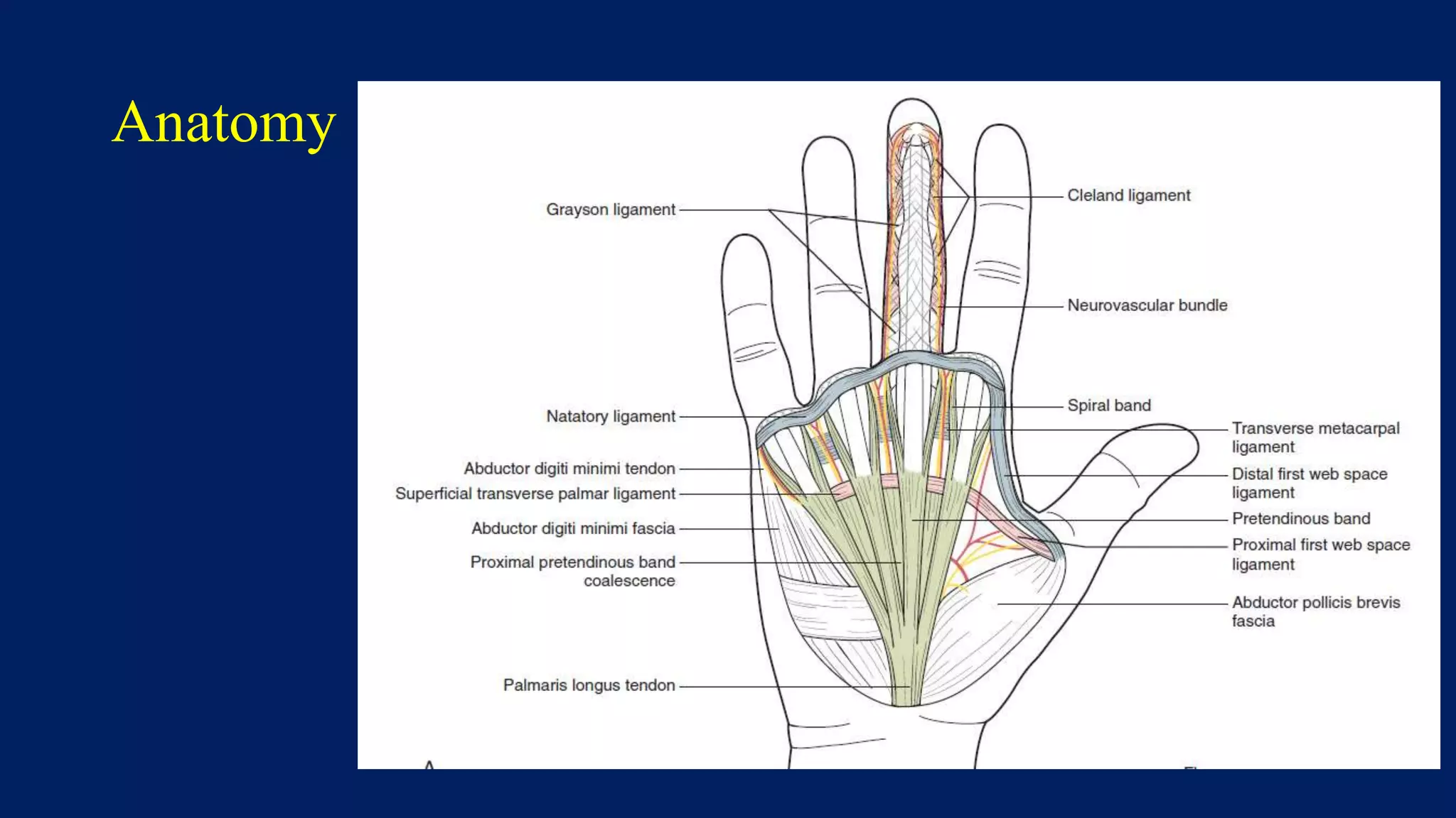
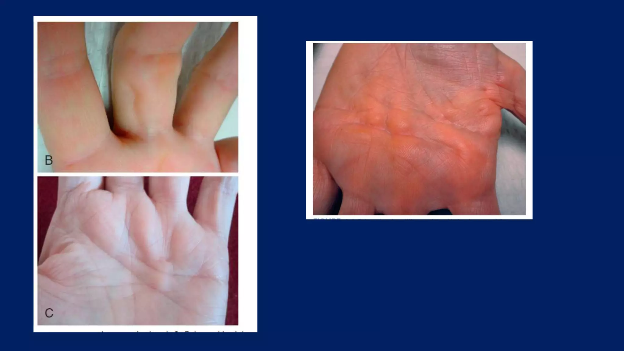
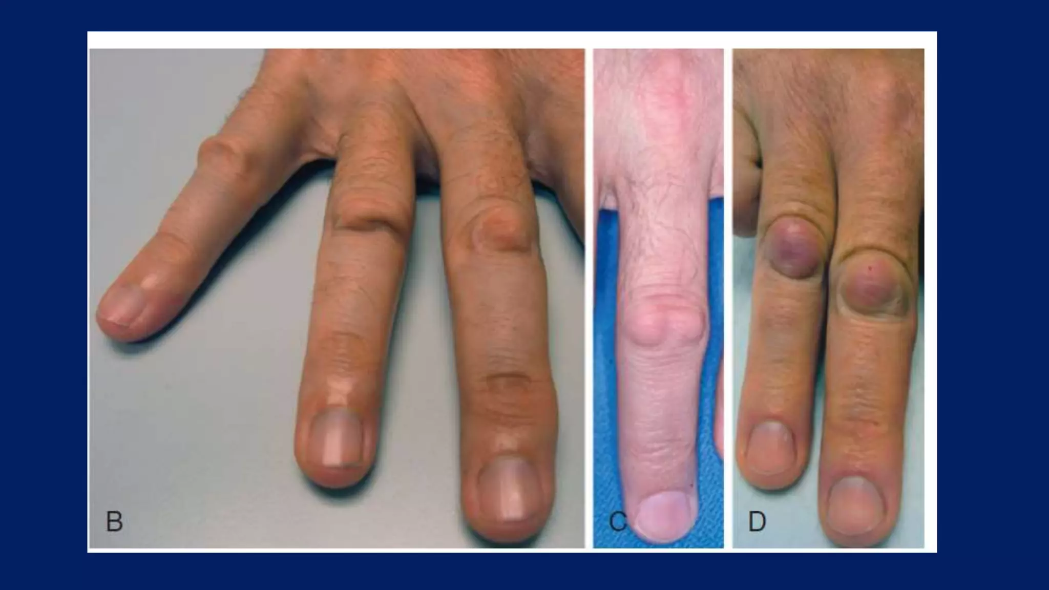
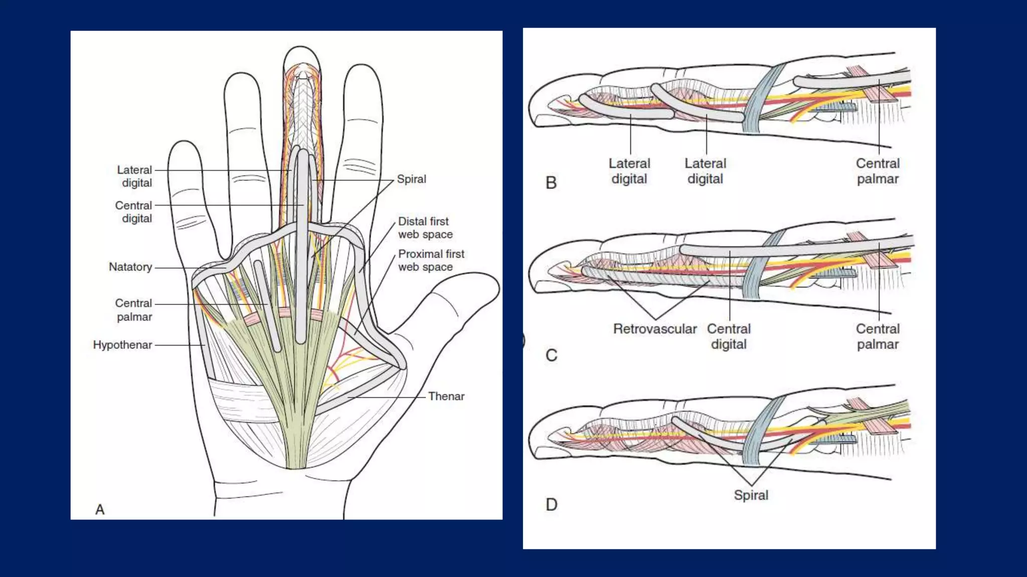
Dupuytren's contracture is a progressive fibroproliferative disease affecting the palmar fascia that can cause fingers to bend into the palm. It typically affects middle-aged Caucasian men and has genetic and environmental risk factors. Physical exam reveals nodules and cords in the palm which can cause finger contractures over time. Treatment options range from minimally invasive needle fasciotomy or collagenase injections to more extensive fasciectomy surgery to remove diseased fascia. Recurrence is common, especially in patients with more severe disease.
Apply gentle pressure proximally
Surgeon: Check distal pulses and capillary refill
If no improvement:
Consider temporary arteriotomy or venous shunt
Delay closure and observe
Flap or graft may be needed
Dupuytren's contracture is a condition causing the fingers to bend towards the palm due to fibrosis of the palmar fascia. It typically affects men over 50 years old and has an autosomal dominant inheritance pattern. Clinically, patients present with nodules and cords in the palm leading to finger contractures. Treatment options include observation for slow cases, radiotherapy in early stages, and surgery involving fasciotomy or fasciectomy. Complete fasciectomy has high recurrence rates while partial fasciectomy balances effectiveness and risk of recurrence. Post-operative splinting and exercises are needed to regain finger extension.
This document provides an overview of hallux valgus, including its anatomy, causes, symptoms, diagnosis, and treatment options. Key points include:
- Hallux valgus is a lateral deviation of the great toe and medial deviation of the first metatarsal. It can cause pain over the bunion.
- Risk factors include heredity, footwear, ligament laxity, and pes planus. Diagnosis involves examining range of motion, deformity, and taking x-rays to measure angles.
- Treatment progresses from footwear modifications and stretching to various surgical procedures depending on severity, including distal soft tissue procedures, osteotomies, and joint fusion or replacement in severe cases.
This case report describes a 21-year old female patient with a 10-year history of habitual left patellar dislocation. Examination found lateral subluxation of the patella in flexion and reduced range of motion. X-rays showed patella baja on the left side and lateral dislocation of the patella. The patient underwent a proximal and distal soft tissue realignment procedure involving lateral release, medial reinforcement, and partial medialization of the patellar tendon. Post-operatively, the patella was centrally located with improved range of motion and stability. Habitual patellar dislocation is rare in adults and can be treated with soft tissue realignment surgery to reinforce the medial structures and release tight
This document discusses outcomes of treating distal tibia fractures using minimally invasive plate osteosynthesis (MIPO) technique. It provides an overview of the MIPO surgical procedure and reviews several studies comparing MIPO to traditional open reduction and internal fixation. The studies found MIPO resulted in high union rates, shorter healing times, and fewer complications like infection compared to open reduction. MIPO preserves the fracture site's blood supply and limits soft tissue damage, allowing for better callus formation and healing.
different type of lower limb amputation with indication, peri-operative care, surgical steps, post op care complication and different type of prosthesis
Tendoachilles rupture and its managementRohan Vakta
Achilles tendon is the strongest tendon of body. There are many causes of its rupture. It can be acute or chronic rupture. Management of chronic rupture by semitendinosus tendon is mentioned here.
Bone growth occurs through two mechanisms: endochondral ossification and intramembranous ossification. Physeal injuries represent 15-30% of fractures in children and commonly involve the phalanges, wrist, and distal tibia. Physeal fractures are classified using the Salter-Harris system from Type I to V based on the location of the fracture line and potential for growth disturbance. Types I-III involve the physis while Types IV-V cross into the epiphysis, increasing the risk of growth arrest or deformity. Proper classification guides treatment to restore anatomy and minimize long term sequelae.
This document discusses congenital vertical talus (CVT), a rare foot deformity. It begins by defining CVT and providing background information. It then describes the anatomy and pathoanatomy of CVT. Key points include that CVT results in an almost vertical talus bone and rigid flatfoot deformity. Treatment involves serial casting and manipulation to prepare for surgery, with the goal of restoring normal anatomical relationships in the foot. Surgical techniques described include open reduction and percutaneous fixation of the talonavicular joint with K-wires. Complications of surgery can include wound issues and stiffness.
This document provides information on fractures of the distal end of the radius bone. It discusses the history, incidence, anatomy, classification, diagnosis, and treatment options for these fractures. Distal radius fractures most commonly result from falls on an outstretched hand and occur in three main age groups. Treatment depends on factors like fracture pattern and stability, and may involve closed reduction with casting or surgical options like percutaneous pinning, plating, or external fixation. The goals of treatment are to restore function, alignment, and stability while avoiding complications.
The document discusses the anatomy and biomechanics of the ankle joint. It describes the ankle joint as a three bone joint composed of the tibia, fibula, and talus. It notes that the talus articulates superiorly with the tibial plafond and posteriorly with the posterior malleolus of the tibia. The lateral articulation is with the malleolus of the fibula. The joint is saddle-shaped and wider anteriorly to accommodate dorsiflexion. Disruption of the ankle mortise can decrease contact area by 42%.
This document provides information on congenital pseudarthrosis of the tibia (CPT), including:
- CPT is a nonunion of the tibia that develops spontaneously in early life and is associated with bowing of the tibia. It occurs more commonly in patients with neurofibromatosis type 1.
- Surgical treatment aims to completely excise fibrous tissue, correct deformity, stimulate bone healing, and properly fix bone fragments. Options include bone grafting, internal fixation, Ilizarov fixation, vascularized fibular grafting, and amputation in severe cases.
- Classification systems divide CPT into types based on clinical features and radiographic findings to help determine prognosis and guide treatment
Dupuytren's contracture is a condition causing the fingers to bend towards the palm. It is caused by a thickening of the fascia in the palm. The initial symptom is a nodule in the palm that develops into cords pulling the fingers into the palm. Surgery is the main treatment and involves excising the diseased tissue and splinting the fingers in extension post-operatively to prevent recurrence of contractures. Complications can include complex regional pain syndrome or recurrence of contractures if splinting is not continued long-term.
Rheumatoid arthritis is a common inflammatory joint disease that can lead to joint deformities. The document discusses common thumb deformities caused by RA like boutonniere and swan neck deformities. Treatment options range from splinting and synovectomy for mild cases to joint fusion or arthroplasty for more severe deformities. Tendon ruptures are also addressed along with surgical techniques like tendon transfers to restore function.
Chondromalacia patellae, also known as runner's knee, is a softening and roughening of the cartilage under the kneecap caused by mechanical overload of the patellofemoral joint. Symptoms include pain in front of or beneath the kneecap that is aggravated by activity like climbing stairs. Examination may reveal tenderness under the kneecap edge or crepitus with knee movement. Conservative treatments include rest, ice, strengthening exercises, and anti-inflammatory medication. Surgery to realign or elevate the patella may be considered if conservative treatments fail after 6 months.
Principle of tension band wiring n its applicationRohit Kansal
1. The tension band technique converts tensile forces into compressive forces through the application of a tension band on the tension side of a bone.
2. Examples of where tension band fixation is commonly used include patella and olecranon fractures, as well as fractures of the greater tuberosity and greater trochanter.
3. Tension band wiring, plating, and external fixation can all function as tension bands by applying a compressive force across a fracture to promote healing.
Charcot joint or neuropathic joint are destructed joint occurs in Diabetes, syphilis, syringomyelia , leprosy, AMLS, Peripheral neuropathy and any condition leads to impair sensation of peripheral part of body
This document discusses radial nerve injury, including its anatomy, causes, clinical presentation, diagnostic workup, and management. It describes the radial nerve's course from the brachial plexus into the arm and forearm. Radial nerve injuries can be caused by fractures, compression, or traction injuries. Clinical examination involves assessing motor function of wrist and finger extensors and sensory function on the back of the hand. Management includes nonsurgical treatment, nerve repair or grafting, and tendon transfers in chronic cases. The goal of treatment is to restore wrist and finger extension through nerve regeneration or reconstruction of function.
Dupuytren's contracture is characterized by abnormal thickening of the fascia in the palm, which forms nodules and cords that cause fingers to bend into the palm. It is most common in Caucasian males over 50. The contracture is caused by an excess of myofibroblasts that synthesize collagen and contract tissues. The disease follows the anatomical planes of the palmar fascia. Treatment involves surgical release of the tightened fascia.
This document discusses mallet finger injuries, which involve disruption of the extensor tendon mechanism at the distal interphalangeal joint. It covers the anatomy of the finger extensor mechanism, classification of mallet finger injuries, clinical evaluation, treatment options including nonsurgical management with splinting and surgical repair or fixation, and management approaches for different types of acute mallet finger injuries.
This document provides an overview of clubfoot (congenital talipes equinovarus), including:
1. The definition, incidence, causes, and typical deformities seen in clubfoot.
2. Evaluation methods like the Pirani scoring system and radiographic assessment.
3. Treatment approaches like the Ponseti method of serial casting and bracing, as well as surgical options like the McKay procedure when non-operative treatment fails.
4. Post-operative casting protocols and complications that can arise from treatment.
De Quervain's tenosynovitis is an inflammation of the tendon sheaths of the abductor pollicis longus and extensor pollicis brevis muscles in the wrist. It commonly affects women ages 30-50 and is caused by repetitive motions like knitting or computer use that strain the thumb and wrist. Symptoms include pain on the radial side of the wrist worsened by thumb movement. Conservative treatment involves splinting, anti-inflammatories, corticosteroid injections, and physical therapy exercises. Surgery may be considered if symptoms persist after several weeks of conservative care.
Carpal tunnel syndrome is caused by compression of the median nerve as it passes through the carpal tunnel in the wrist. It affects around 10% of the general population. Symptoms include numbness, tingling, and pain in the hand and fingers innervated by the median nerve. Diagnosis is based on physical exam findings like Tinel's sign and Phalen's maneuver, as well as electrodiagnostic testing. Conservative treatment involves splinting and anti-inflammatory medications while surgical treatment involves cutting the transverse carpal ligament to relieve pressure on the median nerve. Recurrence can occur due to incomplete release of pressure or scarring.
The document discusses ulnar nerve palsy and tendon transfers used to treat it. It begins by describing the anatomy of the ulnar nerve and its motor and sensory functions. It then discusses clinical findings associated with ulnar nerve injuries at different locations. Various tendon transfers are summarized that aim to restore small and ring finger flexion, key pinch, correct clawing, and improve grip strength for patients with ulnar nerve palsy. These include transferring forearm muscles like the ECRB to restore key pinch or correct clawing. The modified Stiles-Bunnell procedure is also summarized, which uses the middle finger superficialis tendon to dynamically correct clawing during finger flexion.
(9)external fixation indications and techniques(bonatus)Drpraveen Kumar
External fixation involves placing pins or wires connected to bars outside the skin to stabilize bone fragments. It provides relative stability and healing with callus formation. Advantages include minimal damage to soft tissues and blood supply. Disadvantages include restricted motion and pin tract infections. Indications include open fractures, fractures with soft tissue compromise, periarticular fractures, polytrauma, pelvic fractures, and children's fractures. Constructs can be uni-plane, bi-plane, multi-plane, or rings. Stability increases with larger pins, more pins closer to fractures, more bars, and smaller rings. Complications include neurovascular injury, pin loosening, pin tract infections, joint stiffness, malalignment, and malunion/non
De Quervain's tenosynovitis is a condition affecting the tendons that control thumb movement at the wrist. It causes pain and swelling on the outer side of the wrist. Nonsurgical treatments include splinting, anti-inflammatory medication, and corticosteroid injections into the affected tendon sheath. If conservative treatments fail after 6 months, surgical release of the tendon sheath is recommended to relieve pressure and promote healing. The surgery involves cutting the fibrous roof of the tendon compartment to decompress the tendons.
This document provides information about Perthes' disease, including:
- It is characterized by avascular necrosis of the femoral head in children.
- Risk factors include being male and between ages 5-10 years old.
- Imaging studies like x-rays are used to diagnose and monitor the stages of avascular necrosis, fragmentation, ossification, and remodeling.
- Differential diagnosis depends on whether the condition is unilateral or bilateral.
- Treatment aims to prevent deformity through nonsurgical or surgical methods depending on the severity.
Dupuytren's disease is a condition where the normal palmar and digital fascia is replaced by abnormal fibrous tissue containing immature fibroblasts. It commonly affects the ring finger. Risk factors include family history, smoking, and diabetes. In advanced stages, it can cause hand contractures. Treatment options range from observation to corticosteroid injections to surgery like fasciectomy or dermofasciectomy to release contractures. Post-surgical complications can include recurrence, nerve damage, or wound healing issues.
This document provides information on Dupuytren's disease, including:
- It is a progressive fibromatosis of the palmar fascia that causes nodule formation and finger contractures.
- Risk factors include Northern European ancestry, male sex, alcohol abuse, liver disease, and smoking.
- Treatment options range from observation for mild cases to needle aponeurotomy, collagenase injections, fasciectomies, and dermatofasciectomies depending on the severity and location of contractures.
- The goal of treatment is to release contractures through minimally invasive or surgical techniques while preventing recurrence through splinting and rehabilitation.
This document provides information about Dupuytren's contracture, including its history, definition, epidemiology, associated conditions, pathogenesis, treatment options, and complications. Specifically, it describes how Dupuytren's contracture results from abnormal proliferation of fibroblasts in the palmar fascia, which can cause fingers to bend into the palm. Treatment involves nonsurgical options like collagenase injections or surgery to release contracted tissues through techniques like fasciectomy. Postoperative rehabilitation with splinting and range of motion exercises aims to prevent recurrence of contractures.
Dupuytren's disease is a condition where the palmar fascia becomes contracted, most commonly causing fingers to bend towards the palm. It is more common in older men of Northern European descent and risk factors include alcohol use, smoking, diabetes and manual labor. The disease involves proliferation of fibroblasts and collagen deposition within the fascia. Surgical treatment aims to release the contracted fascia and can include techniques such as fasciectomy, dermofasciectomy with skin grafting or flaps to replace skin if shortening has occurred due to the disease. Nonsurgical options provide more limited benefit.
Dupuytren's disease is a condition characterized by abnormal thickening and contraction of the palmar fascia in the hand. It commonly affects middle-aged men of northern European descent and is associated with conditions like alcohol use, smoking, and diabetes. The disease pathology involves proliferation of fibroblasts and myofibroblasts that deposit excess collagen in the palmar fascia, leading to nodule formation and cord-like contractures of the fingers.
This document discusses diseases of the salivary glands, focusing on inflammatory disorders of the parotid gland. It describes various causes of parotid inflammation including viral infections like mumps, bacterial infections, HIV-associated sialadenitis, and obstructive disorders. It also discusses benign conditions such as sialadenosis and Sjogren's syndrome. The document outlines the indications for and steps of parotid surgery including superficial parotidectomy, total parotidectomy, and radical parotidectomy. Post-surgical complications like Frey's syndrome are also mentioned.
The parapharyngeal space is a potential space located in the neck that contains important structures like the carotid artery and cranial nerves. Tumors in this space can be benign or malignant, with the most common types being salivary gland tumors in the prestyloid space and neurogenic tumors in the retrostyloid space. Imaging like CT and MRI are used to determine the location and characteristics of the tumor. Surgical excision is typically the primary treatment, with the surgical approach depending on factors like size and involvement of surrounding structures. Observation or radiation therapy may be options for certain patients who cannot undergo surgery.
Detail case discussion of dupuytren's contracture including clinical aspect and theoretical aspects.
Dupuytren's is progressive superficial palmar fibromatosis, involves superficial palmar fascia of hand and causes contracture and severe disability in advance disease.
Case discussion done under Platiquest channel.
dupuytrens contracture and its interventionSundasIrshad1
Dupuytren's disease is characterized by thickening and contracture of the palmar fascia. It commonly affects the hands of older white males, causing fingers to bend into the palm. Surgical treatment involves selectively removing diseased fascia through small incisions while preserving healthy tissue and skin integrity. Post-operative management depends on the surgical technique used and skin condition. Recurrence remains a challenge due to residual diseased fascia and skin contractures.
Thyroid swelling and management. In detail case discussion of thyroid swelling and its management. Details of examination as well included in the slide.
This document discusses various benign, premalignant, and malignant lesions of the penis. It covers the etiology, risk factors, diagnosis, staging, and management options for penile cancer including surgery, radiotherapy, and chemotherapy. The main types of penile cancer are squamous cell carcinoma (>95%) and mesenchymal tumors (<3%). Surgical options range from circumcision to partial or total penectomy. Radiotherapy can be delivered via brachytherapy or external beam radiation. Management depends on tumor stage, size, histology, and patient preferences regarding organ preservation.
This document summarizes various benign, premalignant, and malignant lesions of the penis. It discusses the etiology, diagnosis, staging, and management of penile carcinoma. The main types of penile carcinoma are squamous cell carcinoma (>95%) and mesenchymal tumors (<3%). Risk factors include phimosis, number of sexual partners, and HPV infection. Diagnosis involves physical exam, imaging, and histology. Treatment options depend on the location, size, stage, and grade of the tumor, and may include surgery, radiation therapy, chemotherapy, or a combination. Surgery ranges from circumcision to penectomy, while radiation includes brachytherapy and external beam radiation therapy. Prognosis depends on
Uterine fibroids by oouth unit b medical students o&gTolulope Balogun
Mrs. AA is a 42 year old woman presenting with a large abdominal mass but no heavy bleeding symptoms. Examination and ultrasound reveal two large fibroids. Uterine fibroids are benign muscle cell tumors that are very common among women over 25, especially African women. They can cause heavy periods, abdominal pain or pressure, and infertility. Treatment options depend on symptoms and fertility desires, and include medical management to shrink fibroids, myomectomy or hysterectomy for surgical removal, or uterine artery embolization.
Medulloblastoma is the most common malignant pediatric brain tumor. It arises in the midline cerebellum. Diagnosis involves imaging and biopsy. Treatment involves maximal surgical resection followed by craniospinal radiation and chemotherapy for high-risk patients. Prognosis depends on age, extent of resection, and presence of metastasis. Long-term surveillance is needed due to risk of recurrence within several years of diagnosis.
a comprehensive presentation on the subject of spinal dysraphism and spina bifida and its neurosurgical management as well as the management of its various other types
Necrotizing soft tissue infections (NSTIs) are severe infections involving the skin and soft tissues that cause necrosis. They range from potentially life-threatening types caused by streptococci and clostridia to less severe types caused by staphylococci. Clinical features include pain out of proportion to exam findings, swelling beyond the erythema, and systemic signs of sepsis. Diagnosis involves a high clinical suspicion based on history and exam. Treatment requires aggressive surgical debridement of necrotic tissue, broad-spectrum antibiotics, and supportive care. The prognosis depends on how quickly treatment is started, with early diagnosis and intervention being crucial to survival.
Cutaneous fibromatosis.pptx (Clinical, Histopathological and Treatment)DR. MOHNISH SEKAR
Fibromatoses are a group of benign connective tissue tumors characterized by the infiltrative, aggressive proliferation of well-differentiated fibroblasts, leading to frequent local recurrence. Within this heterogeneous disease group, superficial fibromatoses show slower growth and more benign infiltration of surrounding tissues than deep fibromatoses. Superficial fibromatoses relevant to dermatology include palmar, plantar, and penile fibromatosis, knuckle pads, pachydermodactyly and infantile digital fibromatosis.
Clinical features,presentation,clinical and surgical management of TMJ disloc...EUROUNDISA
This document discusses various types of temporomandibular joint (TMJ) disorders including dislocation, arthritis, and infection. It provides details on the classification, causes, clinical presentation, diagnosis, and treatment of TMJ dislocation. It also discusses the epidemiology, clinical manifestations, radiographic changes, and treatment of rheumatoid, psoriatic, juvenile, and septic arthritis of the TMJ. Gout and pseudogout arthritis are also briefly mentioned.
This document discusses tenosynovitis, including its definition, etiology, prognosis, pathophysiology, history, physical examination findings, workup, treatment, and postoperative care. Tenosynovitis is inflammation of the tendon sheath that can be caused by overuse, infection, or inflammatory conditions like rheumatoid arthritis. Physical exam may reveal tenderness, swelling, or limited range of motion. Treatment depends on the cause but may include rest, splinting, anti-inflammatories, corticosteroid injections, or surgery. Prognosis is generally good if treated early without comorbidities, while complications can include adhesion formation or tendon rupture if left untreated.
This document summarizes benign breast disorders. It begins with embryology and anatomy of the breast. It then discusses various benign breast conditions such as fibroadenomas, breast cysts, periductal mastitis, papillomas and sclerosing adenosis. It provides details on clinical features, investigations, diagnosis and management of these common benign breast disorders. Specific imaging findings and histopathological characteristics are also highlighted. The document serves as a comprehensive review of benign breast conditions for medical residents.
This document discusses contralateral C7 transfer, a surgical procedure where the unaffected C7 nerve root is used to reinnervate nerves on the affected side for patients with brachial plexus injuries and total root avulsions. It can be done in a single stage by directly coapting the C7 to the recipient nerve, or in a staged procedure using a vascularised ulnar nerve graft to allow for nerve growth. The C7 is used because its muscles are cross-innervated, minimizing function loss on the normal side. Careful patient selection and post-operative physical therapy are important for achieving reinnervation and return of motor function on the affected side.
Thumb hypoplasia - congenital hand IIISatish Kumar
This document discusses different types of congenital thumb hypoplasia, including 10 specific types. It covers the development and anatomy of the thumb, provides detailed descriptions of each type of hypoplasia, and discusses approaches to management and treatment, including surgical techniques to address deficiencies in the first webspace, instability of joints, and lack of mobility. The goal of treatment is to reconstruct a functional thumb with a stable joint, adequate webspace, and motor function for pinching and grasping.
Craniofacial Microsomia and Hemifacial AtrophySatish Kumar
Craniofacial microsomia involves congenital malformations of structures arising from the first and second branchial arches. It can cause deformities of the orbit, mandible, ear, nerves and soft tissues that range from mild to severe. The etiology is unclear but may involve vascular perturbations, teratogen exposure or neural crest pathology. Treatment involves surgery to correct deformities such as mandibular distraction, commissuroplasty or facial reanimation procedures.
Cephalometry involves taking standardized lateral radiographs of the head to measure and analyze the relationships between craniofacial structures and teeth. Key points analyzed include angles relating the maxilla and mandible to cranial landmarks, dental angulation and relationships, and facial heights and proportions. Occlusion refers to the contact relationship between teeth during jaw movements. Normal occlusion involves the maxillary first molar cusps fitting into grooves of the mandibular molars. Malocclusions are classified as Class I, II, or III depending on the anteroposterior positioning of the maxillary molars relative to the mandibular molars and jaws.
Botulinum toxin is produced by Clostridium botulinum bacteria. It works by blocking the release of acetylcholine at neuromuscular junctions, preventing muscle contraction. It has been used since the 1980s to treat medical conditions involving muscle overactivity like strabismus and dystonia. In the 1990s, its use was explored for cosmetic purposes to reduce facial wrinkles. The FDA approved its use for frown lines in 2002 and excessive underarm sweating in 2004. It is injected into specific facial muscles to weaken them and smooth wrinkles. Common sites include the glabella, forehead, crow's feet, bunny lines, marionette lines and platysmal bands. Potential complications include
This document provides information on facelift procedures. It discusses facial aging changes and facelift anatomy. Several facelift techniques are described including subcutaneous, superficial musculoaponeurotic system (SMAS), extended SMAS, lateral SMAS-ectomy, platysma-SMAS plication, deep plane, short scar, and secondary facelifts. Neck rejuvenation techniques like submental dissection and platysmaplasty are also outlined. Potential complications are noted. The goal of facelifts is to lift tissues while avoiding an operated look through careful surgical planning and technique.
This document provides an overview of liposuction procedures. It discusses the basic science and anatomy related to liposuction, patient assessment and selection criteria, surgical techniques including tumescent liposuction and different modalities like ultrasound-assisted and laser-assisted liposuction. Post-operative care and potential complications are also reviewed. The document aims to inform practitioners on best practices for liposuction to achieve optimal contouring results.
This document summarizes the anterolateral thigh (ALT) flap, including:
1) The ALT flap has reliable blood supply from branches of the lateral circumflex femoral artery and can provide skin, muscle, fascia and a long pedicle.
2) The anatomy is variable but the flap is typically based on musculocutaneous perforators from the descending branch of the lateral circumflex femoral artery.
3) The ALT flap has been used successfully for reconstruction of the head and neck, esophagus, abdomen, breast, extremities and perineum, with success rates over 95% and versatility in design.
4) Advantages include reliable anatomy, long pedicle, versatility
Anatomy of eyelid and eyelid reconstructionSatish Kumar
The document provides an overview of eyelid anatomy and techniques for eyelid reconstruction. It describes the layers, muscles, nerves and blood supply of the eyelid. Functions include spreading secretions, tears and blinking. Reconstruction methods are classified by defect size and include direct closure, lateral cantholysis, semicircular flaps, lid switch flaps, Cutler-Beard procedure, forehead flaps, Tripier flaps, Hughes flaps and cheek rotation flaps to reconstruct the anterior or posterior lamella. The goal is to reestablish eyelid function and protection of the eye while achieving reasonable cosmesis.
1. The gastrocnemius muscle consists of medial and lateral heads that are supplied by the medial and lateral sural arteries respectively.
2. The medial gastrocnemius flap is most commonly used due to its large size and reliable vascular pedicle. It can be raised as a muscle or musculocutaneous flap to cover defects of the upper leg and knee.
3. The lateral gastrocnemius flap is smaller but can be used for smaller defects of the upper lateral leg and knee. Both flaps have consistent anatomy and can be reliably elevated based on the dominant sural artery pedicles.
1) Fractures of the zygoma are commonly caused by road traffic accidents and assaults, with the left side more frequently involved than the right.
2) Classification systems categorize fractures based on the location and degree of displacement. Surgical approaches are determined by the fracture pattern and may involve extraoral or intraoral incisions.
3) Treatment involves closed or open reduction and fixation using methods like K-wires, plates and screws to properly align the zygomatic arch, frontal bone sutures, and orbital rim/floor. Complications can include sinusitis, malunion, diplopia or nerve numbness if not addressed correctly.
Wound healing involves three overlapping phases: inflammatory, proliferative, and remodeling. In the inflammatory phase, fibrin and a provisional matrix form over the wound to prevent infection. In the proliferative phase, re-epithelialization occurs to regrow skin cells over the wound. Granulation tissue forms below the wound as new blood vessels and extracellular matrix develop. Collagen is deposited and remodeled. Finally, in the remodeling phase, collagen is reorganized and cross-linked to increase wound strength as it matures over 6-12 months. Factors like age, nutrition, smoking, and infection can impact wound healing.
Transplantation involves grafting tissue from one site to another or from one individual to another. Major advances include the first kidney transplant in 1955 and hand transplant in 1998. Survival depends on preventing rejection through immunosuppression. The goal is transplantation is to replace failed organs and tissues. Rejection occurs via the major histocompatibility complex and immune cells such as T cells, B cells, macrophages, and natural killer cells. Immunosuppression drugs help prevent rejection and include corticosteroids, calcineurin inhibitors, antiproliferatives, monoclonal antibodies, and mTOR inhibitors. Future areas of focus include inducing immunological tolerance through clonal deletion, anergy, or suppression.
Basavarajeeyam is a Sreshta Sangraha grantha (Compiled book ), written by Neelkanta kotturu Basavaraja Virachita. It contains 25 Prakaranas, First 24 Chapters related to Rogas& 25th to Rasadravyas.
These lecture slides, by Dr Sidra Arshad, offer a quick overview of the physiological basis of a normal electrocardiogram.
Learning objectives:
1. Define an electrocardiogram (ECG) and electrocardiography
2. Describe how dipoles generated by the heart produce the waveforms of the ECG
3. Describe the components of a normal electrocardiogram of a typical bipolar lead (limb II)
4. Differentiate between intervals and segments
5. Enlist some common indications for obtaining an ECG
6. Describe the flow of current around the heart during the cardiac cycle
7. Discuss the placement and polarity of the leads of electrocardiograph
8. Describe the normal electrocardiograms recorded from the limb leads and explain the physiological basis of the different records that are obtained
9. Define mean electrical vector (axis) of the heart and give the normal range
10. Define the mean QRS vector
11. Describe the axes of leads (hexagonal reference system)
12. Comprehend the vectorial analysis of the normal ECG
13. Determine the mean electrical axis of the ventricular QRS and appreciate the mean axis deviation
14. Explain the concepts of current of injury, J point, and their significance
Study Resources:
1. Chapter 11, Guyton and Hall Textbook of Medical Physiology, 14th edition
2. Chapter 9, Human Physiology - From Cells to Systems, Lauralee Sherwood, 9th edition
3. Chapter 29, Ganong’s Review of Medical Physiology, 26th edition
4. Electrocardiogram, StatPearls - https://www.ncbi.nlm.nih.gov/books/NBK549803/
5. ECG in Medical Practice by ABM Abdullah, 4th edition
6. Chapter 3, Cardiology Explained, https://www.ncbi.nlm.nih.gov/books/NBK2214/
7. ECG Basics, http://www.nataliescasebook.com/tag/e-c-g-basics
Histololgy of Female Reproductive System.pptxAyeshaZaid1
Dive into an in-depth exploration of the histological structure of female reproductive system with this comprehensive lecture. Presented by Dr. Ayesha Irfan, Assistant Professor of Anatomy, this presentation covers the Gross anatomy and functional histology of the female reproductive organs. Ideal for students, educators, and anyone interested in medical science, this lecture provides clear explanations, detailed diagrams, and valuable insights into female reproductive system. Enhance your knowledge and understanding of this essential aspect of human biology.
Here is the updated list of Top Best Ayurvedic medicine for Gas and Indigestion and those are Gas-O-Go Syp for Dyspepsia | Lavizyme Syrup for Acidity | Yumzyme Hepatoprotective Capsules etc
Clinic ^%[+27633867063*Abortion Pills For Sale In Tembisa Central19various
Clinic ^%[+27633867063*Abortion Pills For Sale In Tembisa Central Clinic ^%[+27633867063*Abortion Pills For Sale In Tembisa CentralClinic ^%[+27633867063*Abortion Pills For Sale In Tembisa CentralClinic ^%[+27633867063*Abortion Pills For Sale In Tembisa CentralClinic ^%[+27633867063*Abortion Pills For Sale In Tembisa Central
TEST BANK For Community Health Nursing A Canadian Perspective, 5th Edition by...Donc Test
TEST BANK For Community Health Nursing A Canadian Perspective, 5th Edition by Stamler, Verified Chapters 1 - 33, Complete Newest Version Community Health Nursing A Canadian Perspective, 5th Edition by Stamler, Verified Chapters 1 - 33, Complete Newest Version Community Health Nursing A Canadian Perspective, 5th Edition by Stamler Community Health Nursing A Canadian Perspective, 5th Edition TEST BANK by Stamler Test Bank For Community Health Nursing A Canadian Perspective, 5th Edition Pdf Chapters Download Test Bank For Community Health Nursing A Canadian Perspective, 5th Edition Pdf Download Stuvia Test Bank For Community Health Nursing A Canadian Perspective, 5th Edition Study Guide Test Bank For Community Health Nursing A Canadian Perspective, 5th Edition Ebook Download Stuvia Test Bank For Community Health Nursing A Canadian Perspective, 5th Edition Questions and Answers Quizlet Test Bank For Community Health Nursing A Canadian Perspective, 5th Edition Studocu Test Bank For Community Health Nursing A Canadian Perspective, 5th Edition Quizlet Test Bank For Community Health Nursing A Canadian Perspective, 5th Edition Stuvia Community Health Nursing A Canadian Perspective, 5th Edition Pdf Chapters Download Community Health Nursing A Canadian Perspective, 5th Edition Pdf Download Course Hero Community Health Nursing A Canadian Perspective, 5th Edition Answers Quizlet Community Health Nursing A Canadian Perspective, 5th Edition Ebook Download Course hero Community Health Nursing A Canadian Perspective, 5th Edition Questions and Answers Community Health Nursing A Canadian Perspective, 5th Edition Studocu Community Health Nursing A Canadian Perspective, 5th Edition Quizlet Community Health Nursing A Canadian Perspective, 5th Edition Stuvia Community Health Nursing A Canadian Perspective, 5th Edition Test Bank Pdf Chapters Download Community Health Nursing A Canadian Perspective, 5th Edition Test Bank Pdf Download Stuvia Community Health Nursing A Canadian Perspective, 5th Edition Test Bank Study Guide Questions and Answers Community Health Nursing A Canadian Perspective, 5th Edition Test Bank Ebook Download Stuvia Community Health Nursing A Canadian Perspective, 5th Edition Test Bank Questions Quizlet Community Health Nursing A Canadian Perspective, 5th Edition Test Bank Studocu Community Health Nursing A Canadian Perspective, 5th Edition Test Bank Quizlet Community Health Nursing A Canadian Perspective, 5th Edition Test Bank Stuvia
2. Dupuytren Disease
• Most common heritable disorder affecting connective tissues
• Inherited, benign, chronic progressive condition that results in fibrotic
changes of the palmar and digital fascia and adjacent soft tissues
• tissues shorten along lines of mechanical tension, limiting digit extension.
• Dupuytren contracture (DC) is the end result of DD.
3. Epidemiology
• Senior Caucasian men with a family history of the condition
• Early fifties to early sixties.
• Presents bilaterally in only 20% of patients, but over time increases to at
least 70%
• Autosomal dominant
• Specific genetic locus yet to be identified
• Positive family history is the single strongest predictor of the disease and
is associated with earlier age of onset
4. Associated conditions
• Hypercholesterolemia, diabetes, smoking tobacco, excessive alcohol use,
antiepileptic medication, regional trauma, chronic heavy manual labor and
a lower-than-average body mass index.
• GENETIC BIOMARKERS
• Genetic markers specific to DD - identified in profibrotic pathways involving
regulation of TGFβ1; cell differentiation; proliferation and apoptosis;
metalloproteinase activity; fibroblast growth factor; vascular endothelial
growth factor; hypoxia inducible factor alpha.
• HLA studies
• Increased incidence of HLA-DRB1 genotype in Caucasians
• HLA-B7 haplotype in both Peyronie disease and DD.
7. Longitudinal fibres
• Superficial layer
• Into undersurface of the dermis in the distal palm
• Intermediate layer
• Spiral bands of Glosset
• Goes deep to N-V bundle and natatory lig
• Attaches to lateral digital sheath
• Deep layer
• pierce the transverse Metacarpal ligament to merge with fibers of the sagittal bands of the
extensor mechanism.
12. Ligaments in the digits
• Lateral digital sheet
• Thickening of the superficial fascia in the lateral aspect of digits
• Cleland’s ligament
• Dorsal to the N-V bundle
• Grayson’s ligament
• Volar to the N-V bundle
• “ligaments” - loose meshwork of multiple layers of crossing oblique
curved fibers.
13.
14.
15. Physical Examination
• The earliest signs -
• skin tightness
• contour changes
• Nodules
• cords without contractures
• prominence of the palmar monticule
16.
17. • Nodules and cords are usually arranged as beads on a string.
• Palpated cords feel like strings beneath the skin
• feel firm only when placed under tension, have well-defined
margins, and are not fixed to the dermis
• Nodules are flattened round or ovoid areas of subdermal
firmness, fixed to the dermis, typically 0.5 to 1.5 cm in diameter
with indistinct peripheral margins
• DD of nodules - fibrosarcoma, fibrous histiocytoma, giant cell
tumor, synovial sarcoma, calcifying aponeurotic fibroma,
epithelioid sarcoma
18. • Dorsal Dupuytren nodules (Garrod pads or knuckle pads)
• firm masses on the extensor aspect of the digital joints
• most commonly affect the proximal interphalangeal (PIP) joints
• fixed to the superficial paratenon of the extensor mechanism
and involve overlying subcutaneous tissue
• DDN are found in one in five DD patients
• often precede palmar DD and associated with more aggressive
biology.
19.
20. Dupuytren contracture
• Passive extension deficit due to a contracted cord
• most often affects MCP and PIP joints of the fingers
• Thumb involvement
• CMC, MCP or IP flexion contractures and palmar adduction contractures
• Contractures on the ulnar border of the palm
• Little finger MCP flexion/abduction contractures
21. Staging
• Luck Classification
• 3 histologic stages
• Proliferative – nodules
• Involutional - nodular cords
• Residual - nonnodular cords
• key cell - myofibroblast
• Normal palmar fascia - little to no type III collagen.
• Abnormally high levels of type III collagen - palmar fascia of DD patients even in the absence of
contracture
• ratio of type III to type I collagen highest in proliferative stage (>35%)
23. Diathesis Score and Severity
• Diathesis factors
• family history of DD in siblings or parents
• gender
• age of onset of DD
• current age
• age of first treatment
• bilaterality of DD
• number of digits involved
• thumb involvement
• presence of nodules
• presence of DDNs
• presence of Ledderhose disease; Peyronie disease
• history of frozen shoulder
24. • Diathesis factors predict biologic severity
• Biologic severity affects the clinical course both before and after
treatment
• Greater the number of diathesis factors, the higher the
recurrence rate after surgery.
• The strongest predictor - younger age of onset
25. Diagnostic Imaging
• Diagnosis – physical examination
• Hueston's tabletop test
• Plain x-rays
• Presence and extent of degenerative joint changes
or heterotopic ossification of cords.
• Ultrasound imaging or Doppler ultrasound
• identify neurovascular displacement from a spiral cord.
26. Pathology
• Fibroblasts subjected to mechanical stress and in presence of
TGFβ1 > differentiate into myofibroblasts.
• Cell-matrix attachments of collagen strands to myofibroblast
signal myofibroblasts to contract in response to mechanical
stress on ECM
• stiffens the ECM through collagen crosslinking
• stiffened matrix transmits mechanical forces to adjacent tissues
which then undergo the same process.
27. • Dupuytren-type abnormalities
• Abnormally increased levels of type III collagen
• abnormal mechanical stress–strain curves
• abnormal tension-related contraction
• DNA alterations
• Dupuytren-related gene expression markers.
28. • Common central palm cords
• Central palmar
• Spiral
• Proximal first web.
• Common border palm cords
• Natatory
• Distal first web
• Hypothenar
• Thenar
• Thenar and hypothenar cords - uncommon and associated with
diffuse disease or aggressive biology
29.
30. •spiral cord
•cause of PIP contracture
•typically inserts distally into the lateral digital sheet then into Grayson's
ligament
•components
•pretendinous band
•spiral band
•lateral digital sheet
•Grayson's ligament
•travels under the neurovascular bundle displacing it central and
superficial
•at risk during surgical resection
31. •central cord
•inserting into flexor sheath at PIPJ level
•causes MCP contracture
•forms palmar nodules and pits between distal palmar crease and palmar
digital crease
•NOT involved with neurovascular bundle
•retrovascular cord
•runs dorsal to the neurovascular bundle distally
•originates from proximal phalanx, inserts on distal phlanx
•causes DIP contracture
•natatory cord (from natatory ligament)
•causes web space contracture
32. Secondary pathology
• Flexed posture results in anatomic changes of joints and tendons
• Central slip attenuation leads to extension deficit for PIP contractures >60
degrees
• Boutonnière, sagittal band rupture, or mallet deformity develop
secondary to chronic contractures.
• Little finger PIP joint contractures have worse prognosis and
higher recurrence
35. Percutaneous needle fasciotomy
• Requirements –
• Cooperative patient with joint contracture due to palpable, tensionable
cord with adequate skin reserve.
• Contraindications –
• patients who cannot tolerate an awake procedure or who have tight skin or
scars preventing extension, diffuse skin involvement
• lack of a palpable cord
• infection in the area of the procedure.
36. • Procedure-
• Under local anesthesia, small gauge hypodermic needle
inserted through a portal
• cord maintained under tension, the needle tip is used to
progressively sever cord fibers until the cord “gives” at that level
• After palpable cords released, final passive extension
performed and light bandages applied
• no splinting necessary
37.
38. Enzymatic Fasciotomy With Collagenase
Clostridium Histolyticum
• Contraindications -
• allergic reaction to CCH or to collagenase
• Pregnant / breastfeeding
• bleeding problem
• on anticoagulant medications
• < 18 years old
• easiest and least time-consuming option
39. • Procedure –
• central substance of the chosen cord
segment injected at three closely
spaced points
• Soft immobilizing bandage given
• Manipulation done by the physician
on postinjection days 1 through 4
• Night time static extension splint to
wear for 1 month
• Active exercises during the day
40. Fasciectomy
• Segmental fasciectomy involves minimal dissection to remove short segments of cords.
• Regional fasciectomy (local fasciectomy) removal of all diseased fascia
• Both similar outcomes
• Radical fasciectomy - removal of all palmar fascia, including paratendinous septa,
subcutaneous and subfascial fatty tissue, and fat pads of the palmar monticuli.
• no longer recommended
• Indications
• failed minimally invasive treatment
• diffuse disease
• concurrent treatment of secondary pathology
41. • Procedure -
• Segmental Fasciectomy
• multiple short transverse or longitudinal “C”-shaped incisions
• incisions planned directly over the nodules.
• Nodules and cord segments excised through these incisions to restore extension.
• entire cord pathology not removed.
• skin closed, soft bandage applied.
• Regional Fasciectomy.
• longitudinal, longitudinal zigzag, or transverse incisions.
• Combinations of incisions for exposure of multiple fingers
• Local flaps (Z- or Y–V-plasty) incorporated into the primary skin incisions
42. • all visibly diseased tissue removed
• Follow neurovascular structures from uninvolved areas toward the
diseased areas (“known to unknown”).
• Use sharp dissection
• Do not skeletonize the neurovascular bundles.
• Soft bandage is applied and splinting during the first postoperative week.
• Open palm technique of McCash is regional fasciectomy through
transverse incisions, which are not closed.
• healing by wound contracture over the next 3 to 4 weeks
43.
44. Dermofasciectomy
• Functional unit replacement of skin and regional nonessential palmar soft
tissues with full thickness skin graft.
• Recurrence rates are lower
• Replacement of skin with full thickness skin graft both changes soft tissue
mechanics and inhibits myofibroblast activity.
• Indications –
• Longitudinal skin shortage
• Recurrent contracture with diffuse skin involvement or extensive scarring
• Skin irretrievably devascularized during surgery
• young patients with strong diathesis profile
45. Procedure
• skin excision in palm - truncated ellipse centered over the distal palmar
crease
• fingers as a rectangular palmar hemicircumference of the pulp space
• skin graft avoided over the palmar prominences of metacarpal heads to
avoid durability and sensitivity issues
• Incisions planned such that the healed junctions of graft and normal skin
follow tension-free lines
• In digits, all lateral digital fascia including Cleland ligament excised
• For little finger involvement, the abductor digiti minimi fascia also excised.
46.
47. Dorsal Dupuytren Nodules - Rx
• Do not require treatment
• Steroid injections
• helpful for relieving symptoms of pain, tenderness, and extensor tightness
• 5 mg triamcinolone
• lateral approach
• aiming for the center of the nodule
• excision – risk of extensor tendon injury
• Recurrence or persistent tenderness is common.
48. Severe PIP Contracture Management
• Skeletal shortening
procedures
• Amputation – DC most
common indication for
elective finger
amputation
• PIP + MCP contracture
– Ray amputation
• Isolated PIP
contracture – PIP
disarticulation
palmaris longus tendon four central bands of fascia extend distally
toward each of the fingers pretendinous bands
At the level
of the distal palmar crease, the central bands are bridged transversely
by the superficial transverse palmar ligament
superficial transverse
palmar ligament is in continuity with the proximal firstweb-
space ligament
subdermal fascial layer borders the periphery of the web
spaces from thumb to little finger natatory ligament
continuation
across the first-web space is referred to as the distal first-webspace
ligament.
Superficial fibres
Intermediate
deep
S skin
T tendon
M metacarpal
Lf longitudinal fibres
Tf transverse fibres
Vertical fibres
Dtl deep transverse metacarpal ligament
Landsmeer ligaments
Transverse ret lig a3
Oblique ret lig a2
Cleland lig
Vertucal fibres
Long fibres
Sup fibres to skin
Interm fibres spiral band
Deep fibres to trans mc lig
Percutaneous fasciotomy and open fasciotomy most common procedures early 1800s
Fasciectomy dominant procedure in the late 1800s
Radical fasciectomy, developed early 1900s, recommended procedure until the mid- 1900s,
Percutaneous needle fasciotomy and enzymatic fasciotomy, developed mid-1900s
Common fasciectomy incisions. For flaps, initial incisions are red; blue lines are final closure. A, Incisions without skin
rearrangement: zigzag (little finger), C (ring finger), transverse (middle finger); the C and transverse incisions are typical for segmental fasciectomy.
B, Z-plasty (little finger), Y–V-plasty (middle finger). C, Combined incisions for multiple fingers: in continuity, with closure (ring and
little fingers), discontinuous, with open palm technique (index and middle fingers
palm - truncated ellipse
fingers as a rectangular palmar
Digit including Cleland ligament excised
Extension osteotomy results in increased PIP joint flexion,