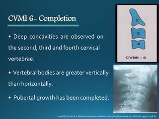
Downloaded 184 times

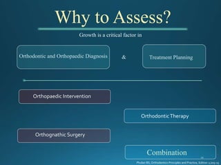
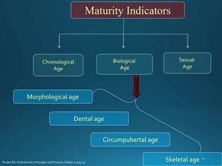
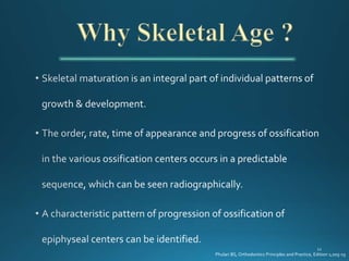
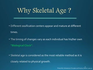
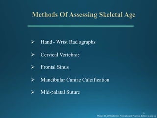
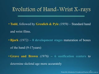
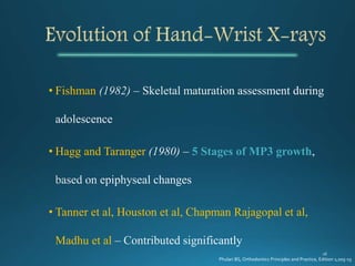
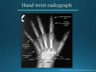
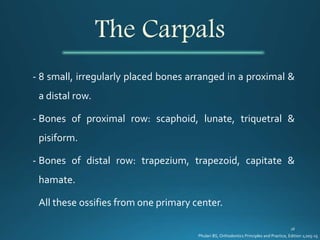
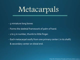
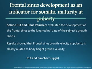
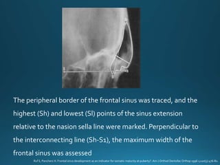
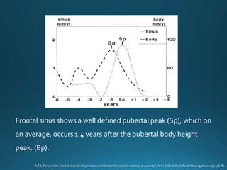


The document discusses various methods for assessing skeletal maturity and growth, including hand-wrist radiographs. It describes the bones seen in hand-wrist radiographs and several methods for analyzing skeletal maturity based on stages of ossification, including the Greulich and Pyle atlas method, Bjork method, Fishman method, and Hagg and Taranger method. The document also discusses other indicators of skeletal maturity such as cervical vertebrae and their relationship to skeletal age assessment.


