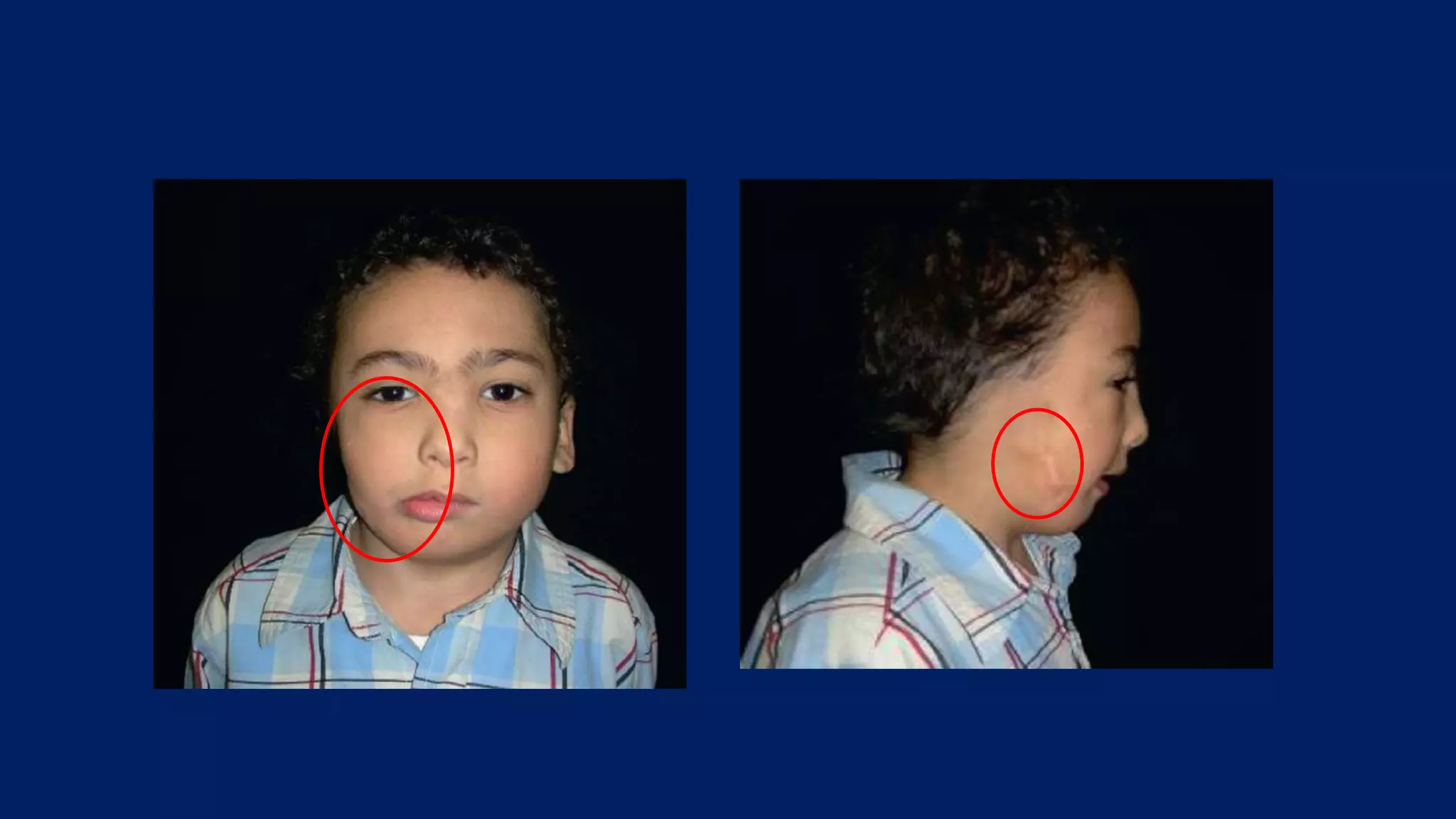
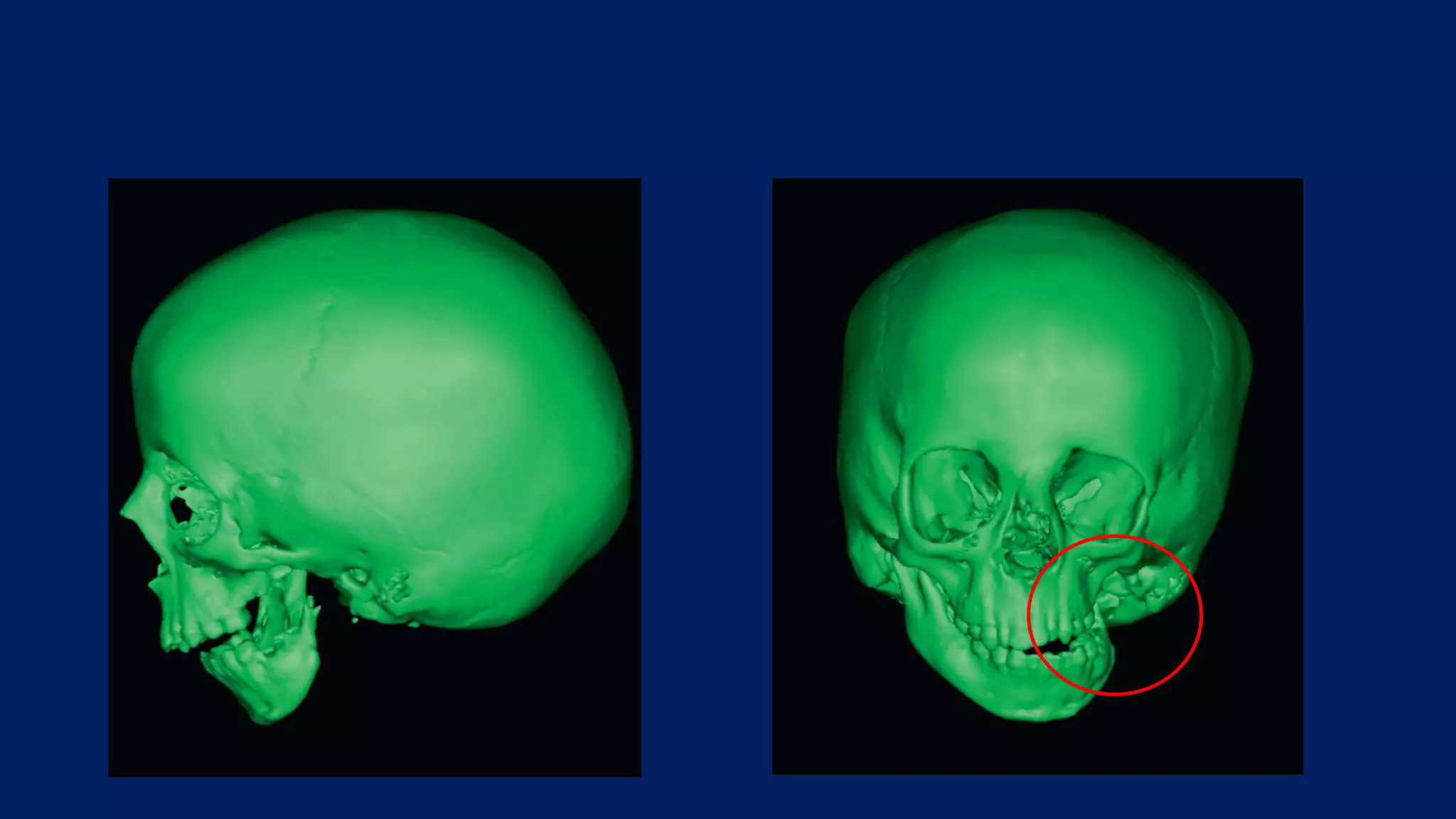
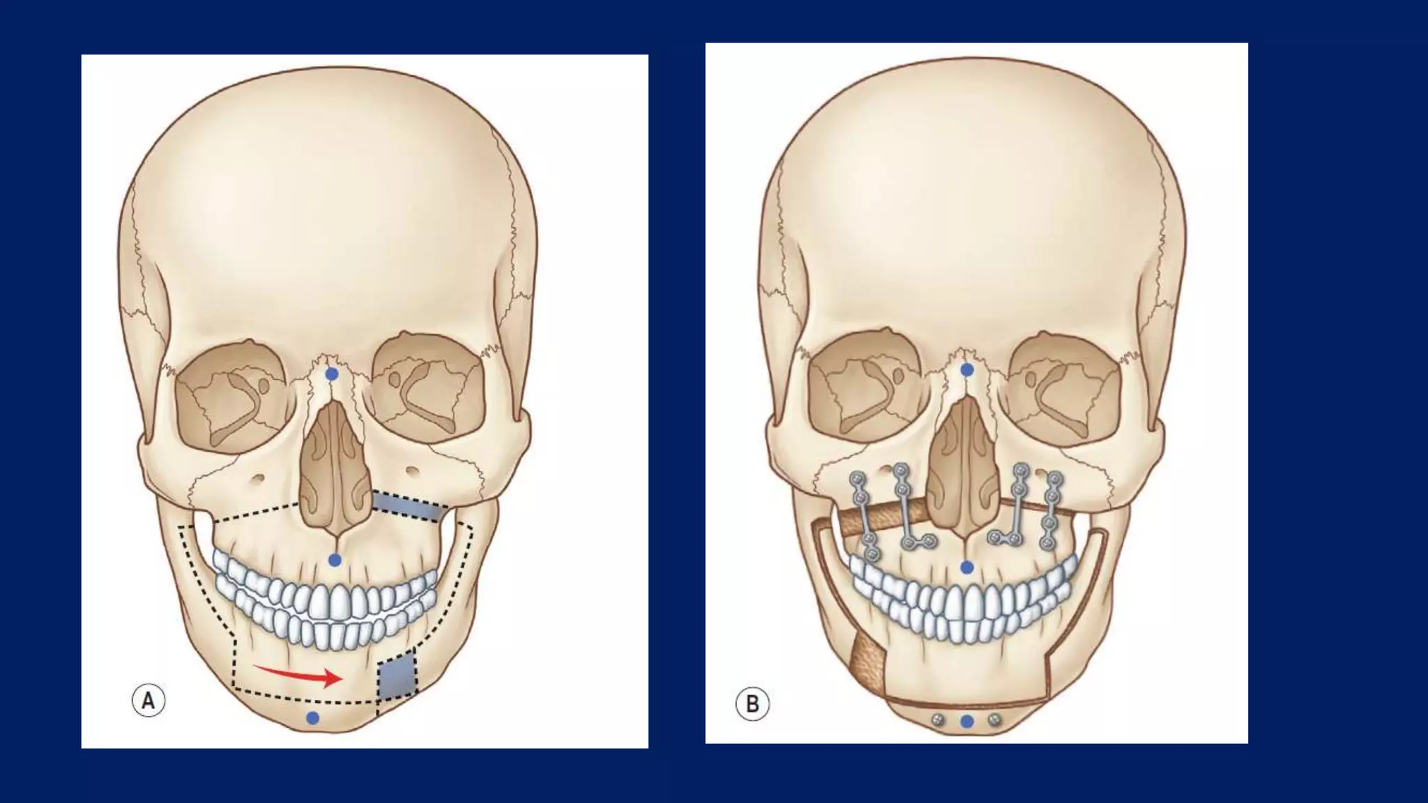
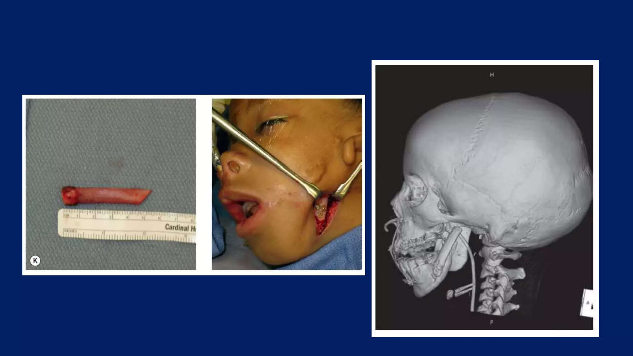
Lecture class
Dr. S.Satish Kumar
CRANIO FACIAL MICROSOMIA
• Craniofacial microsomia (CFM) involves a spectrum of congenital
malformations of craniofacial structures that arise from or are
intimately related to the first and second branchial arches
• one of the most common congenital malformations of the head and
neck, second only to cleft lip and palate
• may be unilateral or bilateral
• bilateral hypoplasia has been noted in 5– 30% of cases
Embryology
• The first branchial arch (mandibular arch) gives rise to the maxilla,
mandible, zygoma, trigeminal nerve, muscles of mastication,
connective tissue of the face, and a minority of the external ear (tragus,
root of helix, superior portion of the helix).
• The second branchial arch (hyoid arch) gives rise to the stapes, styloid
process, portions of hyoid bone, facial nerve, facial musculature, and
the majority of the external ear (inferior portion helix, antihelix,
antitragus and lobule).
Embryology
• During the first trimester all branchial arches have their own artery arising
from the aortic arch.
• In the third week of gestation, the internal and external carotid arteries
develop from the third branchial arch
• The first and second aortic arch become less necessary.
• The remnant of the second aortic arch is the stapedial artery, which forms
the anastomosis between the internal and external carotid arteries in the
fourth week of gestation.
• In the fifth week the stapedial artery atrophies and disappears
• Around day 40 of gestation the external carotid artery supplies the blood for
the first and second branchial arches.
Etiology
• Etiology of CFM remains unclear
• Associated with
• vascular perturbation
• teratogen exposure
• neural crestopathy
• combination of the three.
Terminology
• Canton and Von Arlt in 1861 and 1881
• Craniofacial microsomia
• Hemifacial microsomia
• First and second branchial arch syndrome
• Otomandibular dysostosis
• Auriculobranchiogenic dysplasia
• Intrauterine facial necrosis
• Lateral facial dysplasia
• Hemignathia and microtia
syndrome
• Necrotic facial dysplasia
• Oto-mandibular-facial
dysmorphogenesis
• Mandibular laterognathism
• Oculoauriculovertebral
spectrum
• Facioauriculovertebral
malformation complex
Pathology
Orbit
• Smaller size and/or abnormal position
• Periocular abnormalities may range from mild inferior displacement of
the lateral canthus and/or palpebral fissure to microphthalmia/
anophthalmia
• Rarely, colobomas of the iris or upper lid with absence of the
eyelashes.
Mandibular deformity
• always involved
• range from mild hypoplasia or flattening of the condylar head to
complete agenesis of the condyle, ramus, and glenoid fossa
• temporomandibular joint abnormalities lead to deformities ranging
from mild malpositioning with aberrant cranial base articulation to
complete obliteration.
• The mandibular body may also be reduced in all dimensions, with an
increase in gonial angle.
Maxillary deformity
• Previously, maxillary hypoplasia.
• Now, findings suggest that the maxillary anomaly is not a true
volumetric deficiency.
• The maxillary cant observed is secondary to vertical growth inhibition
by the pathologically small mandible.
• no differences noted between the affected and unaffected sides with
regards to maxillary shape and volume.
Ear
• ear anomalies associated with CFM can be categorized into external ear
malformations , middle ear malformations and the presence of branchial
remnants and sinus tracts.
• Hypoplasia of the external ear can range from mild effacement of auricular
architecture to complete auricular agenesis and external auditory canal
atresia.
• In severe cases, only observable evidence of external ear development is
represented by a primitive auricular remnant and located caudally and
ventrally
• In very rare instances, no remnant is observable.
• hearing loss predominantly conductive in nature seen in 75% of patients
Nervous system
• Ipsilateral cerebral hypoplasia
• hypoplasia of the corpus callosum
• hydrocephalus
• intracranial lipoma
• hypoplasia and malformation of the brainstem and cerebellum
• cognitive delay
• epilepsy
Cranial nerve abnormalities
• Unilateral agenesis and hypoplasia of the optic nerve
• congenital ophthalmoplegia
• hypoplasia of the trochlear and abducens nuclei and nerves
• congenital trigeminal anesthesia
• aplasia of the trigeminal nerve
Soft tissue deficiency
• Most evident in the malar and masseteric region of the face as well as
in the region of the external ear, orbit, and the temporal region
• Malar flattening and temporal hollowing
• Macrostomia or clefting through the oral commissure and hypoplasia
of the parotid gland may also be present
• Tongue dysmorphogenesis (mild asymmetry to severe hypoplasia)
may contribute to the feeding and speech difficulties
• Ipsilateral palatal anomalies including weakness or paralysis of the
velum have been found
Diagnosis
• Cousley and Calvert suggested minimal diagnostic criteria for CFM
• 1) ipsilateral mandibular and ear defects or
• (2) asymmetric mandibular or ear defects
• in association with either
• (a) two or more indirectly associated anomalies or
• (b) a positive family history of CFM.
• Indirectly associated anomalies were defined as those ‘not normally
related either in terms of developmental fields or function’ ”.
Classification systems
• In late1960s, Pruzansky (later modified by Kaban and colleagues)
• Segregated the mandibular anomalies of craniofacial microsomia into
three grades (types I through III) of increasing hypoplasia
• Based largely on the morphology of the ramus and condyle
• Most comprehensive classification scheme is the OMENS (Orbital
Mandible Ear Nerve Soft tissue) classification, later modified to the
OMENS+ to include extracranial manifestations
• Vento et al. in 1991
• Scoring was done on the basis of conventional radiographs including
posterior/anterior, lateral, submental, and panoramic views, and
physical examination and photographs.
• The pictorial representation of the OMENS+ system was described by
Gougoutas et al
• Scoring of the orbital deformity reflects orbital size and position.
• Scoring within the mandible category is done so on the basis of radiographs and
uses the system of Pruzansky, later modified by Kaban
• Scoring of external ear anomalies uses the systems of Marx and Meurman, with
the addition of the grade 0 category representingthe absence of any observable
external ear malformation
• E I: Mild hypoplasia, with obvious malformation, but with all structures present
• E II: Atresia of the external auditory canal, with a vertically oriented
cartilaginous remnant
• E III: Absent auricle, the lobular remnant is anteriorly and inferiorly displaced
• Scoring within the facial nerve category groups, the zygomatic and
temporal branches into one group and the buccal, marginal mandibular, and
cervical branches into another, divides facial nerve involvement into upper
and lower halves.
• scoring of soft-tissue deficiencies grades subcutaneous/muscular deficiency
as either absent, mild, moderate, or severe.
• Classification for each side of the face is done separately in cases of
bilateral CFM.
• To include significant extracraniofacial anomalies, the OMENS
classification was modified by Horgan et al. in 1995 to allow for the
optional addition of a plus sign (OMENS+)
• Bartlett and associates developed an anatomical classification utilizing
3D computed tomography (CT) scans and guiding treatment,
specifically of the mandibular deformity.
• At present, the Pruzansky/Kaban classification remains the most
commonly used system.
Treatment/surgical technique
• Tracheostomy/gastrostomy
• In the neonatal period, tracheostomy can be a lifesaving maneuver in
patients with severe respiratory distress.
• In severe cases of CFM ( type III mandibular deformity), in which
mandibular distraction cannot be performed, tracheostomy may be
necessary.
• For infants with severe swallowing/eating problems, a gastrostomy is
indicated to improve the nutritional status of the child
Commissuroplasty
• Clefts of the lip and/or commissure are typically repaired in infancy to
increase feeding efficiency.
• Goals
• (1) normalize the oral commissure position,
• (2) reconstruct the orbicularis oris
• (3) minimize scar and create normal vermillion structures
Commissuroplasty
• Vermilion and oral mucosal flaps are designed.
• The flaps are approximated and sutured with resorbable sutures.
• The orbicularis muscle stumps are skeletonized and closed in a vest-
over-pants fashion with resorbable sutures.
• The cutaneous closure may incorporate a Z-plasty designed to
simulate the nasolabial fold
Nerve palsy management
• Patients who cannot move their mouth due to deficiencies of the buccal and
marginal mandibular branches can be offered a facial reanimation procedure
• Functional muscle transfer remains the gold standard for facial reanimation.
• Transfer of the temporalis muscle.
• The temporalis muscle can be detached from the coronoid and advanced to
the commissure to provide movement of the lateral mouth.
• Advantages -
• ease of surgery, ease of recovery, and reliability of establishing movement.
• Disadvantages -
• weak strength of pull, limitations on vector of pull, and the need to activate
cranial nerve V to stimulate a smile.
• Two-stage approach with a cross-face nerve graft and muscle free flap
• First stage, a sural nerve graft is harvested and attached to the cut end
of a redundant buccal branch on the functioning side.
• The graft is then passed through the upper lip to the paralyzed side of
the face, where it sits until axonal ingrowth has occurred
• Second stage, a muscle free flap (gracilis) is harvested and attached to
4 points:
• The commissure, the upper lip, the lower lip, and the zygomatic arch
in a direction that mimics the smile vector on the contralateral normal
side.
• Microscopic anastomosis of the vein, artery, and nerve are then
performed.
• Disadvantages -
• length of surgery, recovery time, and need for microsurgical skills
• Timing -
• Facial reanimation surgery better done after microtia reconstruction
and major craniofacial and/or orthognathic surgery
Mandibular reconstruction
• Treatment of Pruzansky/Kaban types I and IIA mandibular deformity
patients can consist of mandibular lengthening by distraction
osteogenesis or conventional osteotomy.
• In type IIB and III patients, both function and position of the
temporomandibular joint are inadequate, often requiring
reconstruction of the mandible to improve function
• The goal of treatment in each patient is to achieve appropriate
symmetry and occlusion with an adequate aesthetic outcome
Mandibular reconstruction
• Pruzansky type I (Bartlett type 1)
• In mild cases, the occlusal relationship can be managed with orthodontics.
• In more severe type I cases, orthognathic surgery may be necessary to improve an
occlusal cant and facial symmetry
• Pruzansky type IIA (Bartlett type 2)
• Vertical lengthening of the mandible.
• This can be done with either distraction osteogenesis or an osteotomy and interposed bone
graft performed after skeletal maturity
• Pruzansky type IIB (Bartlett type 3)
• Distraction osteogenesis as well as rib grafting have been used alone or in combination
for the treatment
• severe Pruzansky type III (Bartlett type 4)
• bone grafting to create a functional ramus and condyle unit and to restore mandibular
height and facial symmetry.
Orthognatic surgery
• Obwegeser combined the Le Fort I maxillary osteotomy with bilateral
sagittal split of the mandibular ramus and genioplasty to ensure
leveling of the occlusal plane and establishment of the optimal
occlusal relationships
• The ultimate goals of orthognathic surgery include the correction of
the occlusal cant while at the same time optimizing the lip–incisor
relationship.
Mandibular distraction osteogenesis
• Distraction osteogenesis - for augmentation of hypoplastic bony
structures using autologous tissue.
• Distraction osteogenesis was initially described by Ilizarov, then
applied to the craniofacial skeleton by Snyder and popularized by
McCarthy
• MDO increases the vertical length of the mandible, produces greater
bone stock and improves soft tissue asymmetry.
• Advantages - shorter operative time, less blood loss, greater vector
control of advancement and the ability to lengthen the mandible at a
younger
• MDO can be used to treat type IIa and IIb mandibles
• MDO combined with a bone graft for treatment of type III mandibles.
MDO devices
• Both single and multivector external devices and semi-buried internal
devices are available
• External devices allow greater freedom to mold the regenerate by changing
the vector of distraction after the osteotomy is made.
• pin placement requires little bone stock, which allows for accurate
placement of the devices in hypoplastic mandibles and with minimal
disruption of the periosteum.
• Disadvantages - external devices create unsightly scars, dislodge easily,
significantly alter the patient’s appearance , prone to pin site infections.
• Internal devices are less visible, create less scarring, and are less prone to
trauma and infection.
• Disadvantages - vector cannot be altered once the device is fixated, greater
preoperative planning is necessary with internal devices, additional surgery
to remove internal device.
Mandibular distraction osteogenesis
• Following a latency period of 2 to 5 days , the distraction phase is
started at a rate of 1 mm per day.
• Patients are followed up weekly with X-rays to confirm the device
functionality and distance of distraction.
• The endpoints of distraction -
• alignment of the maxillary and mandibular dental midlines with the
mid-sagittal plane
• correction/leveling of the mandibular cant
• restoration of facial symmetry.
Bone/cartilage grafting
• Gillies described the first costochondral graft for temporomandibular
joint (TMJ) reconstruction in 1920.
• Clavicle and sternoclavicular joint, fibula, iliac bone and metatarsal
bone
• Autogenous costochondral rib graft remains the method of choice for
mandibular/TMJ reconstruction in children with Pruzansky/Kaban
type IIB and III
• Goal of the procedure
• Reconstruct a new ramus and pseudocondyle with the rib graft
• Procedure done in patients older than 5 year of age.
• The quality of the rib graft in patients less than 5 years of age is
suboptimal.
Orbital reconstruction
• Stimulation of the visual cortex in infants to avoid amblyopia is of prime
importance.
• Epibulbar dermoids require treatment to prevent disruption of the visual
axis.
• Eyelid colobomas for protection of the cornea.
• Orbital bony reconstruction for asymmetry is corrected only if severe and
typically is postponed until the orbital growth is complete, which is
approximately at age 4 or 5 years.
• Orbital repositioning can be approached simply with a simple split calvarial
bone graft
• In more severely asymmetric cases with a 4-wall box osteotomy performed
via an intracranial approach.
Soft-tissue reconstruction
• Fat grafting
• This technique requires
• (1) fat harvest from the abdomen, flanks, thighs, or buttocks
• (2) purification by centrifuge
• (3) injection of small aliquots (<0.1 cc) in multiple planes within the
areas of facial deficiency.
• The benefits of fat injection are precision of delivery, minimal
scarring, and minimal donor-site morbidity.
• The small aliquots do not disrupt the connecting ligaments of the face,
so the fat is less likely to droop or disrupt normal facial movement.
• Microvascular free flap
• An adipofascial free flap is the best way to provide a large amount of
soft tissue in a single surgical procedure for patients with severe
deficiencies.
• Scapular, parascapular, groin, omentum, anterolateral thigh and deep
inferior epigastric perforator (DIEP).
• Need of debulking procedure.
• Other drawbacks include donor-site morbidity and scarring, length of
procedure, and the need for microsurgical skills.
• Generally performed after the skeletal anomalies of CFM have been
corrected.
Auricular reconstruction
• E1 - surgical treatment involves cartilage folding, scoring, or weakening
techniques to restore normal shape and anatomy.
• When the hypoplasia is more severe (E2 and E3), an auricular
reconstruction with a new framework is done.
• Autogenous reconstruction – rib cartilage
• Brent technique or Nagata technique
• Alloplastic reconstruction –
• Porous polyethylene’s inert nature and pore size provides the best safety
profile and allows for some tissue ingrowth
• John Reinisch
• Advantages - rigidity of construct, lack of donor site morbidity, and the
ability to reconstruct younger, smaller patients.
• Disadvantages - extrusion and infection
HEMIFACIALATROPHY
• Idiopathic progressive hemifacial atrophy (PHA)
• Parry–Romberg syndrome
• Typically characterized by slow progressive unilateral atrophy of the
skin and soft tissue of the face, with deeper involvement of the
underlying muscles and osteocartilaginous structures resulting in both
aesthetic and functional orofacial issues.
Epidemiology
• Incidence of 5 per 100000 people and prevalence of 8 per 100000
people
• There is no racial predilection of PHA.
• There is a slight female predominance with most studies having
female to male ratios 3 : 1.
• The median age of onset is 10 years old with a general range of 5–15
years of age
Etiopathogenesis
• Autoimmune process
• PHA is a likely variant of the autoimmune disease “localized
scleroderma”, specifically the subtype of linear scleroderma that
affects the head (en coup de sabre, ECDS).
• It is often hard to differentiate these two entities, as ECDS often leads
to atrophy of subcutaneous tissue and facial bones, causing hemifacial
atrophy later in the disease course.
• Both share similar characteristics, including age of onset, female
preponderance, neurologic involvement, lymphocytic infiltrate on
biopsy, and a clinical course of evolution for several years followed by
stabilization.
• Neurogenic process
• The distribution of facial atrophy typically follows a dermatome of the
trigeminal nerve
• Neuritis of the trigeminal nerve in patients experiencing episodes of
pain in the involved area prior to the onset of tissue atrophy
• The dermal lymphocytic infiltrate centered around neurovascular
bundles in the dermis on histology also supports a neurologic target
• Clinical, radiological, and CSF laboratory findings of patients with
PHA highly support that the disease affects the central nervous system,
likely in an autoimmune fashion.
• The clinical manifestations of CNS involvement - 8–20% of the
patients with PHA
• Seizures, chronic headaches, optic neuritis and less commonly as
neuropsychiatric disorders, deterioration of intelligence and ischemic
stroke.
• Infection hypothesis
• Most notorious suspect for both PHA and ECDS was Borrelia
burgdorferi.
• The viral infections such as Epstein Barr virus, correlate to the typical
exposure to these infectious agents in first and second decades of life.
Clinical manifestations
• Subcutaneous atrophy
• Typically evolves first on the cheek or temple, and later extends to the
brow, angle of mouth, and neck.
• Later in the disease course, atrophy of the underlying bone and
cartilage can occur, causing further facial deformity.
• Facial muscles may become atrophic, but they tend to maintain their
normal function.
• The disease typically progresses slowly over several years (2–10
years) and then tends to enter a stable phase
Clinical manifestations
• Cutaneous and subcutaneous involvement
• Cutaneous disease include -
• Dyspigmentation (both hyperpigmentation and hypopigmentation)
• Dermal atrophy (signified by shiny skin and visible veins)
• Subcutaneous atrophy (described as a flattening or concavity of the
subcutaneous tissue)
• Skin thickening/fibrosis at the center of the lesion
• Dyspigmentations are typically distributed in a dermatomal
distribution along the trigeminal nerve.
Clinical manifestations
• Musculoskeletal involvement
• Masseteric muscle, tongue and palatal muscles
• Maxilla and mandible hypoplasia - with both sagittal and vertical
undergrowth, causing cosmetic and dental abnormalities and a
deficiency in posterior facial height
• Enophthalmos is common.
• More related to the atrophy of the periorbital subcutaneous tissues
rather than skeletal hypoplasia
Clinical manifestations
• Central nervous system involvement
• 8–21% of the patients with PHA and include seizures, hemiparesis,
migraine headaches, neuropsychiatric disturbances, ischemic stroke,
and intellectual deterioration.
• CNS symptoms occur years after the onset of cutaneous or
subcutaneous findings, with a mean of 4.3 years
• The most common CNS manifestation is localization related seizures
• All of these patients had cortical or subcortical white matter T2
hyperintensities in MRI.
Clinical manifestations
• Ocular involvement
• Uveitis, optic neuritis, and globe retraction being the most common
• Ocular muscle paralysis, ptosis, Horner syndrome, heterochromia
iridis, and dilated fixed pupil have also been reported
• Ophthalmologic examination using a slit-lamp is recommended in
patients with PHA and/or ECDS to assess for an inflammatory-fibrotic
process, which may be arrested by immunosuppressive therapy.
Clinical manifestations
• Oral involvement
• The tongue and upper lip on the affected side of the face are often markedly
atrophic.
• The maxilla and mandible may be underdeveloped (hypoplastic), resulting
in malocclusion and altered dentition.
• As a result of jaw hypoplasia, there is often a unilateral posterior crossbite
and occlusal canting, and an abnormally skewed high arched palate may
also be observed.
• As the active stage of PHA coincides with the period of root formation and
the eruption of permanent teeth, deficient root development and delayed
tooth eruption also occur.
Laboratory findings
• anti-nuclear antibody (ANA)
• anti-single stranded DNA (ss-DNA)
• anti-histone
• anti-doublestranded DNA
• anti-centromere
• anti-Scl-70 antibodies.
Differential diagnosis
• congenital hemifacial or craniofacial microsomia (CFM)
• ECDS subtype of localized scleroderma
• CFM
• present at birth
• not progressive
• ECDS
• scalp and forehead involvement and induration of the skin and subcutaneous
tissues during the acute phase
• There is considerable overlap between the two entities, with 30–40% of
patients being classified as having coexistent ECDS and PHA
Patient selection
• Several factors need be considered-
• (1) the age of the patient
• (2) the nature and the complexity of the deformity (i.e., which tissue
types are affected)
• (3) the presence of associated disorders and conditions
• (4) patient’s understanding of the problem and what are the options for
• Treatment
• (5) the timing for surgery
• Reconstructive surgery is best done only after the disease has “burned
itself out
• For both mild and moderate deformities, the use of fat injections
(structural fat grafting) and buried dermal fat grafts have a significant
role.
• In severe deformities, free tissue transfers are an option
Role of immunosuppression
• Patients with PHA that have any cutaneous features are treated with
immunosuppression.
• Typically corticosteroids in combination with a disease-modifying
agent such as methotrexate, have been found to have cessation of
disease progression and reversal/ improvement of disease damage
• hyperpigmented skin becomes lighter
• sclerotic skin softens
• subcutaneous atrophy is less noticeable with some “filling in” of fat
• hair growth is observed in areas of alopecia
• tongue atrophy is less dramatic.
Role of immunosuppression
• PHA patients with neurologic manifestations, such as seizures and
optic neuritis, have shown benefit from immunosuppressive therapy
• After a period of 3–5 years on immunosuppressive therapy ,
immunosuppression is weaned off as the disease is felt to have
stabilized at that point
Non-surgical intervention
• Oral rehabilitation
• Osseous defects are usually seen when the atrophy manifests before 15
years of age.
• Fronto-maxillary defects are seen in onset before 5 years of age
• Mandibular defects onset is between 5 and 15 years of age
• Later onset (>15 years of age) has almost exclusively soft tissue
changes
Oral rehabilitation
• Orthodontic treatment options include:
• (1) Orthodontics to manage crossbite correction with orthopedic
expansion of the maxillary mid-palatal suture and dental eruption
guidance
• (2) Functional appliance therapy while the disease is in its active phase
and facial skeletal growth is occurring
• (3) After disease progression and facial/skeletal growth to complete
before addressing the resultant malocclusion with definitive combined
orthodontic and orthognathic surgical intervention
Oral rehabilitation
• The objective of functional appliance treatment in growing patients
with PHA is maintenance of parallelism of the facial planes,
specifically bilateral symmetry of mandibular ramus height and
resultant mandibular plane.
• The intention of treatment is to stimulate condylar growth of the
affected side in a vertical direction to obtain equal vertical
development of the mandible and minimize progressive atrophy of
facial growth
• Generally, appliance worn for 6 yrs, 12–14 hours a day; monthly
orthodontic visits for appliance adjustment made during this period.
Non-surgical intervention
• Alloplastic fillers
• non-surgical alloplastic filling agents is advantageous for facial
contour reconstruction because of the absence of a donor site and their
abundant supply
• silicone gel
• hydroxyapatite beads
• hyaluronic acid
Surgical intervention
• Dermal fat grafts, local/pedicles flaps, and free tissue transfer.
• Local pedicled flaps have been described to reconstruct deficits and
deformities in the head and neck.
• Their lack of significant bulk has limited their role in cases of
extensive soft tissue deficit
• Soft tissue based on the superficial temporal pedicle may be rotated
inferiorly to fill more superficial depressions
Surgical intervention
• Smaller deficits may be reconstructed with smaller muscle or fascial
flaps, such as the gracilis and radial forearm adipofascial flap
• The deep inferior epigastric perforator (DIEP) flap can provide more
soft tissue bulk in patients with significant deformities.
• In the heavier patient, the omentum, supplied by either the right or left
gastroepiploic arteries, provides an adequate source of fat for soft
tissue volume
• Disadvantages - harvest requires entering the peritoneal cavity and the
lack of internal structure of the free flap often leads to soft tissue
descent with time.
Surgical intervention
• Anterolateral thigh adipofascial flap in 1984 by Song et al
• Advantages –
• the ability to harvest the flap in the supine position away from the area of
inset
• the large reliable skin flap that is available
• its proximity to larger muscles that may be incorporated into the flap for
bulk
• its relatively long vascular pedicle
• Donor site defect may be closed directly in the absence of sufficient tension,
• Disadvantages
• Variability of the skin perforators and the potentially tedious dissection of
the pedicle if it courses within the muscle for an extended length
Surgical intervention
• The scapular and parascapular adipofascial flaps, based on the circumflex
scapular pedicle, are among the most useful flaps for restoring facial
volume
• Advantages
• Relatively straightforward harvest
• posterior torso donor scar
• minimal functional deficit.
• Disadvantages
• the need to position the patient in either a prone or lateral decubitus
position in order to harvest the flap
• Superficial inferior epigastric artery (SIEA) flap, the transverse rectus
abdominis muscle (TRAM) flap ,the deltopectoral flap, LD flap with
vascularized costochondral graft have also been described.
Orthognathic surgical treatment
• In severe cases, soft tissue augmentation may not be sufficient, treated
with combined orthodontic and orthognathic surgery
• The problems addressed include:
• (1) hypoplasia of the zygomatic complex and maxilla which can cause
orbital dystopia and depressive deformities in the
zygomaticomaxillary region
• (2) hypoplasia of the mandible, especially the ramus, which causes
obvious deviation of the chin and occlusal plane
Orthognathic surgical treatment
• Pre-surgical fixed orthodontic appliances (braces)
• Surgical procedures utilized to address the maxillary and mandibular
dysmorphologies of PHA include -
• LeFort I osteotomy
• MEDPOR implants
• Bone grafting of osseous defects
• Rib grafting
• Genioplasty
• Mandibular Distraction
• Period of postoperative orthodontic treatment is also required
Secondary procedures
• Debulking of the flap is usually one component of the revision, along
with further addition of soft tissue to areas that remain deficient.
• Flap tissue from overly bulky areas may be rotated and transferred to
areas with a persistent lack of volume.
• Additional autogenous fat or alloplastic filler may be used for smaller
areas of need
Outcome
• Facial symmetry as the ultimate goal
• Success is predicated on –
• Identifying the tissue types involved,
• The precise location of the deficit
• Choosing an appropriate intervention strategy
• At present, free tissue survival is highly successful especially in
younger patients with healthy donor and recipient vascularity.
THANK YOU

Craniofacial Microsomia and Hemifacial Atrophy

  • 1.
  • 2.
    CRANIO FACIAL MICROSOMIA •Craniofacial microsomia (CFM) involves a spectrum of congenital malformations of craniofacial structures that arise from or are intimately related to the first and second branchial arches • one of the most common congenital malformations of the head and neck, second only to cleft lip and palate • may be unilateral or bilateral • bilateral hypoplasia has been noted in 5– 30% of cases
  • 4.
    Embryology • The firstbranchial arch (mandibular arch) gives rise to the maxilla, mandible, zygoma, trigeminal nerve, muscles of mastication, connective tissue of the face, and a minority of the external ear (tragus, root of helix, superior portion of the helix). • The second branchial arch (hyoid arch) gives rise to the stapes, styloid process, portions of hyoid bone, facial nerve, facial musculature, and the majority of the external ear (inferior portion helix, antihelix, antitragus and lobule).
  • 5.
    Embryology • During thefirst trimester all branchial arches have their own artery arising from the aortic arch. • In the third week of gestation, the internal and external carotid arteries develop from the third branchial arch • The first and second aortic arch become less necessary. • The remnant of the second aortic arch is the stapedial artery, which forms the anastomosis between the internal and external carotid arteries in the fourth week of gestation. • In the fifth week the stapedial artery atrophies and disappears • Around day 40 of gestation the external carotid artery supplies the blood for the first and second branchial arches.
  • 6.
    Etiology • Etiology ofCFM remains unclear • Associated with • vascular perturbation • teratogen exposure • neural crestopathy • combination of the three.
  • 7.
    Terminology • Canton andVon Arlt in 1861 and 1881 • Craniofacial microsomia • Hemifacial microsomia • First and second branchial arch syndrome • Otomandibular dysostosis • Auriculobranchiogenic dysplasia • Intrauterine facial necrosis • Lateral facial dysplasia • Hemignathia and microtia syndrome • Necrotic facial dysplasia • Oto-mandibular-facial dysmorphogenesis • Mandibular laterognathism • Oculoauriculovertebral spectrum • Facioauriculovertebral malformation complex
  • 8.
    Pathology Orbit • Smaller sizeand/or abnormal position • Periocular abnormalities may range from mild inferior displacement of the lateral canthus and/or palpebral fissure to microphthalmia/ anophthalmia • Rarely, colobomas of the iris or upper lid with absence of the eyelashes.
  • 9.
    Mandibular deformity • alwaysinvolved • range from mild hypoplasia or flattening of the condylar head to complete agenesis of the condyle, ramus, and glenoid fossa • temporomandibular joint abnormalities lead to deformities ranging from mild malpositioning with aberrant cranial base articulation to complete obliteration. • The mandibular body may also be reduced in all dimensions, with an increase in gonial angle.
  • 11.
    Maxillary deformity • Previously,maxillary hypoplasia. • Now, findings suggest that the maxillary anomaly is not a true volumetric deficiency. • The maxillary cant observed is secondary to vertical growth inhibition by the pathologically small mandible. • no differences noted between the affected and unaffected sides with regards to maxillary shape and volume.
  • 12.
    Ear • ear anomaliesassociated with CFM can be categorized into external ear malformations , middle ear malformations and the presence of branchial remnants and sinus tracts. • Hypoplasia of the external ear can range from mild effacement of auricular architecture to complete auricular agenesis and external auditory canal atresia. • In severe cases, only observable evidence of external ear development is represented by a primitive auricular remnant and located caudally and ventrally • In very rare instances, no remnant is observable. • hearing loss predominantly conductive in nature seen in 75% of patients
  • 14.
    Nervous system • Ipsilateralcerebral hypoplasia • hypoplasia of the corpus callosum • hydrocephalus • intracranial lipoma • hypoplasia and malformation of the brainstem and cerebellum • cognitive delay • epilepsy
  • 15.
    Cranial nerve abnormalities •Unilateral agenesis and hypoplasia of the optic nerve • congenital ophthalmoplegia • hypoplasia of the trochlear and abducens nuclei and nerves • congenital trigeminal anesthesia • aplasia of the trigeminal nerve
  • 16.
    Soft tissue deficiency •Most evident in the malar and masseteric region of the face as well as in the region of the external ear, orbit, and the temporal region • Malar flattening and temporal hollowing • Macrostomia or clefting through the oral commissure and hypoplasia of the parotid gland may also be present • Tongue dysmorphogenesis (mild asymmetry to severe hypoplasia) may contribute to the feeding and speech difficulties • Ipsilateral palatal anomalies including weakness or paralysis of the velum have been found
  • 18.
    Diagnosis • Cousley andCalvert suggested minimal diagnostic criteria for CFM • 1) ipsilateral mandibular and ear defects or • (2) asymmetric mandibular or ear defects • in association with either • (a) two or more indirectly associated anomalies or • (b) a positive family history of CFM. • Indirectly associated anomalies were defined as those ‘not normally related either in terms of developmental fields or function’ ”.
  • 19.
    Classification systems • Inlate1960s, Pruzansky (later modified by Kaban and colleagues) • Segregated the mandibular anomalies of craniofacial microsomia into three grades (types I through III) of increasing hypoplasia • Based largely on the morphology of the ramus and condyle
  • 21.
    • Most comprehensiveclassification scheme is the OMENS (Orbital Mandible Ear Nerve Soft tissue) classification, later modified to the OMENS+ to include extracranial manifestations • Vento et al. in 1991 • Scoring was done on the basis of conventional radiographs including posterior/anterior, lateral, submental, and panoramic views, and physical examination and photographs. • The pictorial representation of the OMENS+ system was described by Gougoutas et al • Scoring of the orbital deformity reflects orbital size and position.
  • 22.
    • Scoring withinthe mandible category is done so on the basis of radiographs and uses the system of Pruzansky, later modified by Kaban • Scoring of external ear anomalies uses the systems of Marx and Meurman, with the addition of the grade 0 category representingthe absence of any observable external ear malformation • E I: Mild hypoplasia, with obvious malformation, but with all structures present • E II: Atresia of the external auditory canal, with a vertically oriented cartilaginous remnant • E III: Absent auricle, the lobular remnant is anteriorly and inferiorly displaced
  • 23.
    • Scoring withinthe facial nerve category groups, the zygomatic and temporal branches into one group and the buccal, marginal mandibular, and cervical branches into another, divides facial nerve involvement into upper and lower halves. • scoring of soft-tissue deficiencies grades subcutaneous/muscular deficiency as either absent, mild, moderate, or severe. • Classification for each side of the face is done separately in cases of bilateral CFM. • To include significant extracraniofacial anomalies, the OMENS classification was modified by Horgan et al. in 1995 to allow for the optional addition of a plus sign (OMENS+)
  • 26.
    • Bartlett andassociates developed an anatomical classification utilizing 3D computed tomography (CT) scans and guiding treatment, specifically of the mandibular deformity. • At present, the Pruzansky/Kaban classification remains the most commonly used system.
  • 28.
    Treatment/surgical technique • Tracheostomy/gastrostomy •In the neonatal period, tracheostomy can be a lifesaving maneuver in patients with severe respiratory distress. • In severe cases of CFM ( type III mandibular deformity), in which mandibular distraction cannot be performed, tracheostomy may be necessary. • For infants with severe swallowing/eating problems, a gastrostomy is indicated to improve the nutritional status of the child
  • 29.
    Commissuroplasty • Clefts ofthe lip and/or commissure are typically repaired in infancy to increase feeding efficiency. • Goals • (1) normalize the oral commissure position, • (2) reconstruct the orbicularis oris • (3) minimize scar and create normal vermillion structures
  • 30.
    Commissuroplasty • Vermilion andoral mucosal flaps are designed. • The flaps are approximated and sutured with resorbable sutures. • The orbicularis muscle stumps are skeletonized and closed in a vest- over-pants fashion with resorbable sutures. • The cutaneous closure may incorporate a Z-plasty designed to simulate the nasolabial fold
  • 32.
    Nerve palsy management •Patients who cannot move their mouth due to deficiencies of the buccal and marginal mandibular branches can be offered a facial reanimation procedure • Functional muscle transfer remains the gold standard for facial reanimation. • Transfer of the temporalis muscle. • The temporalis muscle can be detached from the coronoid and advanced to the commissure to provide movement of the lateral mouth. • Advantages - • ease of surgery, ease of recovery, and reliability of establishing movement. • Disadvantages - • weak strength of pull, limitations on vector of pull, and the need to activate cranial nerve V to stimulate a smile.
  • 33.
    • Two-stage approachwith a cross-face nerve graft and muscle free flap • First stage, a sural nerve graft is harvested and attached to the cut end of a redundant buccal branch on the functioning side. • The graft is then passed through the upper lip to the paralyzed side of the face, where it sits until axonal ingrowth has occurred • Second stage, a muscle free flap (gracilis) is harvested and attached to 4 points: • The commissure, the upper lip, the lower lip, and the zygomatic arch in a direction that mimics the smile vector on the contralateral normal side. • Microscopic anastomosis of the vein, artery, and nerve are then performed.
  • 34.
    • Disadvantages - •length of surgery, recovery time, and need for microsurgical skills • Timing - • Facial reanimation surgery better done after microtia reconstruction and major craniofacial and/or orthognathic surgery
  • 35.
    Mandibular reconstruction • Treatmentof Pruzansky/Kaban types I and IIA mandibular deformity patients can consist of mandibular lengthening by distraction osteogenesis or conventional osteotomy. • In type IIB and III patients, both function and position of the temporomandibular joint are inadequate, often requiring reconstruction of the mandible to improve function • The goal of treatment in each patient is to achieve appropriate symmetry and occlusion with an adequate aesthetic outcome
  • 36.
    Mandibular reconstruction • Pruzanskytype I (Bartlett type 1) • In mild cases, the occlusal relationship can be managed with orthodontics. • In more severe type I cases, orthognathic surgery may be necessary to improve an occlusal cant and facial symmetry • Pruzansky type IIA (Bartlett type 2) • Vertical lengthening of the mandible. • This can be done with either distraction osteogenesis or an osteotomy and interposed bone graft performed after skeletal maturity • Pruzansky type IIB (Bartlett type 3) • Distraction osteogenesis as well as rib grafting have been used alone or in combination for the treatment • severe Pruzansky type III (Bartlett type 4) • bone grafting to create a functional ramus and condyle unit and to restore mandibular height and facial symmetry.
  • 37.
    Orthognatic surgery • Obwegesercombined the Le Fort I maxillary osteotomy with bilateral sagittal split of the mandibular ramus and genioplasty to ensure leveling of the occlusal plane and establishment of the optimal occlusal relationships • The ultimate goals of orthognathic surgery include the correction of the occlusal cant while at the same time optimizing the lip–incisor relationship.
  • 39.
    Mandibular distraction osteogenesis •Distraction osteogenesis - for augmentation of hypoplastic bony structures using autologous tissue. • Distraction osteogenesis was initially described by Ilizarov, then applied to the craniofacial skeleton by Snyder and popularized by McCarthy • MDO increases the vertical length of the mandible, produces greater bone stock and improves soft tissue asymmetry. • Advantages - shorter operative time, less blood loss, greater vector control of advancement and the ability to lengthen the mandible at a younger • MDO can be used to treat type IIa and IIb mandibles • MDO combined with a bone graft for treatment of type III mandibles.
  • 40.
    MDO devices • Bothsingle and multivector external devices and semi-buried internal devices are available • External devices allow greater freedom to mold the regenerate by changing the vector of distraction after the osteotomy is made. • pin placement requires little bone stock, which allows for accurate placement of the devices in hypoplastic mandibles and with minimal disruption of the periosteum. • Disadvantages - external devices create unsightly scars, dislodge easily, significantly alter the patient’s appearance , prone to pin site infections. • Internal devices are less visible, create less scarring, and are less prone to trauma and infection. • Disadvantages - vector cannot be altered once the device is fixated, greater preoperative planning is necessary with internal devices, additional surgery to remove internal device.
  • 42.
    Mandibular distraction osteogenesis •Following a latency period of 2 to 5 days , the distraction phase is started at a rate of 1 mm per day. • Patients are followed up weekly with X-rays to confirm the device functionality and distance of distraction. • The endpoints of distraction - • alignment of the maxillary and mandibular dental midlines with the mid-sagittal plane • correction/leveling of the mandibular cant • restoration of facial symmetry.
  • 43.
    Bone/cartilage grafting • Gilliesdescribed the first costochondral graft for temporomandibular joint (TMJ) reconstruction in 1920. • Clavicle and sternoclavicular joint, fibula, iliac bone and metatarsal bone • Autogenous costochondral rib graft remains the method of choice for mandibular/TMJ reconstruction in children with Pruzansky/Kaban type IIB and III • Goal of the procedure • Reconstruct a new ramus and pseudocondyle with the rib graft • Procedure done in patients older than 5 year of age. • The quality of the rib graft in patients less than 5 years of age is suboptimal.
  • 45.
    Orbital reconstruction • Stimulationof the visual cortex in infants to avoid amblyopia is of prime importance. • Epibulbar dermoids require treatment to prevent disruption of the visual axis. • Eyelid colobomas for protection of the cornea. • Orbital bony reconstruction for asymmetry is corrected only if severe and typically is postponed until the orbital growth is complete, which is approximately at age 4 or 5 years. • Orbital repositioning can be approached simply with a simple split calvarial bone graft • In more severely asymmetric cases with a 4-wall box osteotomy performed via an intracranial approach.
  • 46.
    Soft-tissue reconstruction • Fatgrafting • This technique requires • (1) fat harvest from the abdomen, flanks, thighs, or buttocks • (2) purification by centrifuge • (3) injection of small aliquots (<0.1 cc) in multiple planes within the areas of facial deficiency. • The benefits of fat injection are precision of delivery, minimal scarring, and minimal donor-site morbidity. • The small aliquots do not disrupt the connecting ligaments of the face, so the fat is less likely to droop or disrupt normal facial movement.
  • 47.
    • Microvascular freeflap • An adipofascial free flap is the best way to provide a large amount of soft tissue in a single surgical procedure for patients with severe deficiencies. • Scapular, parascapular, groin, omentum, anterolateral thigh and deep inferior epigastric perforator (DIEP). • Need of debulking procedure. • Other drawbacks include donor-site morbidity and scarring, length of procedure, and the need for microsurgical skills. • Generally performed after the skeletal anomalies of CFM have been corrected.
  • 48.
    Auricular reconstruction • E1- surgical treatment involves cartilage folding, scoring, or weakening techniques to restore normal shape and anatomy. • When the hypoplasia is more severe (E2 and E3), an auricular reconstruction with a new framework is done. • Autogenous reconstruction – rib cartilage • Brent technique or Nagata technique • Alloplastic reconstruction – • Porous polyethylene’s inert nature and pore size provides the best safety profile and allows for some tissue ingrowth • John Reinisch • Advantages - rigidity of construct, lack of donor site morbidity, and the ability to reconstruct younger, smaller patients. • Disadvantages - extrusion and infection
  • 49.
    HEMIFACIALATROPHY • Idiopathic progressivehemifacial atrophy (PHA) • Parry–Romberg syndrome • Typically characterized by slow progressive unilateral atrophy of the skin and soft tissue of the face, with deeper involvement of the underlying muscles and osteocartilaginous structures resulting in both aesthetic and functional orofacial issues.
  • 50.
    Epidemiology • Incidence of5 per 100000 people and prevalence of 8 per 100000 people • There is no racial predilection of PHA. • There is a slight female predominance with most studies having female to male ratios 3 : 1. • The median age of onset is 10 years old with a general range of 5–15 years of age
  • 51.
    Etiopathogenesis • Autoimmune process •PHA is a likely variant of the autoimmune disease “localized scleroderma”, specifically the subtype of linear scleroderma that affects the head (en coup de sabre, ECDS). • It is often hard to differentiate these two entities, as ECDS often leads to atrophy of subcutaneous tissue and facial bones, causing hemifacial atrophy later in the disease course. • Both share similar characteristics, including age of onset, female preponderance, neurologic involvement, lymphocytic infiltrate on biopsy, and a clinical course of evolution for several years followed by stabilization.
  • 53.
    • Neurogenic process •The distribution of facial atrophy typically follows a dermatome of the trigeminal nerve • Neuritis of the trigeminal nerve in patients experiencing episodes of pain in the involved area prior to the onset of tissue atrophy • The dermal lymphocytic infiltrate centered around neurovascular bundles in the dermis on histology also supports a neurologic target
  • 54.
    • Clinical, radiological,and CSF laboratory findings of patients with PHA highly support that the disease affects the central nervous system, likely in an autoimmune fashion. • The clinical manifestations of CNS involvement - 8–20% of the patients with PHA • Seizures, chronic headaches, optic neuritis and less commonly as neuropsychiatric disorders, deterioration of intelligence and ischemic stroke.
  • 55.
    • Infection hypothesis •Most notorious suspect for both PHA and ECDS was Borrelia burgdorferi. • The viral infections such as Epstein Barr virus, correlate to the typical exposure to these infectious agents in first and second decades of life.
  • 56.
    Clinical manifestations • Subcutaneousatrophy • Typically evolves first on the cheek or temple, and later extends to the brow, angle of mouth, and neck. • Later in the disease course, atrophy of the underlying bone and cartilage can occur, causing further facial deformity. • Facial muscles may become atrophic, but they tend to maintain their normal function. • The disease typically progresses slowly over several years (2–10 years) and then tends to enter a stable phase
  • 57.
    Clinical manifestations • Cutaneousand subcutaneous involvement • Cutaneous disease include - • Dyspigmentation (both hyperpigmentation and hypopigmentation) • Dermal atrophy (signified by shiny skin and visible veins) • Subcutaneous atrophy (described as a flattening or concavity of the subcutaneous tissue) • Skin thickening/fibrosis at the center of the lesion • Dyspigmentations are typically distributed in a dermatomal distribution along the trigeminal nerve.
  • 59.
    Clinical manifestations • Musculoskeletalinvolvement • Masseteric muscle, tongue and palatal muscles • Maxilla and mandible hypoplasia - with both sagittal and vertical undergrowth, causing cosmetic and dental abnormalities and a deficiency in posterior facial height • Enophthalmos is common. • More related to the atrophy of the periorbital subcutaneous tissues rather than skeletal hypoplasia
  • 60.
    Clinical manifestations • Centralnervous system involvement • 8–21% of the patients with PHA and include seizures, hemiparesis, migraine headaches, neuropsychiatric disturbances, ischemic stroke, and intellectual deterioration. • CNS symptoms occur years after the onset of cutaneous or subcutaneous findings, with a mean of 4.3 years • The most common CNS manifestation is localization related seizures • All of these patients had cortical or subcortical white matter T2 hyperintensities in MRI.
  • 61.
    Clinical manifestations • Ocularinvolvement • Uveitis, optic neuritis, and globe retraction being the most common • Ocular muscle paralysis, ptosis, Horner syndrome, heterochromia iridis, and dilated fixed pupil have also been reported • Ophthalmologic examination using a slit-lamp is recommended in patients with PHA and/or ECDS to assess for an inflammatory-fibrotic process, which may be arrested by immunosuppressive therapy.
  • 62.
    Clinical manifestations • Oralinvolvement • The tongue and upper lip on the affected side of the face are often markedly atrophic. • The maxilla and mandible may be underdeveloped (hypoplastic), resulting in malocclusion and altered dentition. • As a result of jaw hypoplasia, there is often a unilateral posterior crossbite and occlusal canting, and an abnormally skewed high arched palate may also be observed. • As the active stage of PHA coincides with the period of root formation and the eruption of permanent teeth, deficient root development and delayed tooth eruption also occur.
  • 63.
    Laboratory findings • anti-nuclearantibody (ANA) • anti-single stranded DNA (ss-DNA) • anti-histone • anti-doublestranded DNA • anti-centromere • anti-Scl-70 antibodies.
  • 64.
    Differential diagnosis • congenitalhemifacial or craniofacial microsomia (CFM) • ECDS subtype of localized scleroderma • CFM • present at birth • not progressive • ECDS • scalp and forehead involvement and induration of the skin and subcutaneous tissues during the acute phase • There is considerable overlap between the two entities, with 30–40% of patients being classified as having coexistent ECDS and PHA
  • 65.
    Patient selection • Severalfactors need be considered- • (1) the age of the patient • (2) the nature and the complexity of the deformity (i.e., which tissue types are affected) • (3) the presence of associated disorders and conditions • (4) patient’s understanding of the problem and what are the options for • Treatment • (5) the timing for surgery
  • 66.
    • Reconstructive surgeryis best done only after the disease has “burned itself out • For both mild and moderate deformities, the use of fat injections (structural fat grafting) and buried dermal fat grafts have a significant role. • In severe deformities, free tissue transfers are an option
  • 67.
    Role of immunosuppression •Patients with PHA that have any cutaneous features are treated with immunosuppression. • Typically corticosteroids in combination with a disease-modifying agent such as methotrexate, have been found to have cessation of disease progression and reversal/ improvement of disease damage • hyperpigmented skin becomes lighter • sclerotic skin softens • subcutaneous atrophy is less noticeable with some “filling in” of fat • hair growth is observed in areas of alopecia • tongue atrophy is less dramatic.
  • 68.
    Role of immunosuppression •PHA patients with neurologic manifestations, such as seizures and optic neuritis, have shown benefit from immunosuppressive therapy • After a period of 3–5 years on immunosuppressive therapy , immunosuppression is weaned off as the disease is felt to have stabilized at that point
  • 69.
    Non-surgical intervention • Oralrehabilitation • Osseous defects are usually seen when the atrophy manifests before 15 years of age. • Fronto-maxillary defects are seen in onset before 5 years of age • Mandibular defects onset is between 5 and 15 years of age • Later onset (>15 years of age) has almost exclusively soft tissue changes
  • 70.
    Oral rehabilitation • Orthodontictreatment options include: • (1) Orthodontics to manage crossbite correction with orthopedic expansion of the maxillary mid-palatal suture and dental eruption guidance • (2) Functional appliance therapy while the disease is in its active phase and facial skeletal growth is occurring • (3) After disease progression and facial/skeletal growth to complete before addressing the resultant malocclusion with definitive combined orthodontic and orthognathic surgical intervention
  • 71.
    Oral rehabilitation • Theobjective of functional appliance treatment in growing patients with PHA is maintenance of parallelism of the facial planes, specifically bilateral symmetry of mandibular ramus height and resultant mandibular plane. • The intention of treatment is to stimulate condylar growth of the affected side in a vertical direction to obtain equal vertical development of the mandible and minimize progressive atrophy of facial growth • Generally, appliance worn for 6 yrs, 12–14 hours a day; monthly orthodontic visits for appliance adjustment made during this period.
  • 72.
    Non-surgical intervention • Alloplasticfillers • non-surgical alloplastic filling agents is advantageous for facial contour reconstruction because of the absence of a donor site and their abundant supply • silicone gel • hydroxyapatite beads • hyaluronic acid
  • 73.
    Surgical intervention • Dermalfat grafts, local/pedicles flaps, and free tissue transfer. • Local pedicled flaps have been described to reconstruct deficits and deformities in the head and neck. • Their lack of significant bulk has limited their role in cases of extensive soft tissue deficit • Soft tissue based on the superficial temporal pedicle may be rotated inferiorly to fill more superficial depressions
  • 74.
    Surgical intervention • Smallerdeficits may be reconstructed with smaller muscle or fascial flaps, such as the gracilis and radial forearm adipofascial flap • The deep inferior epigastric perforator (DIEP) flap can provide more soft tissue bulk in patients with significant deformities. • In the heavier patient, the omentum, supplied by either the right or left gastroepiploic arteries, provides an adequate source of fat for soft tissue volume • Disadvantages - harvest requires entering the peritoneal cavity and the lack of internal structure of the free flap often leads to soft tissue descent with time.
  • 76.
    Surgical intervention • Anterolateralthigh adipofascial flap in 1984 by Song et al • Advantages – • the ability to harvest the flap in the supine position away from the area of inset • the large reliable skin flap that is available • its proximity to larger muscles that may be incorporated into the flap for bulk • its relatively long vascular pedicle • Donor site defect may be closed directly in the absence of sufficient tension, • Disadvantages • Variability of the skin perforators and the potentially tedious dissection of the pedicle if it courses within the muscle for an extended length
  • 77.
    Surgical intervention • Thescapular and parascapular adipofascial flaps, based on the circumflex scapular pedicle, are among the most useful flaps for restoring facial volume • Advantages • Relatively straightforward harvest • posterior torso donor scar • minimal functional deficit. • Disadvantages • the need to position the patient in either a prone or lateral decubitus position in order to harvest the flap • Superficial inferior epigastric artery (SIEA) flap, the transverse rectus abdominis muscle (TRAM) flap ,the deltopectoral flap, LD flap with vascularized costochondral graft have also been described.
  • 79.
    Orthognathic surgical treatment •In severe cases, soft tissue augmentation may not be sufficient, treated with combined orthodontic and orthognathic surgery • The problems addressed include: • (1) hypoplasia of the zygomatic complex and maxilla which can cause orbital dystopia and depressive deformities in the zygomaticomaxillary region • (2) hypoplasia of the mandible, especially the ramus, which causes obvious deviation of the chin and occlusal plane
  • 80.
    Orthognathic surgical treatment •Pre-surgical fixed orthodontic appliances (braces) • Surgical procedures utilized to address the maxillary and mandibular dysmorphologies of PHA include - • LeFort I osteotomy • MEDPOR implants • Bone grafting of osseous defects • Rib grafting • Genioplasty • Mandibular Distraction • Period of postoperative orthodontic treatment is also required
  • 81.
    Secondary procedures • Debulkingof the flap is usually one component of the revision, along with further addition of soft tissue to areas that remain deficient. • Flap tissue from overly bulky areas may be rotated and transferred to areas with a persistent lack of volume. • Additional autogenous fat or alloplastic filler may be used for smaller areas of need
  • 82.
    Outcome • Facial symmetryas the ultimate goal • Success is predicated on – • Identifying the tissue types involved, • The precise location of the deficit • Choosing an appropriate intervention strategy • At present, free tissue survival is highly successful especially in younger patients with healthy donor and recipient vascularity.
  • 83.

Editor's Notes

  • #17 Tessier number 7
  • #24 Muray
  • #39 Obwegeser combined the Le Fort I maxillary osteotomy with bilateral sagittal split of the mandibular ramus and genioplasty
  • #44 the rib graft may be very small and thin, thus increasing the risk of splitting and poor fixation
  • #49 E I: Mild hypoplasia, with obvious malformation, but with all structures present • E II: Atresia of the external auditory canal, with a vertically oriented cartilaginous remnant • E III: Absent auricle, the lobular remnant is anteriorly and inferiorly displaced
  • #59 Hyperpigmentation subcut atrophy
  • #72 Orthodontic functional appliance therapy is the application of an oral appliance designed to alter the neuromuscular environment of the orofacial region to improve occlusal development and/or craniofacial skeletal growth.