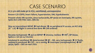
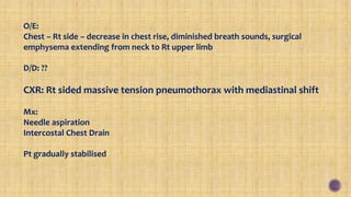
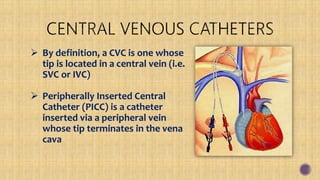
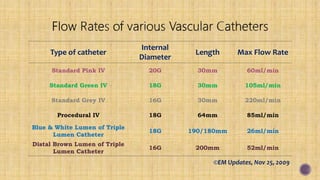
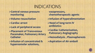
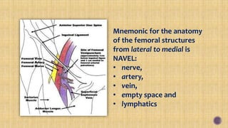
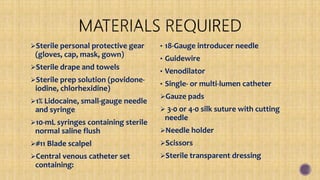
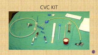
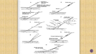
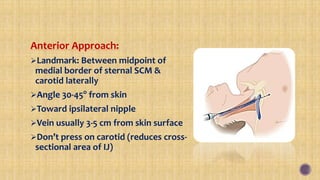
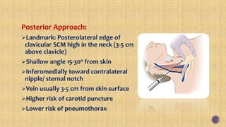
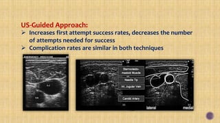
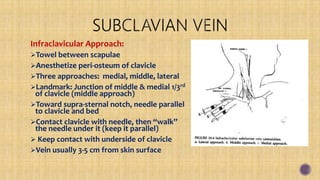
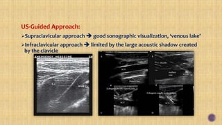
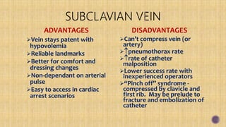
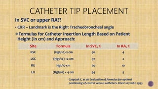
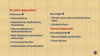
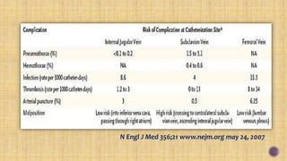
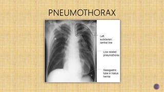
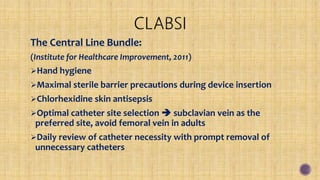
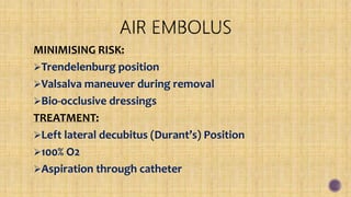
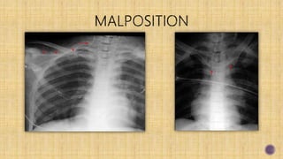
A 72 yrs old male pt in ICU, ventilated, unresponsive
Known C/O COPD, heart failure, hypertension, DM, hypokalaemia
Present vitals: HR-142/min, sinus tachycardia, BP-90/50 on inotropes, RR-24/min,
SpO2-82% with FiO2 100%, CBS-162
Was on intermittent BiPAP  had dinner  no peripheral IV access, on KCl drip
(𝑲+
2.8)  Right IJ central line done  CXR ordered
Became tachypnoeic  put on BiPAP  drowsy, restless  HR, BP 80/40,
SpO2 70% on 14 l O2/min
Intubated & ventilated  deteriorated  BP – NR, very tachypnoeic  IV fluids,
sedated & paralysed  CVP – 10-12 mmHg  Inotropes (norepinephrine)  BP
90/50, SpO2 ~ 80% on 100% FiO2
O/E:
Chest – Rt side – decrease in chest rise, diminished breath sounds, surgical
emphysema extending from neck to Rt upper limb
D/D: ??
CXR: Rt sided massive tension pneumothorax with mediastinal shift
Mx:
Needle aspiration
Intercostal Chest Drain
Pt gradually stabilised
Dr. Bodhisatwa Choudhuri
MBBS, MD, MRCEM (UK), MEM (GWU-USA)
Consultant & HOD, Dept of Emergency & Critical Care, ILS Hospital, Howrah
 By definition, a CVC is one whose
tip is located in a central vein (i.e.
SVC or IVC)
 Peripherally Inserted Central
Catheter (PICC) is a catheter
inserted via a peripheral vein
whose tip terminates in the vena
cava
Infraclavicular (IC) Subclavian venipuncture - first
described by Aubaniac in 1952
In 1965, the supraclavicular (SC) approach was
described
IJ approach (later known as the central approach) was
described in 1966
Later on came the Femoral and cephalic-basilic
approaches
Hagen–Poiseuille equation/Poiseuille’s law:
Rate of flow of an infusion =
𝝅𝒓 𝟒∆𝑷
𝟖𝜼𝑳
where r = catheter
radius, ∆P = pressure gradient, ղ = viscosity, L = length
of catheter
The rate of flow is directly proportional to the catheter
radius and the pressure gradient, and inversely
proportional to the dynamic fluid viscosity and catheter
length
Flow rates are maximized by using the largest internal
diameter catheter possible
Type of catheter
Internal
Diameter
Length Max Flow Rate
Standard Pink IV 20G 30mm 60ml/min
Standard Green IV 18G 30mm 105ml/min
Standard Grey IV 16G 30mm 220ml/min
Procedural IV 18G 64mm 85ml/min
Blue & White Lumen of Triple
Lumen Catheter
18G 190/180mm 26ml/min
Distal Brown Lumen of Triple
Lumen Catheter
16G 200mm 52ml/min
©EM Updates, Nov 25, 2009
Central venous pressure
monitoring
Volume resuscitation
Cardiac arrest
Lack of peripheral access
Placement of Transvenous
Pacemaker, Pulmonary Artery
Catheter
Infusion of concentrated &
hyperosmolar solutions,
vasopressors,
chemotherapeutic agents
Infusion of hyperalimentation
Need of long-term IV
antibiotics
Cardiac Catheterization,
Pulmonary Angiography
Hemodialysis , Plasmapheresis
Aspiration of Air emboli
Distorted local anatomy, No
discernable anatomical
landmarks
Extremes of weight
Vasculitis
Prior long-term venous
cannulation
Prior injection of sclerosis agents
Suspected proximal vascular
injury
Previous radiation therapy
Cellulitis, burns, severe
dermatitis at site
Bleeding disorders
Anticoagulation or thrombolytic
therapy
Combative patients
Inexperienced, unsupervised
physician
Site-specific contraindications
External Jugular Vein
Internal Jugular Vein
 Traditional Approaches (Central/ Anterior/ Posterior)
 US-Guided Approach
Subclavian Vein
 Traditional Approaches (Infraclavicular/ Supraclavicular)
 US-Guided Approach
Femoral vein
 Traditional Approach
 US-Guided Approach
PICC (Peripherally introduced Central Catheters)
Mnemonic for the anatomy
of the femoral structures
from lateral to medial is
NAVEL:
• nerve,
• artery,
• vein,
• empty space and
• lymphatics
Sterile personal protective gear
(gloves, cap, mask, gown)
Sterile drape and towels
Sterile prep solution (povidone-
iodine, chlorhexidine)
1% Lidocaine, small-gauge needle
and syringe
10-mL syringes containing sterile
normal saline flush
#11 Blade scalpel
Central venous catheter set
containing:
• 18-Gauge introducer needle
• Guidewire
• Venodilator
• Single- or multi-lumen catheter
Gauze pads
 3-0 or 4-0 silk suture with cutting
needle
Needle holder
Scissors
Sterile transparent dressing
SELDINGER Technique:
Seldinger originally described this technique in 1953 for
percutaneous arteriography
Use introducing needle to locate vein
Guidewire is threaded through the needle & advanced
Needle is removed over the guidewire
Skin and vessel are dilated using dilator
Catheter is placed over the guidewire
Guidewire is removed
Catheter is secured in place
Easy to find
Angle 10° from skin
Difficulty traversing the EJV-SVC junction
Usually requires J-wire
Excellent site of simple IV cannulation
No risk of pneumothorax
Decreased bleeding risk
Success rates reported 50-90%
Central Approach:
Landmark: Triangle by clavicle & 2
heads of SCM muscle, carotid pulse
Start high in the triangle, 1cm below
apex
Angle 30-45o
from skin
Toward ipsilateral nipple
Vein usually 2-3 cm from skin surface
Anterior Approach:
Landmark: Between midpoint of
medial border of sternal SCM &
carotid laterally
Angle 30-45o
from skin
Toward ipsilateral nipple
Vein usually 3-5 cm from skin surface
Don’t press on carotid (reduces cross-
sectional area of IJ)
Posterior Approach:
Landmark: Posterolateral edge of
clavicular SCM high in the neck (3-5 cm
above clavicle)
Shallow angle 15-30o from skin
Inferomedially toward contralateral
nipple/ sternal notch
Vein usually 3-5 cm from skin surface
Higher risk of carotid puncture
Lower risk of pneumothorax
US-Guided Approach:
 Increases first attempt success rates, decreases the number
of attempts needed for success
 Complication rates are similar in both techniques
ADVANTAGES
Better control of bleeding -
Can compress carotid
RIJ - straight path to SVC/RA
Lower failure rate with
inexperienced operators
Reliable landmarks
 risk of venous thrombosis
in ESRD
DISADVANTAGES
Carotid puncture, hematoma,
airway compression
Higher incidence of arterial
puncture compared with SCV
Difficult with tracheostomy
Vein collapse with hypovolemia
ICP
Difficult for long-term
LIJ may injure thoracic duct
Infraclavicular Approach:
Towel between scapulae
Anesthetize peri-osteum of clavicle
Three approaches: medial, middle, lateral
Landmark: Junction of middle & medial 1/3rd
of clavicle (middle approach)
Toward supra-sternal notch, needle parallel
to clavicle and bed
Contact clavicle with needle, then “walk”
the needle under it (keep it parallel)
 Keep contact with underside of clavicle
Vein usually 3-5 cm from skin surface
Supraclavicular Approach:
Landmark: 1 cm lateral to the clavicular
head of the SCM and 1 cm posterior to
the clavicle
Angle of 10° from skin
Toward the contralateral nipple
Vein usually 2-3 cm from skin surface
Fewer failures, fewer catheter
malpositions, less interference with
CPR
Can be performed in the upright
position
US-Guided Approach:
Supraclavicular approach  good sonographic visualization, ‘venous lake’
Infraclavicular approach  limited by the large acoustic shadow created
by the clavicle
ADVANTAGES
Vein stays patent with
hypovolemia
Reliable landmarks
Better for comfort and
dressing changes
Non-dependant on arterial
pulse
Easy to access in cardiac
arrest scenarios
DISADVANTAGES
Can’t compress vein (or
artery)
pneumothorax rate
rate of catheter
malposition
Lower success rate with
inexperienced operators
“Pinch off” syndrome -
compressed by clavicle and
first rib. May be prelude to
fracture and embolization of
catheter
Traditional Approach:
Find the inguinal ligament,
femoral arterial pulse
Landmark: Medial to femoral
arterial pulse, 1-2cm below inguinal
ligament
Angle 45-60o
from skin surface
Keep hip slightly abducted & leg
slightly externally rotated
US-Guided Approach:
ADVANTAGES
High success rate
No pneumothorax risk
Compressible vessel
DISADVANTAGES
 Infection rate
 Thrombosis rate
Peritoneal cavity entry
Risk of hidden retroperitoneal
haemorrhage
Difficult for PA catheter
insertion
Marked limitation in patient
mobility
Subclavian vein:
Chest wall deformities
Pneumothorax in
contralateral side
Coagulopathy
Chronic obstructive
pulmonary disease
Jugular vein:
Intravenous drug abuse
via the jugular system
Cervical spine injury
Femoral vein:
Need for patient mobility
INDICATIONS
Patient comfort (less pokes)
Convenience
Decreased risk
(pneumothorax or bleeding)
Long term access (0-432 days)
Home therapy (safe, reliable,
easy to manage)
CONTRAINDICATIONS
Peripheral venous obstruction
Inadequate line care
management
Need for extensive blood
products
Septicemia, Coagulopathy
Ipsilateral paralysis or
mastectomy
End stage renal disease
Skin conditions-burns, infections
Hickman Line
Groshong Line
Broviac Catheters
Quinton Catheters
Subcutaneous Ports
In SVC or upper RA??
• CXR – Landmark is the Right Tracheobroncheal angle
Formulas for Catheter Insertion Length Based on Patient
Height (in cm) and Approach:
Site Formula In SVC, % In RA, %
RSC (Hgt/10)-2 cm 96 4
LSC (Hgt/10) +2 cm 97 2
RIJ Hgt/10 cm 90 10
LIJ (Hgt/10) + 4 cm 94 5
Czepizak C, et al: Evaluation of formulas for optimal
positioning of central venous catheters. Chest 107:1662, 1995
Aspirate blood from each port to check viability
Flush with saline or heparin solution to prevent thrombosis
Cap the ports immediately to prevent air-embolism
Secure catheter with sutures
Cover with sterile dressing – Polyurethane transparent dressings
preferred
Reassess the patient clinically, recheck vitals
Look for any signs of known complications
Obtain chest x-ray for IJ and SC lines
Write a procedure note
General:
Vascular
Air embolus
Artery puncture, Local hematoma
Arterial cannulation
Arteriovenous fistula
Pericardial tamponade
Catheter embolus
Mural thrombus formation
Large vein obstruction
Infectious
CLABSI (Central Line Associated
Blood Stream Infections)
Local cellulitis
Osteomyelitis, Septic arthritis
Miscellaneous
Dysrhythmias
Catheter malposition, knotting
SC and IJ approaches:
Pulmonary
Pneumothorax
Haemothorax, Hydrothorax,
Chylothorax
Haemomediastinum,
Hydromediastinum
Neck hematoma and tracheal
obstruction
Tracheal perforation
Endotracheal cuff perforation
Neurologic
Phrenic nerve injury, Brachial plexus
injury
Cerebral infarct
Femoral approach:
Intra-abdominal
Bowel, bladder perforation
Psoas abscess
N Engl J Med 356;21 www.nejm.org may 24, 2007
The Central Line Bundle:
(Institute for Healthcare Improvement, 2011)
Hand hygiene
Maximal sterile barrier precautions during device insertion
Chlorhexidine skin antisepsis
Optimal catheter site selection  subclavian vein as the
preferred site, avoid femoral vein in adults
Daily review of catheter necessity with prompt removal of
unnecessary catheters
MINIMISING RISK:
Trendelenburg position
Valsalva maneuver during removal
Bio-occlusive dressings
TREATMENT:
Left lateral decubitus (Durant’s) Position
100% O2
Aspiration through catheter
Compression of
catheter
between the
clavicle and 1st
rib
Supraclavicular approach is clearly more effective with less complication
rates, even in absence of US-guidance
• Supraclavicular Subclavian Vein Catheterization: The Forgotten Central Line - Shannon P Patrick et al., West
J Emerg Med. 2009 May; 10(2): 110–114
For immunocompromised patients and high-risk neonates, administer
intravenous antibiotic prophylaxis
Use of Chlorhexidine based solution for skin preparation
Use of transparent bio-occlusive dressings
Real-time US-guidance whenever possible
In unintended cannulation of an arterial vessel with a dilator or large-bore
catheter, the dilator or catheter should be left in place and a general
surgeon, a vascular surgeon, or an interventional radiologist should be
immediately consulted regarding surgical or nonsurgical catheter removal
• Practice guidelines for central venous access: a report by the American Society of Anesthesiologists Task Force.
Anesthesiology. 2012 Mar; 116(3):539-73
Cannulation of the central venous circulation is a necessary
skill for emergency clinicians
Inexperienced clinicians should not undertake these
techniques without supervision
Even in experienced hands, complications should be expected
Care should be taken to reassess the patient clinically post-
procedure, to rule out any immediate life-threatening
complications
Clinical Procedures in Emergency Medicine, Roberts and Hedges, 4th ed, 2004
Tintinalli’s Emergency Medicine : A Comprehensive Study Guide, 7th ed, 2011
Atlas of Human Anatomy, Frank Netter, 5th ed, 2010
Systematic review: is real-time ultrasonic-guided central line placement by ED physicians
more successful than the traditional landmark approach? - Ninfa Mehta et al., Emerg Med J
2013;30:355-359
Supraclavicular Subclavian Vein Catheterization: The Forgotten Central Line - Shannon P
Patrick et al., West J Emerg Med. 2009 May; 10(2): 110–114
Implement the IHI Central Line Bundle
http://www.ihi.org/knowledge/Pages/Changes/ImplementtheCentralLineBundle.aspx
Complications of central venous catheters: internal jugular versus subclavian access - a
systematic review – Sibylle Ruesch et al., Critical Care Medicine, 03/2002; 30(2):454-60
Practice guidelines for central venous access: a report by the American Society of
Anesthesiologists Task Force. Anesthesiology. 2012 Mar; 116(3):539-73
Vascular access devices - management of common complications. Richardson D. & Bruso P.,
1993; J Intrav Nurs 16, 44-49
Wikipedia : http://en.wikipedia.org/wiki/Main_Page
"Central Venous Access: Techniques, Complications, and Best Practices" - by Dr Bodhisatwa Choudhuri

"Central Venous Access: Techniques, Complications, and Best Practices" - by Dr Bodhisatwa Choudhuri

  • 1.
    A 72 yrsold male pt in ICU, ventilated, unresponsive Known C/O COPD, heart failure, hypertension, DM, hypokalaemia Present vitals: HR-142/min, sinus tachycardia, BP-90/50 on inotropes, RR-24/min, SpO2-82% with FiO2 100%, CBS-162 Was on intermittent BiPAP  had dinner  no peripheral IV access, on KCl drip (𝑲+ 2.8)  Right IJ central line done  CXR ordered Became tachypnoeic  put on BiPAP  drowsy, restless  HR, BP 80/40, SpO2 70% on 14 l O2/min Intubated & ventilated  deteriorated  BP – NR, very tachypnoeic  IV fluids, sedated & paralysed  CVP – 10-12 mmHg  Inotropes (norepinephrine)  BP 90/50, SpO2 ~ 80% on 100% FiO2
  • 2.
    O/E: Chest – Rtside – decrease in chest rise, diminished breath sounds, surgical emphysema extending from neck to Rt upper limb D/D: ?? CXR: Rt sided massive tension pneumothorax with mediastinal shift Mx: Needle aspiration Intercostal Chest Drain Pt gradually stabilised
  • 3.
    Dr. Bodhisatwa Choudhuri MBBS,MD, MRCEM (UK), MEM (GWU-USA) Consultant & HOD, Dept of Emergency & Critical Care, ILS Hospital, Howrah
  • 4.
     By definition,a CVC is one whose tip is located in a central vein (i.e. SVC or IVC)  Peripherally Inserted Central Catheter (PICC) is a catheter inserted via a peripheral vein whose tip terminates in the vena cava
  • 5.
    Infraclavicular (IC) Subclavianvenipuncture - first described by Aubaniac in 1952 In 1965, the supraclavicular (SC) approach was described IJ approach (later known as the central approach) was described in 1966 Later on came the Femoral and cephalic-basilic approaches
  • 6.
    Hagen–Poiseuille equation/Poiseuille’s law: Rateof flow of an infusion = 𝝅𝒓 𝟒∆𝑷 𝟖𝜼𝑳 where r = catheter radius, ∆P = pressure gradient, ղ = viscosity, L = length of catheter The rate of flow is directly proportional to the catheter radius and the pressure gradient, and inversely proportional to the dynamic fluid viscosity and catheter length
  • 7.
    Flow rates aremaximized by using the largest internal diameter catheter possible
  • 8.
    Type of catheter Internal Diameter LengthMax Flow Rate Standard Pink IV 20G 30mm 60ml/min Standard Green IV 18G 30mm 105ml/min Standard Grey IV 16G 30mm 220ml/min Procedural IV 18G 64mm 85ml/min Blue & White Lumen of Triple Lumen Catheter 18G 190/180mm 26ml/min Distal Brown Lumen of Triple Lumen Catheter 16G 200mm 52ml/min ©EM Updates, Nov 25, 2009
  • 9.
    Central venous pressure monitoring Volumeresuscitation Cardiac arrest Lack of peripheral access Placement of Transvenous Pacemaker, Pulmonary Artery Catheter Infusion of concentrated & hyperosmolar solutions, vasopressors, chemotherapeutic agents Infusion of hyperalimentation Need of long-term IV antibiotics Cardiac Catheterization, Pulmonary Angiography Hemodialysis , Plasmapheresis Aspiration of Air emboli
  • 10.
    Distorted local anatomy,No discernable anatomical landmarks Extremes of weight Vasculitis Prior long-term venous cannulation Prior injection of sclerosis agents Suspected proximal vascular injury Previous radiation therapy Cellulitis, burns, severe dermatitis at site Bleeding disorders Anticoagulation or thrombolytic therapy Combative patients Inexperienced, unsupervised physician Site-specific contraindications
  • 11.
    External Jugular Vein InternalJugular Vein  Traditional Approaches (Central/ Anterior/ Posterior)  US-Guided Approach Subclavian Vein  Traditional Approaches (Infraclavicular/ Supraclavicular)  US-Guided Approach Femoral vein  Traditional Approach  US-Guided Approach PICC (Peripherally introduced Central Catheters)
  • 13.
    Mnemonic for theanatomy of the femoral structures from lateral to medial is NAVEL: • nerve, • artery, • vein, • empty space and • lymphatics
  • 14.
    Sterile personal protectivegear (gloves, cap, mask, gown) Sterile drape and towels Sterile prep solution (povidone- iodine, chlorhexidine) 1% Lidocaine, small-gauge needle and syringe 10-mL syringes containing sterile normal saline flush #11 Blade scalpel Central venous catheter set containing: • 18-Gauge introducer needle • Guidewire • Venodilator • Single- or multi-lumen catheter Gauze pads  3-0 or 4-0 silk suture with cutting needle Needle holder Scissors Sterile transparent dressing
  • 16.
    SELDINGER Technique: Seldinger originallydescribed this technique in 1953 for percutaneous arteriography Use introducing needle to locate vein Guidewire is threaded through the needle & advanced Needle is removed over the guidewire Skin and vessel are dilated using dilator Catheter is placed over the guidewire Guidewire is removed Catheter is secured in place
  • 18.
    Easy to find Angle10° from skin Difficulty traversing the EJV-SVC junction Usually requires J-wire Excellent site of simple IV cannulation No risk of pneumothorax Decreased bleeding risk Success rates reported 50-90%
  • 19.
    Central Approach: Landmark: Triangleby clavicle & 2 heads of SCM muscle, carotid pulse Start high in the triangle, 1cm below apex Angle 30-45o from skin Toward ipsilateral nipple Vein usually 2-3 cm from skin surface
  • 20.
    Anterior Approach: Landmark: Betweenmidpoint of medial border of sternal SCM & carotid laterally Angle 30-45o from skin Toward ipsilateral nipple Vein usually 3-5 cm from skin surface Don’t press on carotid (reduces cross- sectional area of IJ)
  • 21.
    Posterior Approach: Landmark: Posterolateraledge of clavicular SCM high in the neck (3-5 cm above clavicle) Shallow angle 15-30o from skin Inferomedially toward contralateral nipple/ sternal notch Vein usually 3-5 cm from skin surface Higher risk of carotid puncture Lower risk of pneumothorax
  • 22.
    US-Guided Approach:  Increasesfirst attempt success rates, decreases the number of attempts needed for success  Complication rates are similar in both techniques
  • 23.
    ADVANTAGES Better control ofbleeding - Can compress carotid RIJ - straight path to SVC/RA Lower failure rate with inexperienced operators Reliable landmarks  risk of venous thrombosis in ESRD DISADVANTAGES Carotid puncture, hematoma, airway compression Higher incidence of arterial puncture compared with SCV Difficult with tracheostomy Vein collapse with hypovolemia ICP Difficult for long-term LIJ may injure thoracic duct
  • 24.
    Infraclavicular Approach: Towel betweenscapulae Anesthetize peri-osteum of clavicle Three approaches: medial, middle, lateral Landmark: Junction of middle & medial 1/3rd of clavicle (middle approach) Toward supra-sternal notch, needle parallel to clavicle and bed Contact clavicle with needle, then “walk” the needle under it (keep it parallel)  Keep contact with underside of clavicle Vein usually 3-5 cm from skin surface
  • 25.
    Supraclavicular Approach: Landmark: 1cm lateral to the clavicular head of the SCM and 1 cm posterior to the clavicle Angle of 10° from skin Toward the contralateral nipple Vein usually 2-3 cm from skin surface Fewer failures, fewer catheter malpositions, less interference with CPR Can be performed in the upright position
  • 26.
    US-Guided Approach: Supraclavicular approach good sonographic visualization, ‘venous lake’ Infraclavicular approach  limited by the large acoustic shadow created by the clavicle
  • 27.
    ADVANTAGES Vein stays patentwith hypovolemia Reliable landmarks Better for comfort and dressing changes Non-dependant on arterial pulse Easy to access in cardiac arrest scenarios DISADVANTAGES Can’t compress vein (or artery) pneumothorax rate rate of catheter malposition Lower success rate with inexperienced operators “Pinch off” syndrome - compressed by clavicle and first rib. May be prelude to fracture and embolization of catheter
  • 28.
    Traditional Approach: Find theinguinal ligament, femoral arterial pulse Landmark: Medial to femoral arterial pulse, 1-2cm below inguinal ligament Angle 45-60o from skin surface Keep hip slightly abducted & leg slightly externally rotated
  • 29.
  • 30.
    ADVANTAGES High success rate Nopneumothorax risk Compressible vessel DISADVANTAGES  Infection rate  Thrombosis rate Peritoneal cavity entry Risk of hidden retroperitoneal haemorrhage Difficult for PA catheter insertion Marked limitation in patient mobility
  • 31.
    Subclavian vein: Chest walldeformities Pneumothorax in contralateral side Coagulopathy Chronic obstructive pulmonary disease Jugular vein: Intravenous drug abuse via the jugular system Cervical spine injury Femoral vein: Need for patient mobility
  • 33.
    INDICATIONS Patient comfort (lesspokes) Convenience Decreased risk (pneumothorax or bleeding) Long term access (0-432 days) Home therapy (safe, reliable, easy to manage) CONTRAINDICATIONS Peripheral venous obstruction Inadequate line care management Need for extensive blood products Septicemia, Coagulopathy Ipsilateral paralysis or mastectomy End stage renal disease Skin conditions-burns, infections
  • 34.
    Hickman Line Groshong Line BroviacCatheters Quinton Catheters Subcutaneous Ports
  • 35.
    In SVC orupper RA?? • CXR – Landmark is the Right Tracheobroncheal angle Formulas for Catheter Insertion Length Based on Patient Height (in cm) and Approach: Site Formula In SVC, % In RA, % RSC (Hgt/10)-2 cm 96 4 LSC (Hgt/10) +2 cm 97 2 RIJ Hgt/10 cm 90 10 LIJ (Hgt/10) + 4 cm 94 5 Czepizak C, et al: Evaluation of formulas for optimal positioning of central venous catheters. Chest 107:1662, 1995
  • 36.
    Aspirate blood fromeach port to check viability Flush with saline or heparin solution to prevent thrombosis Cap the ports immediately to prevent air-embolism Secure catheter with sutures Cover with sterile dressing – Polyurethane transparent dressings preferred Reassess the patient clinically, recheck vitals Look for any signs of known complications Obtain chest x-ray for IJ and SC lines Write a procedure note
  • 37.
    General: Vascular Air embolus Artery puncture,Local hematoma Arterial cannulation Arteriovenous fistula Pericardial tamponade Catheter embolus Mural thrombus formation Large vein obstruction Infectious CLABSI (Central Line Associated Blood Stream Infections) Local cellulitis Osteomyelitis, Septic arthritis Miscellaneous Dysrhythmias Catheter malposition, knotting
  • 38.
    SC and IJapproaches: Pulmonary Pneumothorax Haemothorax, Hydrothorax, Chylothorax Haemomediastinum, Hydromediastinum Neck hematoma and tracheal obstruction Tracheal perforation Endotracheal cuff perforation Neurologic Phrenic nerve injury, Brachial plexus injury Cerebral infarct Femoral approach: Intra-abdominal Bowel, bladder perforation Psoas abscess
  • 39.
    N Engl JMed 356;21 www.nejm.org may 24, 2007
  • 41.
    The Central LineBundle: (Institute for Healthcare Improvement, 2011) Hand hygiene Maximal sterile barrier precautions during device insertion Chlorhexidine skin antisepsis Optimal catheter site selection  subclavian vein as the preferred site, avoid femoral vein in adults Daily review of catheter necessity with prompt removal of unnecessary catheters
  • 43.
    MINIMISING RISK: Trendelenburg position Valsalvamaneuver during removal Bio-occlusive dressings TREATMENT: Left lateral decubitus (Durant’s) Position 100% O2 Aspiration through catheter
  • 46.
  • 47.
    Supraclavicular approach isclearly more effective with less complication rates, even in absence of US-guidance • Supraclavicular Subclavian Vein Catheterization: The Forgotten Central Line - Shannon P Patrick et al., West J Emerg Med. 2009 May; 10(2): 110–114 For immunocompromised patients and high-risk neonates, administer intravenous antibiotic prophylaxis Use of Chlorhexidine based solution for skin preparation Use of transparent bio-occlusive dressings Real-time US-guidance whenever possible In unintended cannulation of an arterial vessel with a dilator or large-bore catheter, the dilator or catheter should be left in place and a general surgeon, a vascular surgeon, or an interventional radiologist should be immediately consulted regarding surgical or nonsurgical catheter removal • Practice guidelines for central venous access: a report by the American Society of Anesthesiologists Task Force. Anesthesiology. 2012 Mar; 116(3):539-73
  • 48.
    Cannulation of thecentral venous circulation is a necessary skill for emergency clinicians Inexperienced clinicians should not undertake these techniques without supervision Even in experienced hands, complications should be expected Care should be taken to reassess the patient clinically post- procedure, to rule out any immediate life-threatening complications
  • 49.
    Clinical Procedures inEmergency Medicine, Roberts and Hedges, 4th ed, 2004 Tintinalli’s Emergency Medicine : A Comprehensive Study Guide, 7th ed, 2011 Atlas of Human Anatomy, Frank Netter, 5th ed, 2010 Systematic review: is real-time ultrasonic-guided central line placement by ED physicians more successful than the traditional landmark approach? - Ninfa Mehta et al., Emerg Med J 2013;30:355-359 Supraclavicular Subclavian Vein Catheterization: The Forgotten Central Line - Shannon P Patrick et al., West J Emerg Med. 2009 May; 10(2): 110–114 Implement the IHI Central Line Bundle http://www.ihi.org/knowledge/Pages/Changes/ImplementtheCentralLineBundle.aspx Complications of central venous catheters: internal jugular versus subclavian access - a systematic review – Sibylle Ruesch et al., Critical Care Medicine, 03/2002; 30(2):454-60 Practice guidelines for central venous access: a report by the American Society of Anesthesiologists Task Force. Anesthesiology. 2012 Mar; 116(3):539-73 Vascular access devices - management of common complications. Richardson D. & Bruso P., 1993; J Intrav Nurs 16, 44-49 Wikipedia : http://en.wikipedia.org/wiki/Main_Page

Editor's Notes

  • #13 The IJ vein lies lateral to the IC artery inside the carotid sheath. It joins the subclavian vein to form the brachiocephalic vein. The subclavian vein crosses under the clavicle at the medial to proximal third of the clavicle
  • #23 Vascular structures are anechoic (black) in US imaging Veins are more easily compressed, have thinner walls, and have no arterial pulsation Colour flow may also help differentiate between vein and artery
  • #36 Against R-atrial placement: Cardiac performation and tamponade, Cardiac arrhythmias, Catheter induced thrombosis For R-atrial placement: Optimal performance and superior functional durability
  • #41 SCV > IJV Life threatening More dangerous if on positive pressure ventilation Early diagnosis Prompt intervention Mx: Needle aspiration Intercostal chest drain
  • #43 Within days of insertion, most catheters are coated with a fibrin sheath Most clots arise within 30 days These clots can cause pulmonary embolism (most are asymptomatic) Thrombosis of blood vessel increases the risk of infection
  • #45 Differentiation by Colour of blood, pulsatile nature If in any doubt, do not introduce guidewire May lead to perforation of arterial system, haemothorax, haemomediastinum