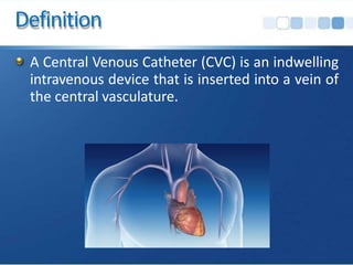
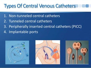
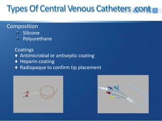
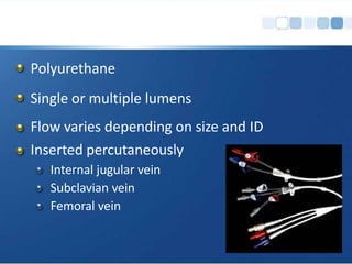
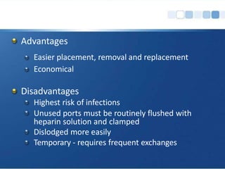
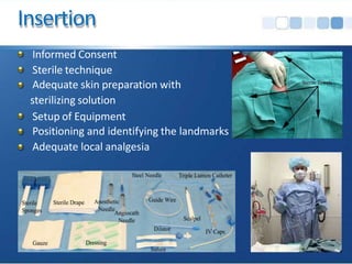
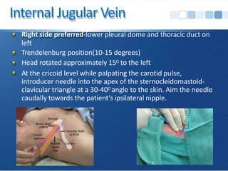
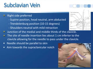
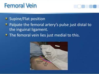
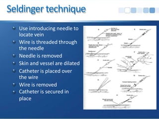
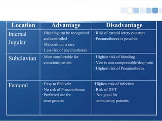
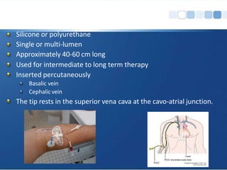
Central Venous Access &
Catheters
Definition
A Central Venous Catheter (CVC) is an indwelling
intravenous device that is inserted into a vein of
the central vasculature.
Uses
1. Difficult Peripheral Vascular Access
Ex - patients with burns, previous vein injuries (such as
IV drug use)
2. Volume Loading
Time-consuming to insert and are associated with
high complication rates.
Flow rate is determined by the calibre and length of
the catheter (Poiseuille’s law)
Shorter and greater calibre catheters delivering
greater volumes over equivalent amounts of time
3. Provision of Caustic Medications or Solutions
Vasoactive medications (vasopressors or inotropes)
Irritant substances (chemotherapeutic agents,
cytotoxic drugs or high concentration solutions)
Total parenteral nutrition
4. Central Venous Pressure Monitoring
The central venous pressure (CVP) is the pressure
measured in the central veins close to Right atrium.
It indicates mean right atrial pressure and is
frequently used as an estimate of right ventricular
preload.
Being used as a guide for fluid management, though
some researches suggest otherwise
(http://www.ncbi.nlm.nih.gov/pubmed/18628220)
5.Repeated Blood Sampling
6.Introduction of Pacemakers or Pulmonary Artery
Catheters
7.For haemodialysis/haemofiltration - For acute and chronic
haemodialysis access
Contraindications
Absolute
Overlying skin or soft tissue infection
Thrombophlebitis
Contraindications .cont
Relative
1. Distorted Anatomy – Trauma, deformity, burns.
2. Infection at the Site of Access – cellulitis
3. Uncooperative patients
4. Proximal Vascular Injury
5. Bleeding disorders & anticoagulation or thrombolytic
therapy.
3% complication rate as long as there are no arterial punctures (Mumatz
et al)
Absolute contraindication for subclavian access
Ultrasound guidance is recommended
Types Of Central Venous Catheters
1. Non-tunneled central catheters
2. Tunneled central catheters
3. Peripherally inserted central catheters (PICC)
4. Implantable ports
TypesOfCentralVenousCatheters.cont
Single & multi-lumen catheters are available in
all catheter types
Each lumen must be treated as a separate
catheter
Types Of CentralVenous Catheters.cont
Open–ended
The catheter is open at the distal tip
The catheter requires clamping before entry into the
system
Clamps are usually built into the catheter
Requires periodic flushing
Types Of CentralVenous Catheters.cont
Closed-ended
A valve is present at the tip of the catheter (eg.
Groshong®) or at the hub of the catheter(eg. PAS-V®)
Clamping is not required as the valve is closed except
during infusion or aspiration
Types Of CentralVenous Catheters.cont
Composition
Silicone
Polyurethane
Coatings
♦ Antimicrobial or antiseptic coating
♦ Heparin coating
♦ Radiopaque to confirm tip placement
The type of CVC inserted depends on the
Type of therapy to be administered
Length of therapy (Short term or Long term)
Previous devices and complications
Patient preference
Non Tunnelled Catheters
Polyurethane
Single or multiple lumens
Flow varies depending on size and ID
Inserted percutaneously
Internal jugular vein
Subclavian vein
Femoral vein
Advantages
Easier placement, removal and replacement
Economical
Disadvantages
Highest risk of infections
Unused ports must be routinely flushed with
heparin solution and clamped
Dislodged more easily
Temporary - requires frequent exchanges
Insertion
Informed Consent
Sterile technique
Adequate skin preparation with
sterilizing solution
Setup of Equipment
Positioning and identifying the landmarks
Adequate local analgesia
Internal Jugular Vein
Right side preferred-lower pleural dome and thoracic duct on
left
Trendelenburg position(10-15 degrees)
Head rotated approximately 150 to the left
At the cricoid level while palpating the carotid pulse,
introducer needle into the apex of the sternocleidomastoid-
clavicular triangle at a 30-400 angle to the skin. Aim the needle
caudally towards the patient’s ipsilateral nipple.
Subclavian Vein
Right side preferred
- Supine position, head neutral, arm abducted
- Trendelenburg position (10-15 degrees)
- Shoulders neutral with mild retraction
Junction of the medial and middle thirds of the clavicle.
The site of needle insertion lies about 1 cm inferior to the
clavicle allowing for the needle to pass under the clavicle.
• Needle should be parallel to skin
• Aim towards the supraclavicular notch
Femoral Vein
Supine/Flat position
Palpate the femoral artery’s pulse just distal to
the inguinal ligament.
The femoral vein lies just medial to this.
Seldinger technique
Use introducing needle to
locate vein
Wire is threaded through
the needle
Needle is removed
Skin and vessel are dilated
Catheter is placed over
the wire
Wire is removed
Catheter is secured in
place
Post-Catheter Placement
Aspirate blood from each port
Flush with saline or sterile water
Secure catheter with sutures
Cover with sterile dressing (tega-derm)
Obtain chest x-ray for IJ and SC lines
Location Advantage Disadvantage
Internal
Jugular
• Bleeding can be recognized
and controlled
• Malposition is rare
• Less risk of pneumothorax
• Risk of carotid artery puncture
• Pneumothorax is possible
Subclavian • Most comfortable for
conscious patient
• Highest risk of bleeding
• Vein is non-compressible/deep vein
• Highest risk of Pneumothorax
Femoral •Easy to find vein
• No risk of Pneumothorax
•Preferred site for
emergencies
•Highest risk of infection
• Risk of DVT
• Not good for
ambulatory patients
Tunnelled Catheters
Single or multiple lumens
Used for long term therapy
Inserted surgically
Small Dacron (Polyethylene terephthalate) cuff sits in
subcutaneous tunnel facilitates anchoring of the catheter
through granulation and acts as a barrier to infection
Advantages
Can be left in place indefinitely (if no infection,
blockage or thrombosis)
Self-care by patient
External portion can be repaired
Disadvantages
Inserted in the OR
Requires a dressing & frequent assessments
External device
Physician must remove
Peripherally Inserted Central
Catheters
Silicone or polyurethane
Single or multi-lumen
Approximately 40-60 cm long
Used for intermediate to long term therapy
Inserted percutaneously
Basalic vein
Cephalic vein
The tip rests in the superior vena cava at the cavo-atrial junction.
Advantages
Can remain in place for several weeks to a year
Can be easily removed
Low infection rates
External portion can be repaired
Disadvantages
Low flows
Requires a dressing & frequent assessments
External device
Small gauge PICC not recommended for blood
sampling
IMPLANTABLE VENOUS ACCESS
DEVICE
(IVAD)
long-term (months to years) single or dual chamber
“port” surgically implanted in the subcutaneous
tissue, usually in the upper chest
Single or double lumen.
Each chamber must be managed separately.
A non-coring point needle is required to access
the device
Unused port is flushed every 28 days with
Heparin solution
Advantages
Internal device, no dressing or site care
Can be permanent
Unrestricted activity
Decreased risk of infection
No external components to break
May be used as long as the device is required,
functional.
Disadvantages
Needle access is required
Surgical procedure required to insert/remove
Cost
Complications Associated
With
Central Venous Catheters
Complications
Acute
Chronic
Complication rate depends on
Site
Patient factors (illnesses, variations in anatomy)
Operator skill and experience.
Acute complications
Cardiac Dysrhythmias
Due to cardiac irritation by the wire or catheter tip.
Withdraw the line into the superior vena cava.
Always use a cardiac monitor.
Haematoma formation – Arterial/Venous puncture
Mechanical injury to nearby structures
Pneumothorax/Haemothoarx
Atrial wall puncture - pericardial tamponade.
Bowel penetration, Bladder puncture, Femoral nerve
injury
Air embolus
Malposition
Lost Guide-wire
Chronic complications
Infections
Catheter fragmentation
Non-function/Blockage - fibrin builds on and
around the catheter and vessel, drug
precipitates, lipid deposits
Thrombosis/Thromboembolism
Air embolism
Deadly complication associated with CVC’s
Signs and Symptoms
Respiratory changes: sudden shortness of breath,
cyanosis
CVS changes: sudden onset of chest pain, ↑HR,
↓BP
CNS changes: altered neurological signs, dizziness,
confusion, loss of Consciousness
Management
Left lateral decubitus with head low Position (Durant
maneuver and Trendelenburg position)
Clamp the Central Venous Catheter
100% O2
Direct removal of air from the venous circulation by
aspiration from a central venous catheter in the right
atrium may be attempted
To minimize the chance of air entering the
system:
Ensure the lumen is clamped prior to opening the
system
Position the patient so that the insertion site is at
or below the level of the heart during insertion
and removal of catheter
Infections
Most frequent and serious complications.
Types
Local infection – Cellulitis
Central Line-Associated Bloodstream Infections
(CLABSI)
Causative Organisms
Staph epidermidis 25-50%
Staph aureus
Candida
25%
5-10%
Risk Factors
Cutaneous colonization of the insertion site
Moisture under the dressing
Prolonged catheter time
Technique of care and placement of the central
line
Evidence-BasedStrategiesSelectedto
Reduce CLA-BSIs
1. Hand hygiene
2. Maximal sterile barriers
3. Chlorhexidine for skin asepsis
4. Avoid femoral lines
5. Avoid/remove unnecessary lines
Thank You
Refenrences
1. http://emedicine.medscape.com/article/80336-overview#a05
2. http://emedicine.medscape.com/article/80355-overview#aw2aab6c10
3. http://emedicine.medscape.com/article/80279-overview
4. Central Venous Access (Ian Rigby, Daniel Howes, Jason Lord, Ian
Walker, Resuscitation Education Consortium/Kingston Resuscitation
Institute)

central lines.pptx

  • 1.
  • 2.
    Definition A Central VenousCatheter (CVC) is an indwelling intravenous device that is inserted into a vein of the central vasculature.
  • 3.
    Uses 1. Difficult PeripheralVascular Access Ex - patients with burns, previous vein injuries (such as IV drug use) 2. Volume Loading Time-consuming to insert and are associated with high complication rates. Flow rate is determined by the calibre and length of the catheter (Poiseuille’s law) Shorter and greater calibre catheters delivering greater volumes over equivalent amounts of time
  • 4.
    3. Provision ofCaustic Medications or Solutions Vasoactive medications (vasopressors or inotropes) Irritant substances (chemotherapeutic agents, cytotoxic drugs or high concentration solutions) Total parenteral nutrition
  • 5.
    4. Central VenousPressure Monitoring The central venous pressure (CVP) is the pressure measured in the central veins close to Right atrium. It indicates mean right atrial pressure and is frequently used as an estimate of right ventricular preload. Being used as a guide for fluid management, though some researches suggest otherwise (http://www.ncbi.nlm.nih.gov/pubmed/18628220)
  • 6.
    5.Repeated Blood Sampling 6.Introductionof Pacemakers or Pulmonary Artery Catheters 7.For haemodialysis/haemofiltration - For acute and chronic haemodialysis access
  • 7.
    Contraindications Absolute Overlying skin orsoft tissue infection Thrombophlebitis
  • 8.
    Contraindications .cont Relative 1. DistortedAnatomy – Trauma, deformity, burns. 2. Infection at the Site of Access – cellulitis 3. Uncooperative patients 4. Proximal Vascular Injury 5. Bleeding disorders & anticoagulation or thrombolytic therapy. 3% complication rate as long as there are no arterial punctures (Mumatz et al) Absolute contraindication for subclavian access Ultrasound guidance is recommended
  • 9.
    Types Of CentralVenous Catheters 1. Non-tunneled central catheters 2. Tunneled central catheters 3. Peripherally inserted central catheters (PICC) 4. Implantable ports
  • 10.
    TypesOfCentralVenousCatheters.cont Single & multi-lumencatheters are available in all catheter types Each lumen must be treated as a separate catheter
  • 11.
    Types Of CentralVenousCatheters.cont Open–ended The catheter is open at the distal tip The catheter requires clamping before entry into the system Clamps are usually built into the catheter Requires periodic flushing
  • 12.
    Types Of CentralVenousCatheters.cont Closed-ended A valve is present at the tip of the catheter (eg. Groshong®) or at the hub of the catheter(eg. PAS-V®) Clamping is not required as the valve is closed except during infusion or aspiration
  • 13.
    Types Of CentralVenousCatheters.cont Composition Silicone Polyurethane Coatings ♦ Antimicrobial or antiseptic coating ♦ Heparin coating ♦ Radiopaque to confirm tip placement
  • 14.
    The type ofCVC inserted depends on the Type of therapy to be administered Length of therapy (Short term or Long term) Previous devices and complications Patient preference
  • 15.
  • 16.
    Polyurethane Single or multiplelumens Flow varies depending on size and ID Inserted percutaneously Internal jugular vein Subclavian vein Femoral vein
  • 17.
    Advantages Easier placement, removaland replacement Economical Disadvantages Highest risk of infections Unused ports must be routinely flushed with heparin solution and clamped Dislodged more easily Temporary - requires frequent exchanges
  • 18.
    Insertion Informed Consent Sterile technique Adequateskin preparation with sterilizing solution Setup of Equipment Positioning and identifying the landmarks Adequate local analgesia
  • 19.
    Internal Jugular Vein Rightside preferred-lower pleural dome and thoracic duct on left Trendelenburg position(10-15 degrees) Head rotated approximately 150 to the left At the cricoid level while palpating the carotid pulse, introducer needle into the apex of the sternocleidomastoid- clavicular triangle at a 30-400 angle to the skin. Aim the needle caudally towards the patient’s ipsilateral nipple.
  • 20.
    Subclavian Vein Right sidepreferred - Supine position, head neutral, arm abducted - Trendelenburg position (10-15 degrees) - Shoulders neutral with mild retraction Junction of the medial and middle thirds of the clavicle. The site of needle insertion lies about 1 cm inferior to the clavicle allowing for the needle to pass under the clavicle. • Needle should be parallel to skin • Aim towards the supraclavicular notch
  • 21.
    Femoral Vein Supine/Flat position Palpatethe femoral artery’s pulse just distal to the inguinal ligament. The femoral vein lies just medial to this.
  • 22.
    Seldinger technique Use introducingneedle to locate vein Wire is threaded through the needle Needle is removed Skin and vessel are dilated Catheter is placed over the wire Wire is removed Catheter is secured in place
  • 23.
    Post-Catheter Placement Aspirate bloodfrom each port Flush with saline or sterile water Secure catheter with sutures Cover with sterile dressing (tega-derm) Obtain chest x-ray for IJ and SC lines
  • 24.
    Location Advantage Disadvantage Internal Jugular •Bleeding can be recognized and controlled • Malposition is rare • Less risk of pneumothorax • Risk of carotid artery puncture • Pneumothorax is possible Subclavian • Most comfortable for conscious patient • Highest risk of bleeding • Vein is non-compressible/deep vein • Highest risk of Pneumothorax Femoral •Easy to find vein • No risk of Pneumothorax •Preferred site for emergencies •Highest risk of infection • Risk of DVT • Not good for ambulatory patients
  • 25.
  • 26.
    Single or multiplelumens Used for long term therapy Inserted surgically Small Dacron (Polyethylene terephthalate) cuff sits in subcutaneous tunnel facilitates anchoring of the catheter through granulation and acts as a barrier to infection
  • 27.
    Advantages Can be leftin place indefinitely (if no infection, blockage or thrombosis) Self-care by patient External portion can be repaired
  • 28.
    Disadvantages Inserted in theOR Requires a dressing & frequent assessments External device Physician must remove
  • 29.
  • 30.
    Silicone or polyurethane Singleor multi-lumen Approximately 40-60 cm long Used for intermediate to long term therapy Inserted percutaneously Basalic vein Cephalic vein The tip rests in the superior vena cava at the cavo-atrial junction.
  • 31.
    Advantages Can remain inplace for several weeks to a year Can be easily removed Low infection rates External portion can be repaired
  • 32.
    Disadvantages Low flows Requires adressing & frequent assessments External device Small gauge PICC not recommended for blood sampling
  • 33.
  • 34.
    long-term (months toyears) single or dual chamber “port” surgically implanted in the subcutaneous tissue, usually in the upper chest Single or double lumen. Each chamber must be managed separately.
  • 35.
    A non-coring pointneedle is required to access the device Unused port is flushed every 28 days with Heparin solution
  • 36.
    Advantages Internal device, nodressing or site care Can be permanent Unrestricted activity Decreased risk of infection No external components to break May be used as long as the device is required, functional.
  • 37.
    Disadvantages Needle access isrequired Surgical procedure required to insert/remove Cost
  • 38.
  • 39.
    Complications Acute Chronic Complication rate dependson Site Patient factors (illnesses, variations in anatomy) Operator skill and experience.
  • 40.
    Acute complications Cardiac Dysrhythmias Dueto cardiac irritation by the wire or catheter tip. Withdraw the line into the superior vena cava. Always use a cardiac monitor. Haematoma formation – Arterial/Venous puncture Mechanical injury to nearby structures Pneumothorax/Haemothoarx Atrial wall puncture - pericardial tamponade. Bowel penetration, Bladder puncture, Femoral nerve injury Air embolus Malposition Lost Guide-wire
  • 41.
    Chronic complications Infections Catheter fragmentation Non-function/Blockage- fibrin builds on and around the catheter and vessel, drug precipitates, lipid deposits Thrombosis/Thromboembolism
  • 42.
    Air embolism Deadly complicationassociated with CVC’s Signs and Symptoms Respiratory changes: sudden shortness of breath, cyanosis CVS changes: sudden onset of chest pain, ↑HR, ↓BP CNS changes: altered neurological signs, dizziness, confusion, loss of Consciousness
  • 43.
    Management Left lateral decubituswith head low Position (Durant maneuver and Trendelenburg position) Clamp the Central Venous Catheter 100% O2 Direct removal of air from the venous circulation by aspiration from a central venous catheter in the right atrium may be attempted
  • 44.
    To minimize thechance of air entering the system: Ensure the lumen is clamped prior to opening the system Position the patient so that the insertion site is at or below the level of the heart during insertion and removal of catheter
  • 45.
    Infections Most frequent andserious complications. Types Local infection – Cellulitis Central Line-Associated Bloodstream Infections (CLABSI)
  • 46.
    Causative Organisms Staph epidermidis25-50% Staph aureus Candida 25% 5-10% Risk Factors Cutaneous colonization of the insertion site Moisture under the dressing Prolonged catheter time Technique of care and placement of the central line
  • 47.
    Evidence-BasedStrategiesSelectedto Reduce CLA-BSIs 1. Handhygiene 2. Maximal sterile barriers 3. Chlorhexidine for skin asepsis 4. Avoid femoral lines 5. Avoid/remove unnecessary lines
  • 48.
    Thank You Refenrences 1. http://emedicine.medscape.com/article/80336-overview#a05 2.http://emedicine.medscape.com/article/80355-overview#aw2aab6c10 3. http://emedicine.medscape.com/article/80279-overview 4. Central Venous Access (Ian Rigby, Daniel Howes, Jason Lord, Ian Walker, Resuscitation Education Consortium/Kingston Resuscitation Institute)