Downloaded 106 times

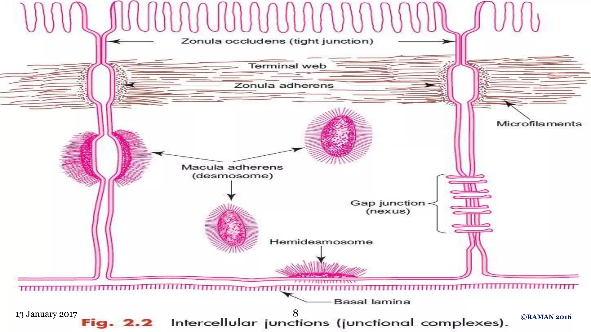
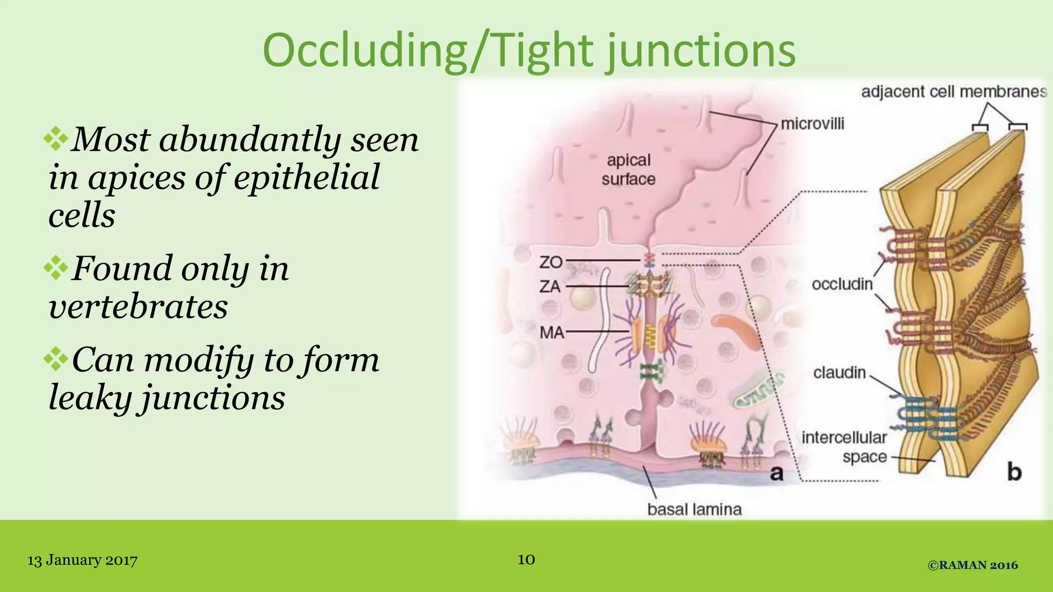
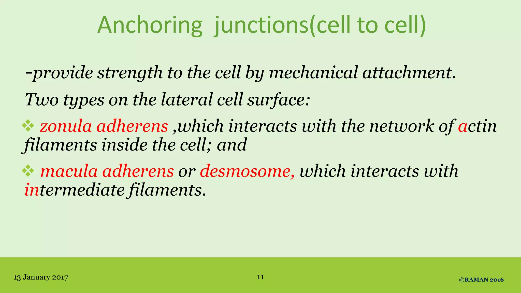
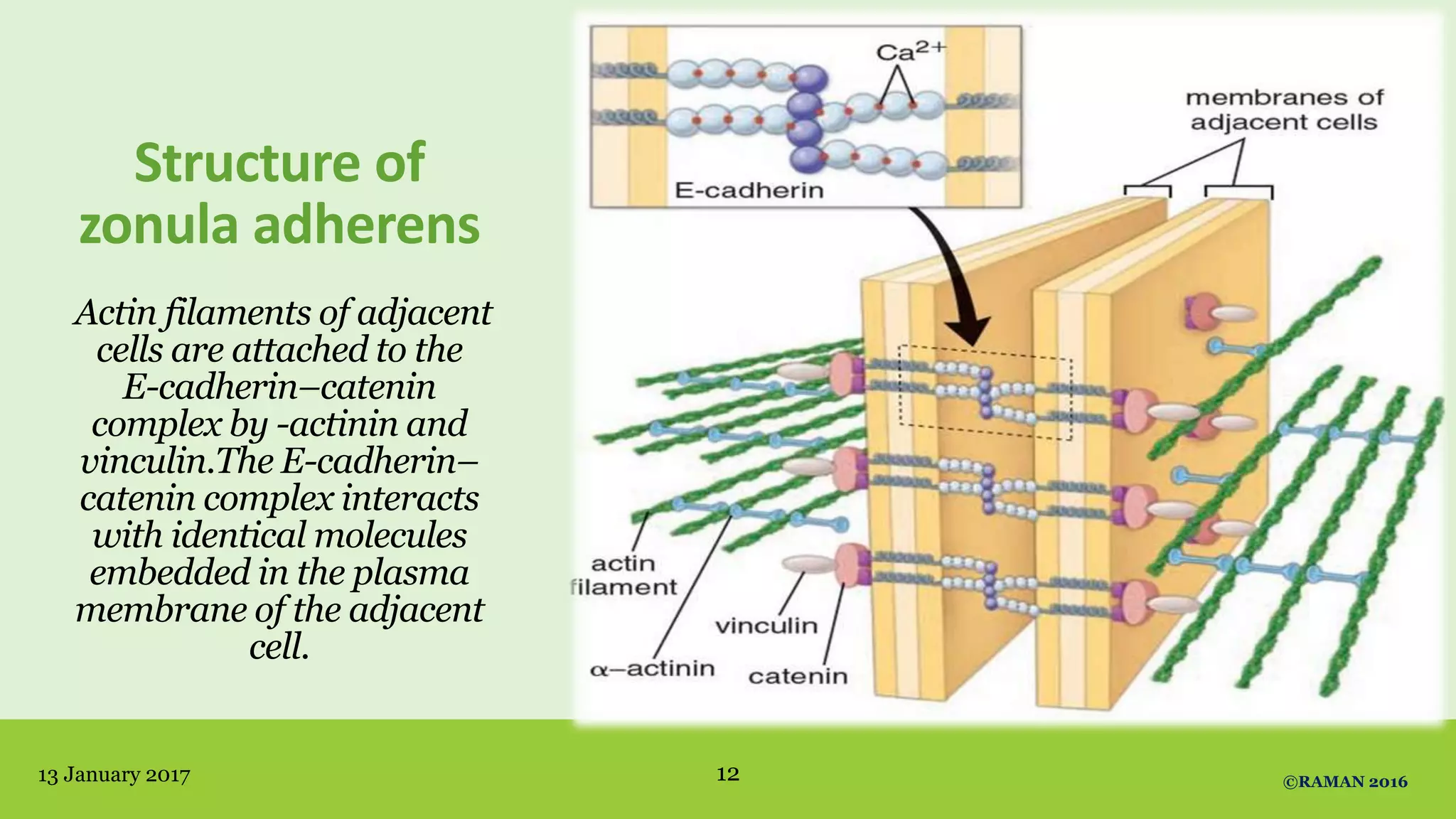
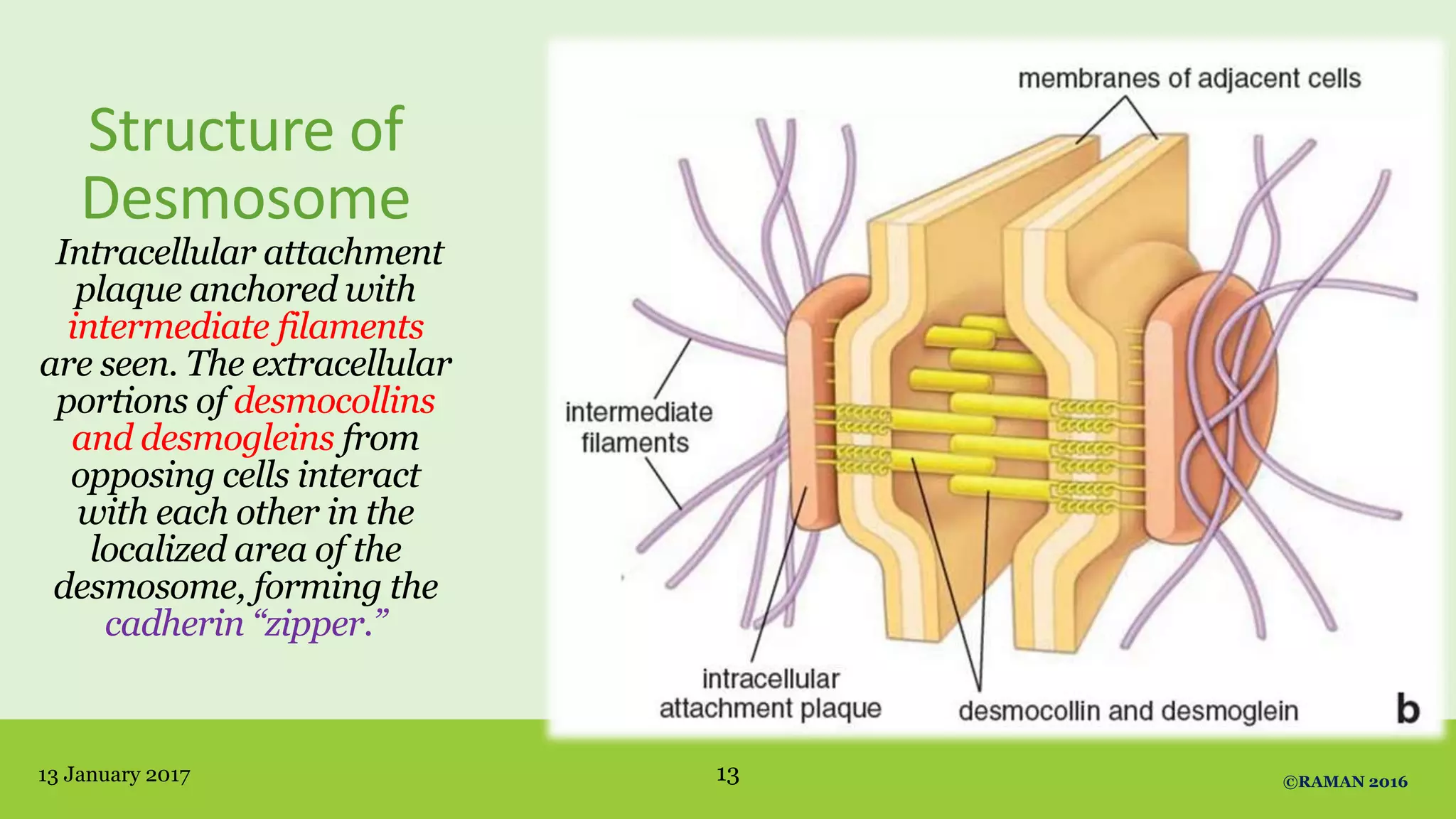
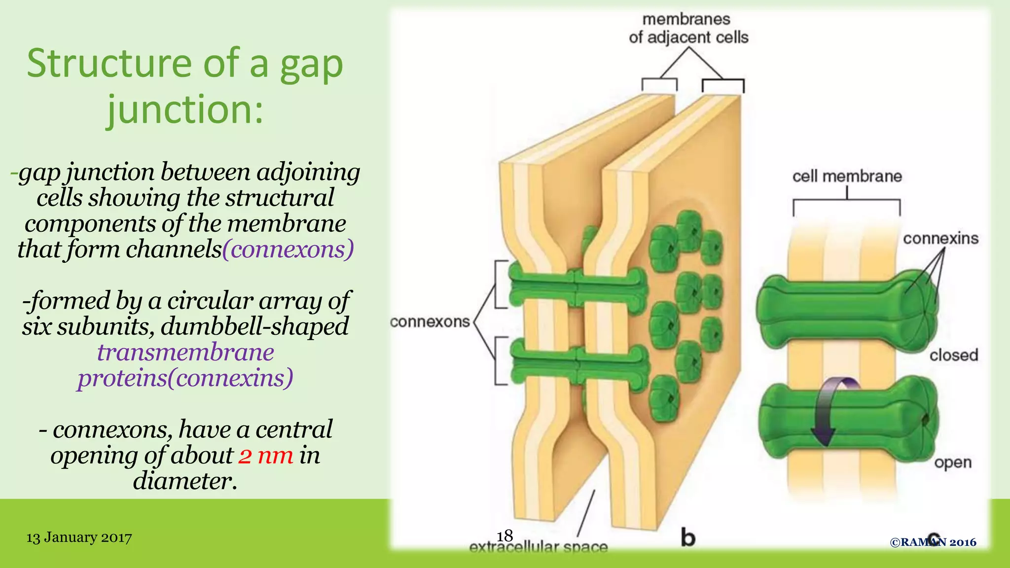

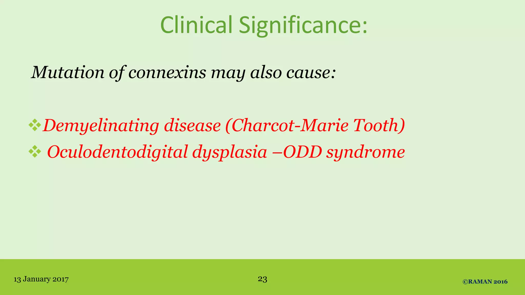
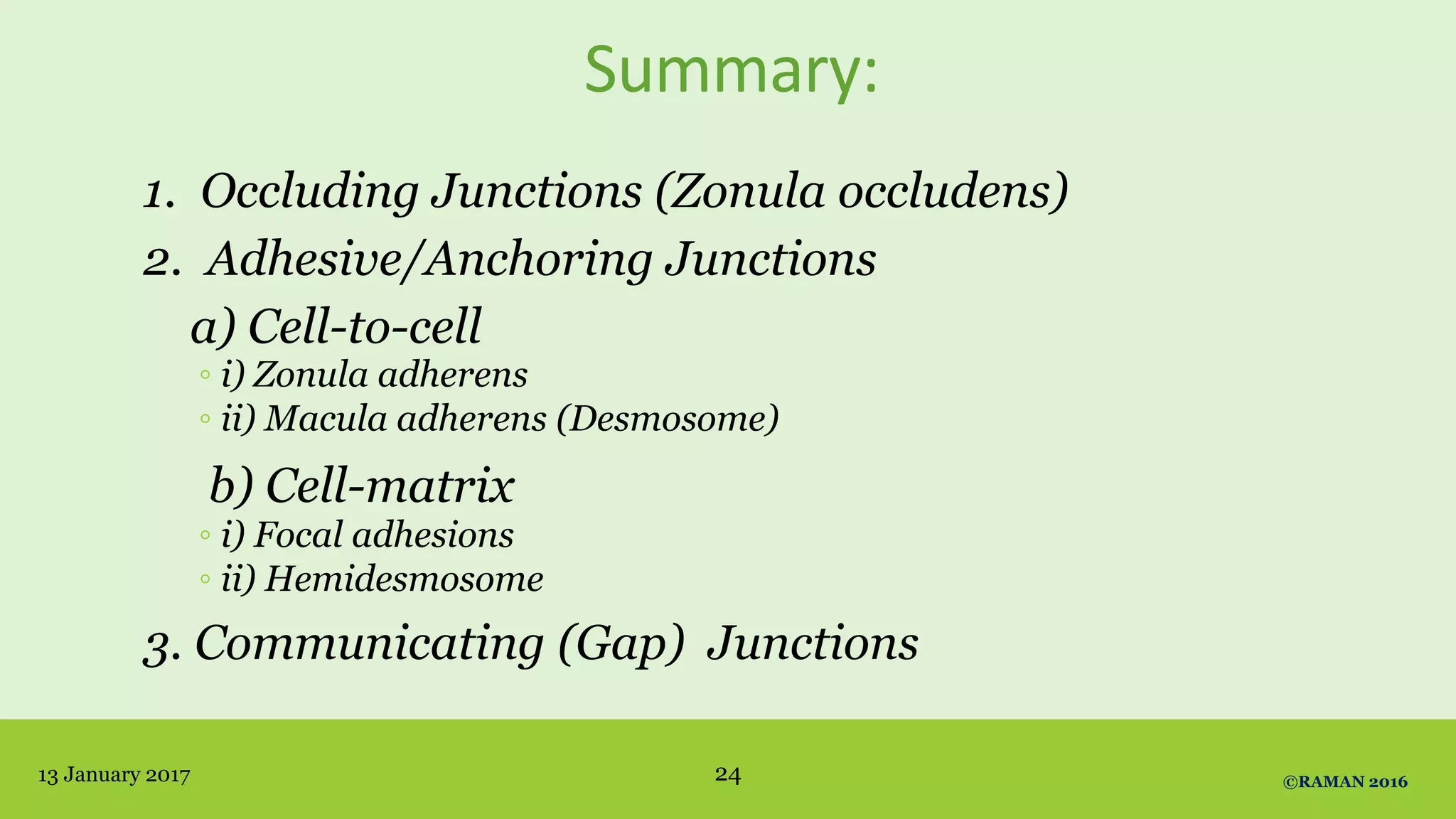
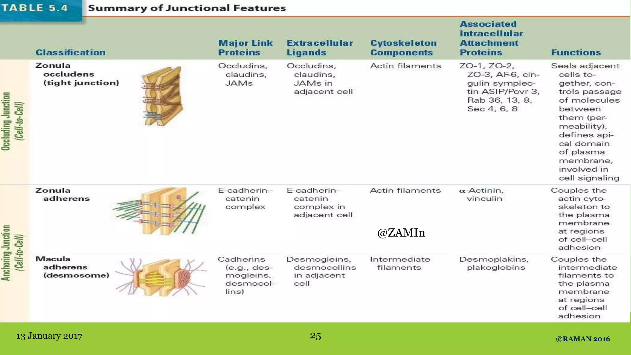
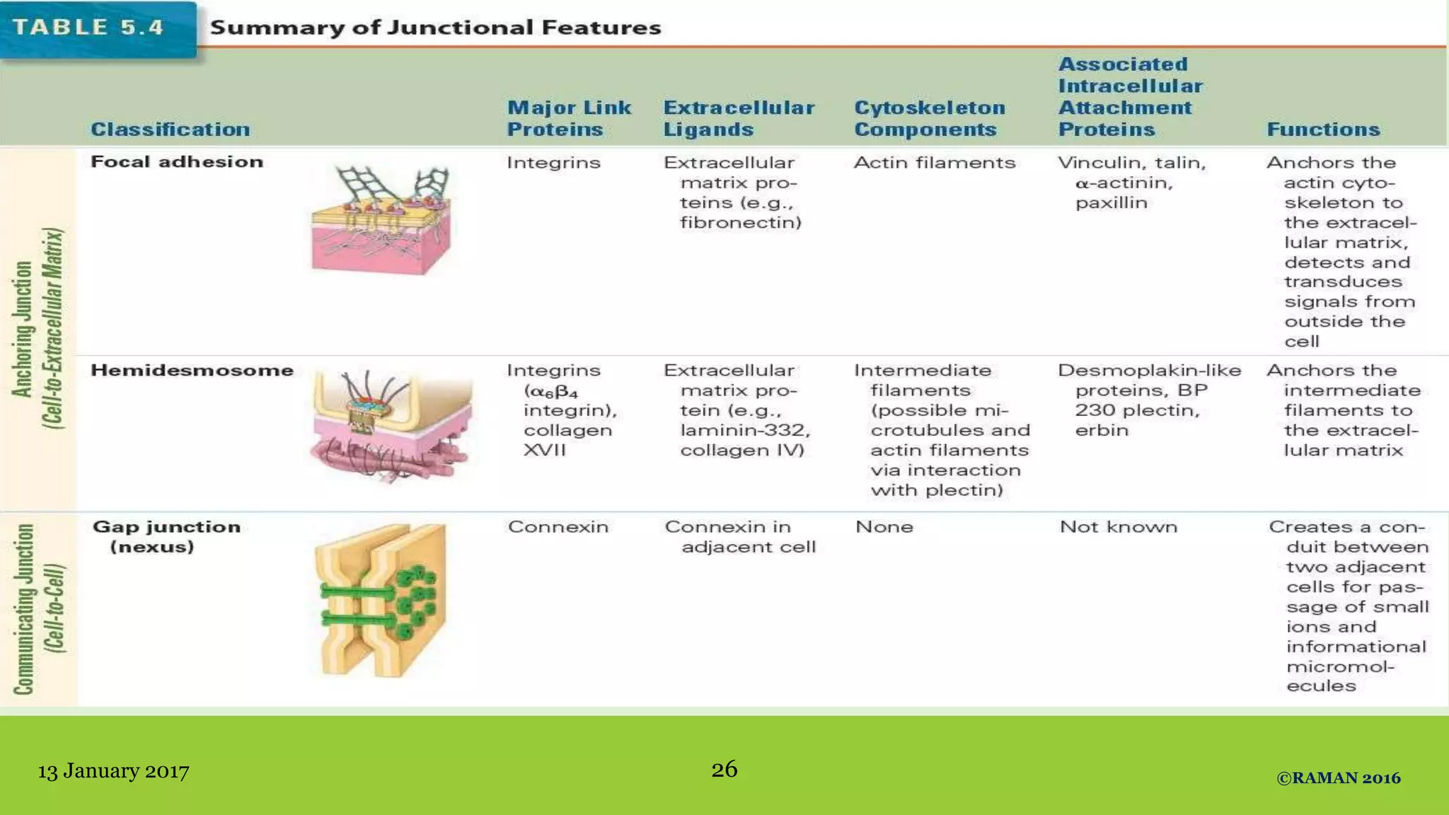
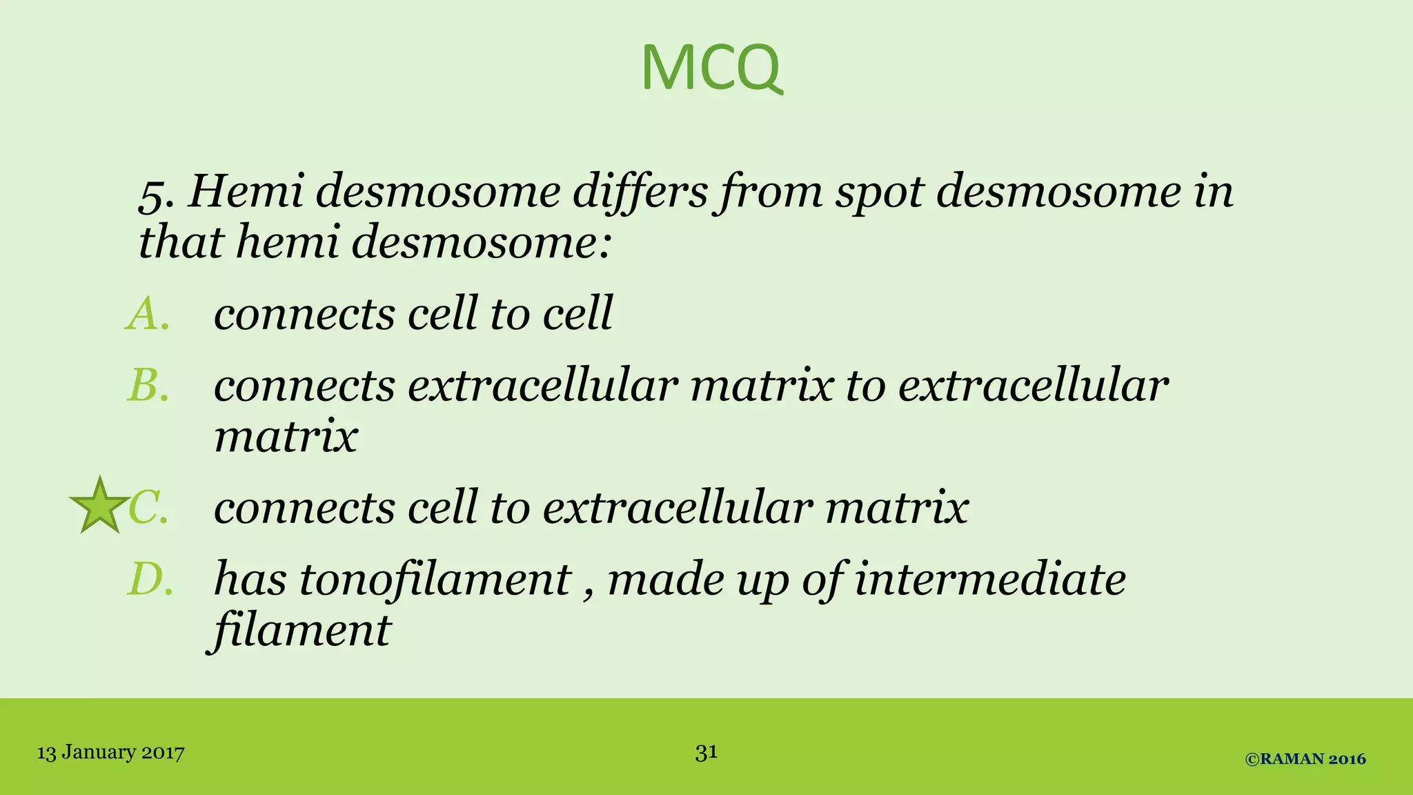


The document discusses intercellular junctions, including their types, functions, and the proteins involved in their structure. It classifies these junctions into occluding, adhesive/anchoring, and communicating types while highlighting their clinical significance related to various disorders. Key examples of clinical abnormalities linked to mutations in junctional proteins are provided, such as pemphigus vulgaris and congenital deafness.


