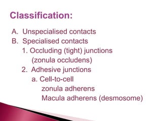
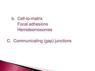
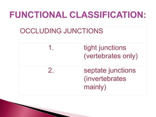
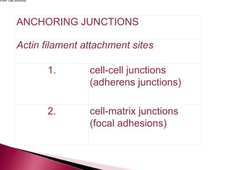
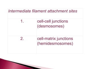
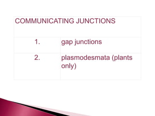
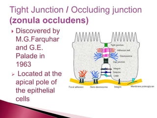
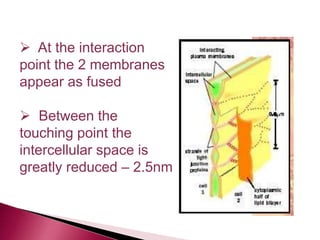
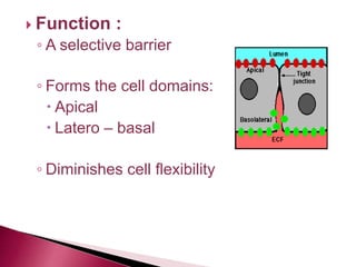
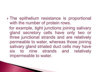
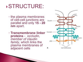
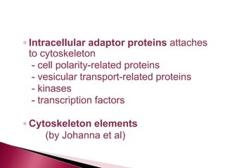
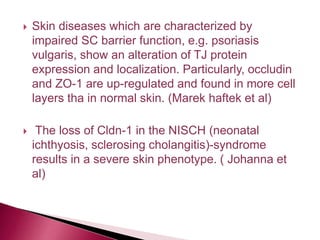
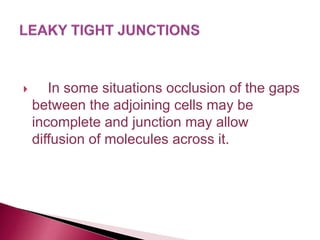
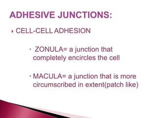
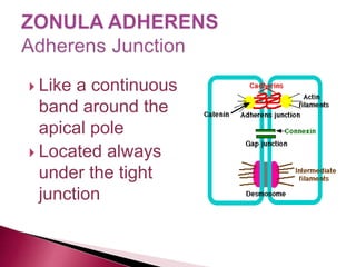
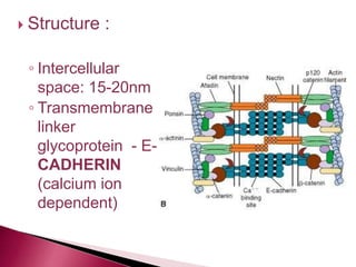
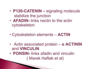
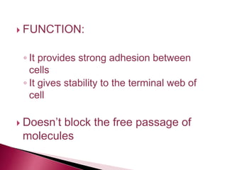
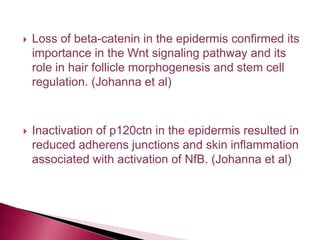
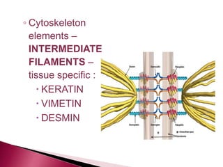
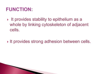
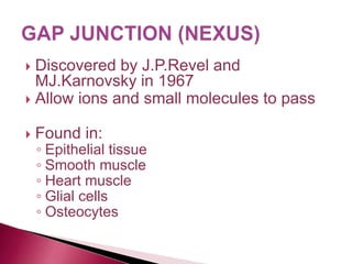
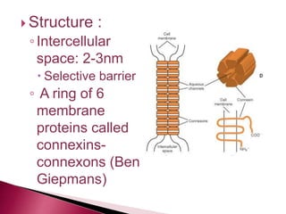
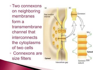
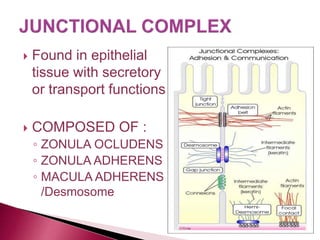
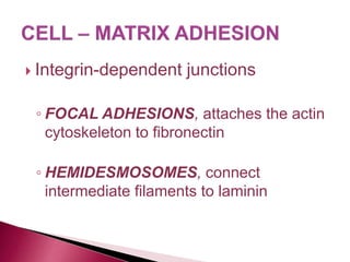
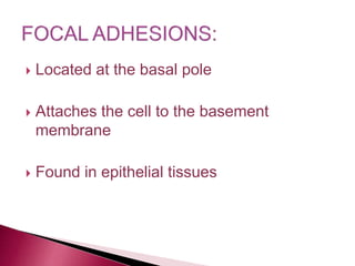
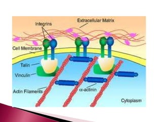
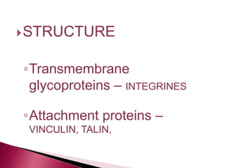
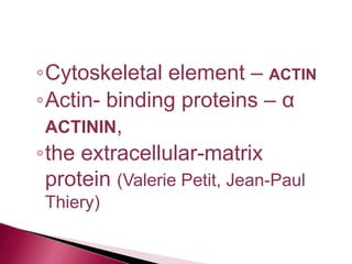
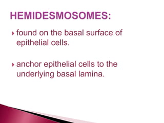
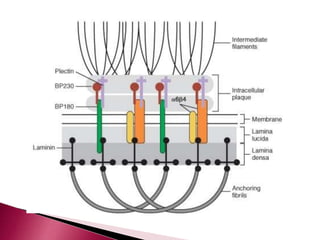
CONTENTS :
1. INTRODUCTION
2. CLASSIFICATION
3. COMPONENTS OF CELL JUNCTION
4. SPECIALISED JUNCTIONS
.
 In tissues in which cells are closely
packed the cell membranes of adjoining
cells are seperated, over most of their
extent by a narrow space (about 20nm).
 this contact is sufficient to bind cells
together , and also allows some degree of
movement of individual cells.
 In some regions the cell membrane
establishes connections with the
neighboring cell or the extracellular matrix
by structures called JUNCTIONS
A. Unspecialised contacts
B. Specialised contacts
1. Occluding (tight) junctions
(zonula occludens)
2. Adhesive junctions
a. Cell-to-cell
zonula adherens
Macula adherens (desmosome)
b. Cell-to-matrix
Focal adhesions
Hemidesmosomes
C. Communicating (gap) junctions
OCCLUDING JUNCTIONS
1. tight junctions
(vertebrates only)
2. septate junctions
(invertebrates
mainly)
ANCHORING JUNCTIONS
Actin filament attachment sites
1. cell-cell junctions
(adherens junctions)
2. cell-matrix junctions
(focal adhesions)
From: Cell Junctions
Intermediate filament attachment sites
1. cell-cell junctions
(desmosomes)
2. cell-matrix junctions
(hemidesmosomes)
COMMUNICATING JUNCTIONS
1. gap junctions
2. plasmodesmata (plants
only)
 On molecular level, cell junctions typically
consists of three components
1. A transmembrane adhesive protein / cell
adhesion molecules (CAM’s)
- glycoprotein in nature
2. A cytoplasmic adapter protein /
intermediate protein
3. A cytoskeletal filament
 Discovered by
M.G.Farquhar
and G.E.
Palade in
1963
 Located at the
apical pole of
the epithelial
cells
 At the interaction
point the 2 membranes
appear as fused
 Between the
touching point the
intercellular space is
greatly reduced – 2.5nm
 Function :
◦ A selective barrier
◦ Forms the cell domains:
 Apical
 Latero – basal
◦ Diminishes cell flexibility
◦ seperates areas of cell membrane that
are specialised for absoption or secretion
from the rest of the cell membrane
◦ prevents lateral migration of proteins
◦ in cells involved in active transport
against a concentration gradient, it
prevents back absorption of transported
substances
 The epithelium resistance is proportional
with the number of protein rows.
for example, tight junctions joining salivary
gland secretory cells have only two or
three junctional strands and are relatively
permeable to water, whereas those joining
salivary gland striated duct cells may have
six to nine strands and relatively
impermeable to water.
STRUCTURE:
◦ the plasma membranes
of cell-cell junctions are
parallel and only 15 - 20
nm apart.
◦ Transmembrane linker
proteins - occludin,
member of claudin
family, which links the
plasma membranes of
adjacent cells
◦ Intracellular adaptor proteins attaches
to cytoskeleton
- cell polarity-related proteins
- vesicular transport-related proteins
- kinases
- transcription factors
◦ Cytoskeleton elements
(by Johanna et al)
 FOUND IN:
Kidney tubules
Intestines
Urinary bladder
 Skin diseases which are characterized by
impaired SC barrier function, e.g. psoriasis
vulgaris, show an alteration of TJ protein
expression and localization. Particularly, occludin
and ZO-1 are up-regulated and found in more cell
layers tha in normal skin. (Marek haftek et al)
 The loss of Cldn-1 in the NISCH (neonatal
ichthyosis, sclerosing cholangitis)-syndrome
results in a severe skin phenotype. ( Johanna et
al)
 In some situations occlusion of the gaps
between the adjoining cells may be
incomplete and junction may allow
diffusion of molecules across it.
 CELL-CELL ADHESION
 ZONULA= a junction that
completely encircles the cell
 MACULA= a junction that is more
circumscribed in extent(patch like)
 Like a continuous
band around the
apical pole
 Located always
under the tight
junction
 Found in:
◦ Epithelial tissue
◦ Digestive tract
◦ Heart muscle
 Structure :
◦ Intercellular
space: 15-20nm
◦ Transmembrane
linker
glycoprotein - E-
CADHERIN
(calcium ion
dependent)
• NECTIN- establish
initial adhesion site and
recruit e-cadherin to the
junction.
• Attachment (Adapter
proteins)
CATENINEαβproteins
link cadherins to the
belt of actin fiament
•
• P120-CATENIN – signaling molecule
stabilize the junction
• AFADIN- links nectin to the actin
cytoskeleton
• Cytoskeleton elements – ACTIN
• Actin associated protein – α ACTININ
and VINCULIN
• PONSIN- links afadin and vinculin
( Marek Haftek et al)
 FUNCTION:
◦ It provides strong adhesion between
cells
◦ It gives stability to the terminal web of
cell
 Doesn’t block the free passage of
molecules
 Loss of beta-catenin in the epidermis confirmed its
importance in the Wnt signaling pathway and its
role in hair follicle morphogenesis and stem cell
regulation. (Johanna et al)
 Inactivation of p120ctn in the epidermis resulted in
reduced adherens junctions and skin inflammation
associated with activation of NfB. (Johanna et al)
 An almost complete loss of adherens junctions
subsequent skin blistering was observed when
beta-catenin was deleted from the epidermis.
(Johanna et al)
 Down regulation of e-cadherin is predictor for
occult metastasis in early SCC.(Gerhard F Huber)
 Discovered by K.R.Porter in 1954
 Disk shape structure
 Under the zonula adherens
 Button like welds joining opposing cell
membranes
 FOUND IN:
◦ Epithelia that are subjected to abrasion
and physical stress
◦ Muscle cells
 STRUCTURE:
◦ Intercellular space: 30nm
◦Transmembrane linker
glycoprotein - CADHERIN
DESMOCOLLINE, DESMOGLEIN
◦ Attachment proteins –
DESMOPLAKIN, PLAKOGLOBIN,
PLAKOPHILIN
form an electron-dense plaque
( Green.K.J, Jones)
◦ Cytoskeleton
elements –
INTERMEDIATE
FILAMENTS –
tissue specific :
 KERATIN
 VIMETIN
 DESMIN
 It provides stability to epithelium as a
whole by linking cytoskeleton of adjacent
cells.
 It provides strong adhesion between cells.
 In several autoimmune diseases autoantibodies
are directed against various desmosome
components, e.g., desmoglein 3 in pemphigus
vulgaris, desmoglein 1 in pemphigus foliaceus, or
several plakins in paraneoplastc pemphigus.
(Johanna et al)
 Hereditary skin diseases provoked by mutations in
genes coding for desmosomal proteins, like
plakophilin 1 which causes an autosomal
recessive skin fragility - ectodermal dysplasia
syndrome with skin, hair and nail defects, or
desmoplakin and desmoglein 1 that underlie the
autosomal dominant striate palmoplantar
keratoderma provide further insights to functions
of various components of these junctions.
(Johanna et al)
 Discovered by J.P.Revel and
MJ.Karnovsky in 1967
 Allow ions and small molecules to pass
 Found in:
◦ Epithelial tissue
◦ Smooth muscle
◦ Heart muscle
◦ Glial cells
◦ Osteocytes
 Structure :
◦Intercellular
space: 2-3nm
 Selective barrier
◦ A ring of 6
membrane
proteins called
connexins-
connexons (Ben
Giepmans)
◦ Two connexons
on neighboring
membranes
form a
transmembrane
channel that
interconnects
the cytoplasms
of two cells
◦ Connexons are
size filters
 FUNCTIONS:
◦ Enable nerve or muscle impulse to
spread rapidly between cells.
◦ Coordinates the activities of cells in
epithelium.
◦ Connexin help in transcriptional and
cytoskeletal regulation.
(Ben N.G.Giepmans)
 Found in epithelial
tissue with secretory
or transport functions
 COMPOSED OF :
◦ ZONULA OCLUDENS
◦ ZONULA ADHERENS
◦ MACULA ADHERENS
/Desmosome
 Integrin-dependent junctions
◦ FOCAL ADHESIONS, attaches the actin
cytoskeleton to fibronectin
◦ HEMIDESMOSOMES, connect
intermediate filaments to laminin
 Located at the basal pole
 Attaches the cell to the basement
membrane
 Found in epithelial tissues
STRUCTURE
◦Transmembrane
glycoproteins – INTEGRINES
◦Attachment proteins –
VINCULIN, TALIN,
◦Cytoskeletal element – ACTIN
◦Actin- binding proteins – α
ACTININ,
◦the extracellular-matrix
protein (Valerie Petit, Jean-Paul
Thiery)
 found on the basal surface of
epithelial cells.
 anchor epithelial cells to the
underlying basal lamina.
 STRUCTURE:
◦ Transmembrane glycoprotein – INTEGRIN-
binds to the laminin and BP 180
◦ Attachment proteins – BP230 and
PLECTIN, form a dense plaque
(Green, K.J.Jones)
◦ Cytoskeletal
element -
INTERMEDIAT
E FILAMENTS
◦ the extracellular-
matrix protein -
LAMININ. (Green,
K.J.Jones)

3. CELL JUNCTIONS.pptx

  • 2.
    CONTENTS : 1. INTRODUCTION 2.CLASSIFICATION 3. COMPONENTS OF CELL JUNCTION 4. SPECIALISED JUNCTIONS
  • 3.
    .  In tissuesin which cells are closely packed the cell membranes of adjoining cells are seperated, over most of their extent by a narrow space (about 20nm).  this contact is sufficient to bind cells together , and also allows some degree of movement of individual cells.
  • 4.
     In someregions the cell membrane establishes connections with the neighboring cell or the extracellular matrix by structures called JUNCTIONS
  • 5.
    A. Unspecialised contacts B.Specialised contacts 1. Occluding (tight) junctions (zonula occludens) 2. Adhesive junctions a. Cell-to-cell zonula adherens Macula adherens (desmosome)
  • 6.
  • 7.
    OCCLUDING JUNCTIONS 1. tightjunctions (vertebrates only) 2. septate junctions (invertebrates mainly)
  • 8.
    ANCHORING JUNCTIONS Actin filamentattachment sites 1. cell-cell junctions (adherens junctions) 2. cell-matrix junctions (focal adhesions) From: Cell Junctions
  • 9.
    Intermediate filament attachmentsites 1. cell-cell junctions (desmosomes) 2. cell-matrix junctions (hemidesmosomes)
  • 10.
    COMMUNICATING JUNCTIONS 1. gapjunctions 2. plasmodesmata (plants only)
  • 11.
     On molecularlevel, cell junctions typically consists of three components 1. A transmembrane adhesive protein / cell adhesion molecules (CAM’s) - glycoprotein in nature
  • 12.
    2. A cytoplasmicadapter protein / intermediate protein 3. A cytoskeletal filament
  • 13.
     Discovered by M.G.Farquhar andG.E. Palade in 1963  Located at the apical pole of the epithelial cells
  • 14.
     At theinteraction point the 2 membranes appear as fused  Between the touching point the intercellular space is greatly reduced – 2.5nm
  • 15.
     Function : ◦A selective barrier ◦ Forms the cell domains:  Apical  Latero – basal ◦ Diminishes cell flexibility
  • 16.
    ◦ seperates areasof cell membrane that are specialised for absoption or secretion from the rest of the cell membrane ◦ prevents lateral migration of proteins ◦ in cells involved in active transport against a concentration gradient, it prevents back absorption of transported substances
  • 17.
     The epitheliumresistance is proportional with the number of protein rows. for example, tight junctions joining salivary gland secretory cells have only two or three junctional strands and are relatively permeable to water, whereas those joining salivary gland striated duct cells may have six to nine strands and relatively impermeable to water.
  • 18.
    STRUCTURE: ◦ the plasmamembranes of cell-cell junctions are parallel and only 15 - 20 nm apart. ◦ Transmembrane linker proteins - occludin, member of claudin family, which links the plasma membranes of adjacent cells
  • 19.
    ◦ Intracellular adaptorproteins attaches to cytoskeleton - cell polarity-related proteins - vesicular transport-related proteins - kinases - transcription factors ◦ Cytoskeleton elements (by Johanna et al)
  • 20.
     FOUND IN: Kidneytubules Intestines Urinary bladder
  • 21.
     Skin diseaseswhich are characterized by impaired SC barrier function, e.g. psoriasis vulgaris, show an alteration of TJ protein expression and localization. Particularly, occludin and ZO-1 are up-regulated and found in more cell layers tha in normal skin. (Marek haftek et al)  The loss of Cldn-1 in the NISCH (neonatal ichthyosis, sclerosing cholangitis)-syndrome results in a severe skin phenotype. ( Johanna et al)
  • 22.
     In somesituations occlusion of the gaps between the adjoining cells may be incomplete and junction may allow diffusion of molecules across it.
  • 23.
     CELL-CELL ADHESION ZONULA= a junction that completely encircles the cell  MACULA= a junction that is more circumscribed in extent(patch like)
  • 24.
     Like acontinuous band around the apical pole  Located always under the tight junction
  • 25.
     Found in: ◦Epithelial tissue ◦ Digestive tract ◦ Heart muscle
  • 26.
     Structure : ◦Intercellular space: 15-20nm ◦ Transmembrane linker glycoprotein - E- CADHERIN (calcium ion dependent)
  • 27.
    • NECTIN- establish initialadhesion site and recruit e-cadherin to the junction. • Attachment (Adapter proteins) CATENINEαβproteins link cadherins to the belt of actin fiament •
  • 28.
    • P120-CATENIN –signaling molecule stabilize the junction • AFADIN- links nectin to the actin cytoskeleton • Cytoskeleton elements – ACTIN • Actin associated protein – α ACTININ and VINCULIN • PONSIN- links afadin and vinculin ( Marek Haftek et al)
  • 29.
     FUNCTION: ◦ Itprovides strong adhesion between cells ◦ It gives stability to the terminal web of cell  Doesn’t block the free passage of molecules
  • 30.
     Loss ofbeta-catenin in the epidermis confirmed its importance in the Wnt signaling pathway and its role in hair follicle morphogenesis and stem cell regulation. (Johanna et al)  Inactivation of p120ctn in the epidermis resulted in reduced adherens junctions and skin inflammation associated with activation of NfB. (Johanna et al)
  • 31.
     An almostcomplete loss of adherens junctions subsequent skin blistering was observed when beta-catenin was deleted from the epidermis. (Johanna et al)  Down regulation of e-cadherin is predictor for occult metastasis in early SCC.(Gerhard F Huber)
  • 32.
     Discovered byK.R.Porter in 1954  Disk shape structure  Under the zonula adherens  Button like welds joining opposing cell membranes
  • 33.
     FOUND IN: ◦Epithelia that are subjected to abrasion and physical stress ◦ Muscle cells
  • 34.
  • 35.
    ◦Transmembrane linker glycoprotein -CADHERIN DESMOCOLLINE, DESMOGLEIN ◦ Attachment proteins – DESMOPLAKIN, PLAKOGLOBIN, PLAKOPHILIN form an electron-dense plaque ( Green.K.J, Jones)
  • 36.
    ◦ Cytoskeleton elements – INTERMEDIATE FILAMENTS– tissue specific :  KERATIN  VIMETIN  DESMIN
  • 37.
     It providesstability to epithelium as a whole by linking cytoskeleton of adjacent cells.  It provides strong adhesion between cells.
  • 38.
     In severalautoimmune diseases autoantibodies are directed against various desmosome components, e.g., desmoglein 3 in pemphigus vulgaris, desmoglein 1 in pemphigus foliaceus, or several plakins in paraneoplastc pemphigus. (Johanna et al)
  • 39.
     Hereditary skindiseases provoked by mutations in genes coding for desmosomal proteins, like plakophilin 1 which causes an autosomal recessive skin fragility - ectodermal dysplasia syndrome with skin, hair and nail defects, or desmoplakin and desmoglein 1 that underlie the autosomal dominant striate palmoplantar keratoderma provide further insights to functions of various components of these junctions. (Johanna et al)
  • 40.
     Discovered byJ.P.Revel and MJ.Karnovsky in 1967  Allow ions and small molecules to pass  Found in: ◦ Epithelial tissue ◦ Smooth muscle ◦ Heart muscle ◦ Glial cells ◦ Osteocytes
  • 41.
     Structure : ◦Intercellular space:2-3nm  Selective barrier ◦ A ring of 6 membrane proteins called connexins- connexons (Ben Giepmans)
  • 42.
    ◦ Two connexons onneighboring membranes form a transmembrane channel that interconnects the cytoplasms of two cells ◦ Connexons are size filters
  • 43.
     FUNCTIONS: ◦ Enablenerve or muscle impulse to spread rapidly between cells. ◦ Coordinates the activities of cells in epithelium. ◦ Connexin help in transcriptional and cytoskeletal regulation. (Ben N.G.Giepmans)
  • 44.
     Found inepithelial tissue with secretory or transport functions  COMPOSED OF : ◦ ZONULA OCLUDENS ◦ ZONULA ADHERENS ◦ MACULA ADHERENS /Desmosome
  • 45.
     Integrin-dependent junctions ◦FOCAL ADHESIONS, attaches the actin cytoskeleton to fibronectin ◦ HEMIDESMOSOMES, connect intermediate filaments to laminin
  • 46.
     Located atthe basal pole  Attaches the cell to the basement membrane  Found in epithelial tissues
  • 48.
  • 49.
    ◦Cytoskeletal element –ACTIN ◦Actin- binding proteins – α ACTININ, ◦the extracellular-matrix protein (Valerie Petit, Jean-Paul Thiery)
  • 50.
     found onthe basal surface of epithelial cells.  anchor epithelial cells to the underlying basal lamina.
  • 52.
     STRUCTURE: ◦ Transmembraneglycoprotein – INTEGRIN- binds to the laminin and BP 180 ◦ Attachment proteins – BP230 and PLECTIN, form a dense plaque (Green, K.J.Jones)
  • 53.
    ◦ Cytoskeletal element - INTERMEDIAT EFILAMENTS ◦ the extracellular- matrix protein - LAMININ. (Green, K.J.Jones)