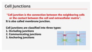
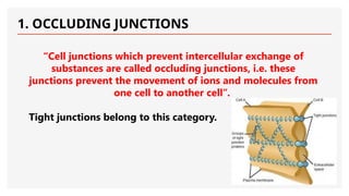
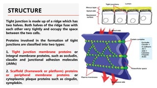
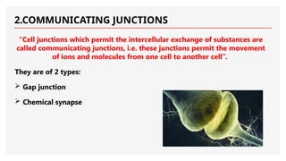
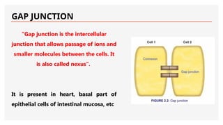
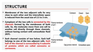
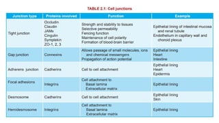
CELL JUNCTIONS
By
Dr. Faraza Javaid
Cell Junctions
“Cell junction is the connection between the neighboring cells
or the contact between the cell and extracellular matrix”.
It is also called membrane junction.
Cell junctions are classified into three types:
1. Occluding junctions
2. Communicating junctions
3. Anchoring junctions
1. OCCLUDING JUNCTIONS
“Cell junctions which prevent intercellular exchange of
substances are called occluding junctions, i.e. these
junctions prevent the movement of ions and molecules from
one cell to another cell”.
Tight junctions belong to this category.
TIGHT JUNCTION
“Tight junction is the intercellular occluding junction that prevents the passage of
large molecules. It is also called zonula occludens. It is the region where the cell
membranes of the adjacent cells fuse together firmly”.
This type of junction is present in the:
 Apical margins of epithelial and endothelial cells in intestinal mucosa,
 Wall of renal tubule
 Capillary wall
 Choroid plexus
STRUCTURE
Tight junction is made up of a ridge which has
two halves. Both halves of the ridge fuse with
each other very tightly and occupy the space
between the two cells.
Proteins involved in the formation of tight
junctions are classified into two types:
1. Tight junction membrane proteins or
integral membrane proteins, such as occludin,
claudin and junctional adhesion molecules
(JAMs)
2. Scaffold (framework or platform) proteins
or peripheral membrane proteins or
cytoplasmic plaque proteins such as cingulin,
symplekin.
Functions of Tight Junction
 Strength and stability
 Selective permeability (gate function)
 Fencing function
 Maintenance of cell polarity
 Blood-brain barrier
2.COMMUNICATING JUNCTIONS
“Cell junctions which permit the intercellular exchange of substances are
called communicating junctions, i.e. these junctions permit the movement
of ions and molecules from one cell to another cell”.
They are of 2 types:
 Gap junction
 Chemical synapse
GAP JUNCTION
“Gap junction is the intercellular
junction that allows passage of ions and
smaller molecules between the cells. It
is also called nexus”.
It is present in heart, basal part of
epithelial cells of intestinal mucosa, etc
STRUCTURE
 Membranes of the two adjacent cells lie very
close to each other and the intercellular space
is reduced from the usual size of 2.5 to 3 nm.
 Cytoplasm of the two cells is connected by the
channels formed by the membranes of both
cells. So, the molecules move from one cell to
another cell directly through these channels,
without having contact with extracellular fluid
(ECF).
 Each channel consists of two halves. Each half
belongs to one of the two adjacent cells. Each
half of the channel is surrounded by 6 subunits
of proteins which are called connexins or
connexons.
Functions of Gap Junction
1.Diameter of the channel in the gap junction is about 1.5 to 3 nm. So,
the channel permits the passage of glucose, amino acids, ions and
other substances, which have a molecular weight less than 1,000
2.It helps in the exchange of chemical messengers between the cells
3.It helps in rapid propagation of action potential from one cell to
another cell.
4.Regulate channel diameter by release of calcium, neurotransmitter,
hormones.
CHEMICAL SYNAPSE
Chemical synapse is the junction
between a nerve fiber and a
muscle fiber or between two
nerve fibers, through which the
signals are transmitted by the
release of chemical transmitter.
3.ANCHORING JUNCTIONS
“Anchoring junctions are the junctions, which provide strength to the
cells by acting like mechanical attachments, i.e. these junctions provide
firm structural attachment between two cells or between a cell and the
extracellular matrix”.
Anchoring junctions are responsible for the structural integrity of the
tissues and are present in the tissues like heart muscle and epidermis of
skin, which are subjected to severe mechanical stress.
The firm attachment between two cells or between a cell and the extracellular
matrix is provided by either actin filaments or the intermediate filaments.
Depending upon this, anchoring junctions are classified into four types:
1. Actin filament attachment
i. Adherens junction (cell to cell)
ii. Focal adhesion (cell to matrix)
2. Intermediate filament attachment
iii. Desmosome (cell to cell)
iv. Hemidesmosome (cell to matrix)
ADHERENS JUNCTION
“Adherens junction is the cell to cell junction, which connects the
actin filaments of one cell to those of another cell”.
 In some places like epithelial linings, this junction forms a
continuous adhesion (zonula adherens) just below the tight
junctions.
 In adherens junction, the membranes of the adjacent cells are held
together by some transmembrane proteins called cadherins.
 Adherens junction provides strong
mechanical attachments of the
adjacent cells.
 Adherens junction is present in the
intercalated disks between the
branches of cardiac muscles and
help in contraction and relaxation
of heart muscle.
 The adherens junction present in
epidermis helps the skin to
withstand the mechanical stress
FOCAL ADHESION
Focal adhesion is the cell to matrix
junctions, which connects the actin
filaments of the cell to the
extracellular matrix.
In epithelia of various organs, this
junction connects the cells with their
basal lamina.
The transmembrane proteins, which
hold the cell membrane and the
matrix are called integrins.
DESMOSOME
Desmosome is a cell to cell junction, where the
intermediate filaments connect two adjacent cells.
Desmosome is also called macula adherens.
Intermediate filaments are attached with the
thickened patches.
Some of these filaments are parallel to the
membrane and others are arranged in radiating
fashion.
Desmosomes function like tight junctions. The
transmembrane proteins involved in desmosome
are mainly cadherins and mainly found in skin.
HEMIDESMOSOME
“Hemidesmosome is a cell to matrix
junction, which connects the
intermediate filaments of the cell to the
extracellular matrix”.
This type of cell junction is like half
desmosome and the thickening of
membrane of only one cell occurs. So,
this is known as hemidesmosome or half
desmosome.
Mostly, the hemidesmosome connects
the cells with their basal lamina. The
proteins involved in this are integrins.
CELL ADHESION MOLECULES
Cell adhesion molecules (CAMs) or cell adhesion proteins are the
protein molecules, which are responsible for the attachment of cells
to their neighbors or to basal lamina (or basal membrane).
CAMs form the important structures of intercellular connections and
are responsible for structural organization of tissues.
TYPES OF CELL ADHESION MOLECULES
1.Cadherins, which form the molecular limbs between neighboring cells.
These CAMs form adherens junction and desomosome
2.Integrins, which form the focal adhesion and hemidesmosome
3.IgG super family, which form the cell adhesion molecules in nervous
system
4.Selectins, which act as receptors for carbohydrates (ligand or mucin)
and are found in platelets and endothelial cells.
Cell junctions and cellular communications .pptx

Cell junctions and cellular communications .pptx

  • 1.
  • 2.
    Cell Junctions “Cell junctionis the connection between the neighboring cells or the contact between the cell and extracellular matrix”. It is also called membrane junction. Cell junctions are classified into three types: 1. Occluding junctions 2. Communicating junctions 3. Anchoring junctions
  • 3.
    1. OCCLUDING JUNCTIONS “Celljunctions which prevent intercellular exchange of substances are called occluding junctions, i.e. these junctions prevent the movement of ions and molecules from one cell to another cell”. Tight junctions belong to this category.
  • 4.
    TIGHT JUNCTION “Tight junctionis the intercellular occluding junction that prevents the passage of large molecules. It is also called zonula occludens. It is the region where the cell membranes of the adjacent cells fuse together firmly”. This type of junction is present in the:  Apical margins of epithelial and endothelial cells in intestinal mucosa,  Wall of renal tubule  Capillary wall  Choroid plexus
  • 5.
    STRUCTURE Tight junction ismade up of a ridge which has two halves. Both halves of the ridge fuse with each other very tightly and occupy the space between the two cells. Proteins involved in the formation of tight junctions are classified into two types: 1. Tight junction membrane proteins or integral membrane proteins, such as occludin, claudin and junctional adhesion molecules (JAMs) 2. Scaffold (framework or platform) proteins or peripheral membrane proteins or cytoplasmic plaque proteins such as cingulin, symplekin.
  • 6.
    Functions of TightJunction  Strength and stability  Selective permeability (gate function)  Fencing function  Maintenance of cell polarity  Blood-brain barrier
  • 7.
    2.COMMUNICATING JUNCTIONS “Cell junctionswhich permit the intercellular exchange of substances are called communicating junctions, i.e. these junctions permit the movement of ions and molecules from one cell to another cell”. They are of 2 types:  Gap junction  Chemical synapse
  • 8.
    GAP JUNCTION “Gap junctionis the intercellular junction that allows passage of ions and smaller molecules between the cells. It is also called nexus”. It is present in heart, basal part of epithelial cells of intestinal mucosa, etc
  • 9.
    STRUCTURE  Membranes ofthe two adjacent cells lie very close to each other and the intercellular space is reduced from the usual size of 2.5 to 3 nm.  Cytoplasm of the two cells is connected by the channels formed by the membranes of both cells. So, the molecules move from one cell to another cell directly through these channels, without having contact with extracellular fluid (ECF).  Each channel consists of two halves. Each half belongs to one of the two adjacent cells. Each half of the channel is surrounded by 6 subunits of proteins which are called connexins or connexons.
  • 10.
    Functions of GapJunction 1.Diameter of the channel in the gap junction is about 1.5 to 3 nm. So, the channel permits the passage of glucose, amino acids, ions and other substances, which have a molecular weight less than 1,000 2.It helps in the exchange of chemical messengers between the cells 3.It helps in rapid propagation of action potential from one cell to another cell. 4.Regulate channel diameter by release of calcium, neurotransmitter, hormones.
  • 11.
    CHEMICAL SYNAPSE Chemical synapseis the junction between a nerve fiber and a muscle fiber or between two nerve fibers, through which the signals are transmitted by the release of chemical transmitter.
  • 12.
    3.ANCHORING JUNCTIONS “Anchoring junctionsare the junctions, which provide strength to the cells by acting like mechanical attachments, i.e. these junctions provide firm structural attachment between two cells or between a cell and the extracellular matrix”. Anchoring junctions are responsible for the structural integrity of the tissues and are present in the tissues like heart muscle and epidermis of skin, which are subjected to severe mechanical stress.
  • 13.
    The firm attachmentbetween two cells or between a cell and the extracellular matrix is provided by either actin filaments or the intermediate filaments. Depending upon this, anchoring junctions are classified into four types: 1. Actin filament attachment i. Adherens junction (cell to cell) ii. Focal adhesion (cell to matrix) 2. Intermediate filament attachment iii. Desmosome (cell to cell) iv. Hemidesmosome (cell to matrix)
  • 14.
    ADHERENS JUNCTION “Adherens junctionis the cell to cell junction, which connects the actin filaments of one cell to those of another cell”.  In some places like epithelial linings, this junction forms a continuous adhesion (zonula adherens) just below the tight junctions.  In adherens junction, the membranes of the adjacent cells are held together by some transmembrane proteins called cadherins.
  • 15.
     Adherens junctionprovides strong mechanical attachments of the adjacent cells.  Adherens junction is present in the intercalated disks between the branches of cardiac muscles and help in contraction and relaxation of heart muscle.  The adherens junction present in epidermis helps the skin to withstand the mechanical stress
  • 16.
    FOCAL ADHESION Focal adhesionis the cell to matrix junctions, which connects the actin filaments of the cell to the extracellular matrix. In epithelia of various organs, this junction connects the cells with their basal lamina. The transmembrane proteins, which hold the cell membrane and the matrix are called integrins.
  • 17.
    DESMOSOME Desmosome is acell to cell junction, where the intermediate filaments connect two adjacent cells. Desmosome is also called macula adherens. Intermediate filaments are attached with the thickened patches. Some of these filaments are parallel to the membrane and others are arranged in radiating fashion. Desmosomes function like tight junctions. The transmembrane proteins involved in desmosome are mainly cadherins and mainly found in skin.
  • 18.
    HEMIDESMOSOME “Hemidesmosome is acell to matrix junction, which connects the intermediate filaments of the cell to the extracellular matrix”. This type of cell junction is like half desmosome and the thickening of membrane of only one cell occurs. So, this is known as hemidesmosome or half desmosome. Mostly, the hemidesmosome connects the cells with their basal lamina. The proteins involved in this are integrins.
  • 19.
    CELL ADHESION MOLECULES Celladhesion molecules (CAMs) or cell adhesion proteins are the protein molecules, which are responsible for the attachment of cells to their neighbors or to basal lamina (or basal membrane). CAMs form the important structures of intercellular connections and are responsible for structural organization of tissues.
  • 20.
    TYPES OF CELLADHESION MOLECULES 1.Cadherins, which form the molecular limbs between neighboring cells. These CAMs form adherens junction and desomosome 2.Integrins, which form the focal adhesion and hemidesmosome 3.IgG super family, which form the cell adhesion molecules in nervous system 4.Selectins, which act as receptors for carbohydrates (ligand or mucin) and are found in platelets and endothelial cells.