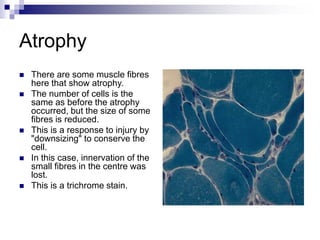
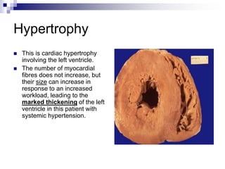
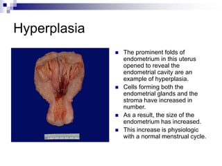
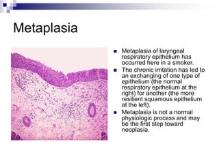
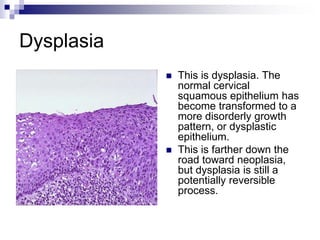
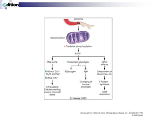
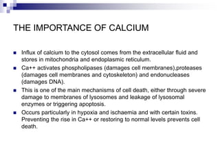
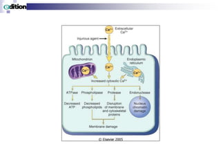
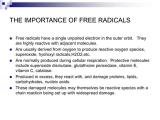
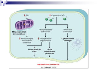
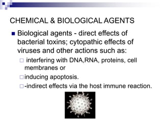
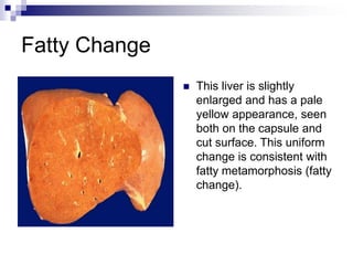
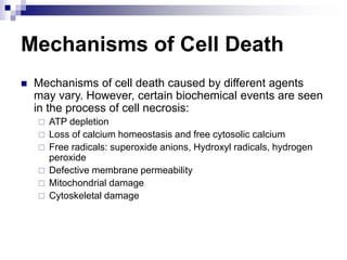
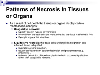
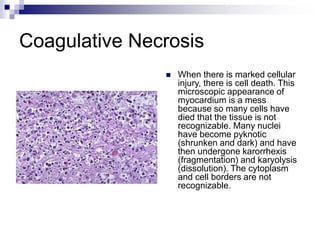
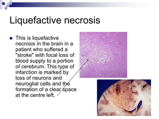
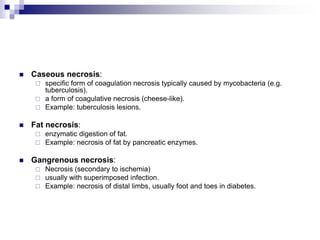
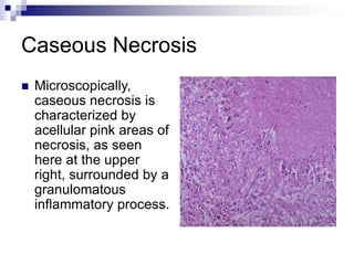
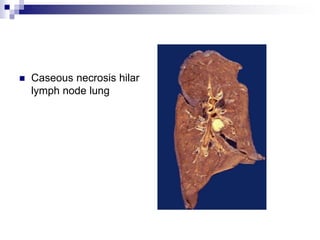
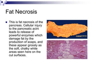
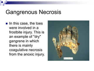
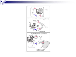
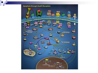
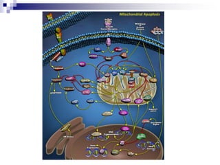
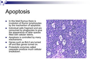
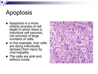
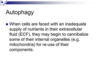
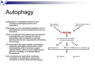
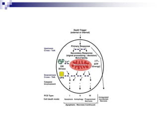
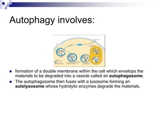
SlideShare a Scribd company logo
1 of 59
Cell Injury and
Cell Death
LMO MUMBA
Objectives
 Upon completion of this lecture you will be able
to:
 Define 4 types of cellular adaptation to physiologic
and pathologic conditions.
 List 7 common causes of cell injury.
 Explain the difference between reversible and
irreversible cell injury.
 Explain the difference between necrosis and
apoptosis.
 Describe patterns of necrosis In tissues or organs.
Important Terms
 Cellular Adaptation
 Cell Injury
 Necrosis
 Apoptosis
 Atrophy
 Hypertrophy
 Hyperplasia
 Metaplasia
 Cells are the structural and functional units of tissues and organs.
They are capable of adjusting their structure and functions in
response to various physiological and pathological conditions. This
capability is called cellular adaptation.
 Cellular adaptations include:
 Atrophy--shrinkage of cells
 Hypertrophy--increase in the size of cells which results in enlargement
of the organs
 Hyperplasia--increased number of cells in an organ or tissue
 Metaplasia--transformation or replacement of one adult cell type with
another
Atrophy
 There are some muscle fibres
here that show atrophy.
 The number of cells is the
same as before the atrophy
occurred, but the size of some
fibres is reduced.
 This is a response to injury by
"downsizing" to conserve the
cell.
 In this case, innervation of the
small fibres in the centre was
lost.
 This is a trichrome stain.
Hypertrophy
 This is cardiac hypertrophy
involving the left ventricle.
 The number of myocardial
fibres does not increase, but
their size can increase in
response to an increased
workload, leading to the
marked thickening of the left
ventricle in this patient with
systemic hypertension.
Hyperplasia
 The prominent folds of
endometrium in this uterus
opened to reveal the
endometrial cavity are an
example of hyperplasia.
 Cells forming both the
endometrial glands and the
stroma have increased in
number.
 As a result, the size of the
endometrium has increased.
 This increase is physiologic
with a normal menstrual cycle.
Metaplasia
 Metaplasia of laryngeal
respiratory epithelium has
occurred here in a smoker.
 The chronic irritation has led to
an exchanging of one type of
epithelium (the normal
respiratory epithelium at the
right) for another (the more
resilient squamous epithelium
at the left).
 Metaplasia is not a normal
physiologic process and may
be the first step toward
neoplasia.
Dysplasia
 This is dysplasia. The
normal cervical
squamous epithelium has
become transformed to a
more disorderly growth
pattern, or dysplastic
epithelium.
 This is farther down the
road toward neoplasia,
but dysplasia is still a
potentially reversible
process.
Various Types of Adaptations
 Cells may undergo various adaptations in
physiological and pathological conditions.
These are controlled by complex
molecular mechanisms.
Atrophy-
 -shrinkage of cells; classified as:
Physiologic--due to decreased work load
(e.g., decreased size of uterus following child
birth)
Pathologic--primarily due to denervation of
muscle, diminished blood supply, nutritional
deficiency
Hypertrophy-
 Increase in the size of cells which results in
enlargement of the organs.
 It is mostly seen in cells that cannot divide, such
as:
 skeletal muscle (pumping iron),
 cardiac muscle (hypertension).
 These changes usually revert to normal if the
cause is removed.
 Hypertrophy is mediated by different
mechanisms.
Hyperplasia-
 Increased number of cells in an organ or tissue.
Hyperplasia may sometimes co-exist with
hypertrophy. Hyperplasia can be classified as:
 physiologic--hormonal (e.g., breast and uterus
during pregnancy)
 compensatory--regeneration of liver following partial
hepatectomy. Various growth factors and interluekins
are important in such hyperplasia.
 pathologic--excessive hormonal stimulation, viral
infection (papilloma viruses); neoplasms
Metaplasia-
 Transformation or replacement of one adult cell type to another
adult cell type
 (e.g., the change from columnar to squamous cells in respiratory
tract, from squamous to columnar in Barrett esophagitis).
 Metaplasia also occurs in mesenchymal tissue (e.g., formation of
bone in skeletal muscle).
 Metaplastic changes usually result from chronic irritation.
 Metaplastic changes seem to precede the development of cancer,
in some instances.
 Metaplasia is thought to arise from reprogramming of stem or
undifferentiated cells that are present in adult tissue.
Cell Injury
 If the cells fail to adapt under stress, they undergo
certain changes called cell injury. The affected cells
may recover from the injury (reversible) or may die
(irreversible).
 Causes of Cell Injury
 oxygen deprivation (anoxia)
 physical agents
 chemical agents
 infections agents
 immunologic reactions
 genetic defects
 nutritional imbalances
IMPORTANT TARGETS OF CELL INJURY
 Aerobic respiration –
 ATP depletion or decreased synthesis.
 Cell membranes - plasma membranes, mitochondrial, lysosomal
and other organelle membranes.
 Protein synthesis.
 Cytoskeleton.
 Genetic apparatus.
Downloaded from: Robbins & Cotran Pathologic Basis of Disease (on 4 April 2005 06:11 PM)
© 2005 Elsevier
ATP DEPLETION - HYPOXIA/ISCHAEMIA
 Mitochondria - reduced oxidative phosphorylation.
 Cell membrane - reduced sodium pump.
 Sodium and water enter the cell; potassium exits.
 Endoplasmic reticulum dilates, the cell swells.
 Anaerobic glycolysis occurs with loss of glycogen, accumulation of
lactic acid, acid pH which interferes with enzymes.
 Failure of the calcium pump leads to influx of Ca++ into the cell,
activate various enzymes to the detriment of the cell.
 RER loses ribosomes and protein synthesis falls - structural proteins
(membranes,cytoskeleton) and enzymes.
 Misfolded proteins lead to the unfolded protein response which may
further injure the cell.
Downloaded from: Robbins & Cotran Pathologic Basis of Disease (on 4 April 2005 06:11 PM)
© 2005 Elsevier
THE IMPORTANCE OF CALCIUM
 Influx of calcium to the cytosol comes from the extracellular fluid and
stores in mitochondria and endoplasmic reticulum.
 Ca++ activates phospholipases (damages cell membranes),proteases
(damages cell membranes and cytoskeleton) and endonucleases
(damages DNA).
 This is one of the main mechanisms of cell death, either through severe
damage to membranes of lysosomes and leakage of lysosomal
enzymes or triggering apoptosis.
 Occurs particularly in hypoxia and ischaemia and with certain toxins.
Preventing the rise in Ca++ or restoring to normal levels prevents cell
death.
THE IMPORTANCE OF FREE RADICALS
 Free radicals have a single unpaired electron in the outer orbit. They
are highly reactive with adjacent molecules.
 Are usually derived from oxygen to produce reactive oxygen species,
superoxide, hydroxyl radicals,H2O2,etc.
 Are normally produced during cellular respiration. Protective molecules
include superoxide dismutase, glutathione peroxidase, vitamin E,
vitamin C, catalase.
 Produced in excess, they react with, and damage proteins, lipids,
carbohydrates, nucleic acids.
 These damaged molecules may themselves be reactive species with a
chain reaction being set up with widespread damage.
FREE RADICALS
 In addition to oxygen-derived free radicals, nitric oxide (NO) can act as
a free radical and be converted to an even more reactive anion.
 Iron and copper catalyze free radical formation and are thus important
in the generation of reactive oxygen species.
 Binding to molecules such as transferrin, ferritin and ceruloplasmin is
protective.
 Free radicals cause lipid peroxidation in cell membranes, oxidation of
amino acids and proteins resulting in fragmentation, and protein-protein
cross linkages. Altered proteins are acted on by the proteosomes with
further cell damage.
Downloaded from: Robbins & Cotran Pathologic Basis of Disease (on 4 April 2005 06:11 PM)
© 2005 Elsevier
FREE RADICALS AND DISEASE
 Free radicals may be a common pathway for most types of cell
damage, particularly oxygen-derived free radicals (oxidative stress).
 Some examples are:-
 oxygen toxicity, ischaemia/reperfusion injury, radiation injury (hydrolyses
H2O to OH & H), metabolism of drugs,toxins,pollutants (eg Paracetamol to
reactive metabolite;CCl4 to CCl3,cigarette smoke);
 leukocyte killing of bacteria or in non-bacterial inflammations,release of iron
in haemorrhages enhances oxidative stress (important in CNS),
 lipid peroxidation of low-density lipoproteins in atherosclerosis,cancer
production (damage to DNA), ageing.
 Therapies for combating oxidative stress are available for prevention or
treatment with antioxidants and/or free-radical scavengers.
ISCHAEMIA/REPERFUSION INJURY
 If cells are reversibly injured due to ischaemia, complete recovery
occurs following restoration of blood flow.
 However, reperfusion can result in more damage including cell death.
 This is due to incompletely metabolised products producing reactive
oxygen species on re-introduction of oxygen
 (especially damaging to mitochondria;
 loss of anti-oxidants during ischaemia;
 inflow of calcium with the renewed blood flow;
 recruitment of leukocytes to the injured area.
 Reperfusion injury is especially important in ischaemic damage to the
heart and brain and in organ transplantation.
 Various therapies and preventive measures are in use.
CHEMICAL & BIOLOGICAL AGENTS
 Biological agents - direct effects of
bacterial toxins; cytopathic effects of
viruses and other actions such as:
 interfering with DNA,RNA, proteins, cell
membranes or
inducing apoptosis.
-indirect effects via the host immune reaction.
Fatty Change
 This liver is slightly
enlarged and has a pale
yellow appearance, seen
both on the capsule and
cut surface. This uniform
change is consistent with
fatty metamorphosis (fatty
change).
Chronic Cell Injury
 Non-lethal injury may cause subcellular changes
some of which are characteristically seen in
certain pathologic conditions. The following are
examples of some of these changes:
 Changes in mitochondria seen in various conditions in
some of which there is an increase in the number of
mitochondria with various morphological
abnormalities.
Cell Death
 Death of cells occurs in two ways:
Necrosis--(irreversible injury) changes
produced by enzymatic digestion of dead
cellular elements
Apoptosis--vital process that helps eliminate
unwanted cells--an internally programmed
series of events effected by dedicated gene
products
Mechanisms of Cell Death
 Mechanisms of cell death caused by different agents
may vary. However, certain biochemical events are seen
in the process of cell necrosis:
 ATP depletion
 Loss of calcium homeostasis and free cytosolic calcium
 Free radicals: superoxide anions, Hydroxyl radicals, hydrogen
peroxide
 Defective membrane permeability
 Mitochondrial damage
 Cytoskeletal damage
Patterns of Necrosis In Tissues
or Organs
 As a result of cell death the tissues or organs display certain
macroscopic changes:
 Coagulative necrosis:
 typically seen in hypoxic environments
 the outline of the dead cells are maintained and the tissue is somewhat firm.
 Example: myocardial infarction
 Liquifactive necrosis: the dead cells undergo disintegration and
affected tissue is liquified.
 Example: cerebral infarction.
 usually associated with cellular destruction and pus formation (e.g.
pneumonia).
 ischemia (restriction of blood supply) in the brain produces liquefactive
rather than coagulative necrosis.
Coagulative Necrosis
 When there is marked cellular
injury, there is cell death. This
microscopic appearance of
myocardium is a mess
because so many cells have
died that the tissue is not
recognizable. Many nuclei
have become pyknotic
(shrunken and dark) and have
then undergone karorrhexis
(fragmentation) and karyolysis
(dissolution). The cytoplasm
and cell borders are not
recognizable.
Liquefactive necrosis
 This is liquefactive
necrosis in the brain in a
patient who suffered a
"stroke" with focal loss of
blood supply to a portion
of cerebrum. This type of
infarction is marked by
loss of neurons and
neuroglial cells and the
formation of a clear space
at the centre left.
 Caseous necrosis:
 specific form of coagulation necrosis typically caused by mycobacteria (e.g.
tuberculosis).
 a form of coagulative necrosis (cheese-like).
 Example: tuberculosis lesions.
 Fat necrosis:
 enzymatic digestion of fat.
 Example: necrosis of fat by pancreatic enzymes.
 Gangrenous necrosis:
 Necrosis (secondary to ischemia)
 usually with superimposed infection.
 Example: necrosis of distal limbs, usually foot and toes in diabetes.
Caseous Necrosis
 Microscopically,
caseous necrosis is
characterized by
acellular pink areas of
necrosis, as seen
here at the upper
right, surrounded by a
granulomatous
inflammatory process.
 Caseous necrosis hilar
lymph node lung
Fat Necrosis
 This is fat necrosis of the
pancreas. Cellular injury
to the pancreatic acini
leads to release of
powerful enzymes which
damage fat by the
production of soaps, and
these appear grossly as
the soft, chalky white
areas seen here on the
cut surfaces.
Gangrenous Necrosis
 In this case, the toes
were involved in a
frostbite injury. This is
an example of "dry"
gangrene in which
there is mainly
coagulative necrosis
from the anoxic injury.
 Gummatous necrosis is restricted to necrosis
involving spirochaetal infections (e.g. syphilis).
 Haemorrhagic necrosis is due to blockage of the
venous drainage of an organ or tissue (e.g. in
testicular torsion).
 Fibrinoid necrosis is caused by immune-mediated
vascular damage. It is marked by deposition of fibrin-
like proteinaceous material in arterial walls, which
appears smudgy and eosinophilic on light microscopy.
Morphological Forms of
Programmed Cell Death
Type I = Apoptosis
Type II = Autophagic Cell Death
Type III = Non-lysosomal
Kerr, Wyllie, and Currie, 1972; Schweicheland Merker, 1973; Clarke
1990Kerr, Wyllie, and Currie, 1972; Schweicheland Merker, 1973
Apoptosis
 This process helps to eliminate unwanted cells by an internally
programmed series of events effected by dedicated gene products.
It serves several vital functions and is seen under various settings.
 During development for removal of excess cells during embryogenesis
 To maintain cell population in tissues with high turnover of cells, such as
skin, bowels.
 To eliminate immune cells after cytokine depletion, and autoreactive T-
cells in developing thymus.
 To remove damaged cells by virus
 To eliminate cells with DNA damage by radiation, cytotoxic agents etc.
 Hormone-dependent involution - Endometrium, ovary, breasts etc.
 Cell death in tumours.
Apoptosis
 In the human body ~ 100,000 cells are
produced every second by mitosis and a
similar number die by apoptosis.
 Development and morphogenesis
 During limb formation separate digits evolve
 Ablation of cells no longer needed (tadpole)
 Homeostasis
 Immune system
 >95% T and B cells die during maturation
(negative selection)
 Deletion of damaged/ dangerous cells
Mechanisms of Apoptosis
 Apoptosis can be induced by various factors
under both physiological and pathological
conditions:
 It is an energy-dependent cascade of molecular
events which include protein cleavage by a
group of enzymes (caspases), protein cross-
linking, DNA breakdown.
 Apoptosis is regulated by a large family of genes
some of which are inhibitory (bcl-2) and some
are stimulatory (bax).
Apoptosis
 There are a number of
mechanisms through which
apoptosis can be induced in
cells.
 The sensitivity of cells to any of
these stimuli can vary
depending on a number of
factors such as:
 the expression of pro- and
anti-apoptotic proteins (eg. the
Bcl-2 proteins or the Inhibitor
of Apoptosis Proteins),
 the severity of the stimulus
and
 the stage of the cell cycle.
Apoptosis
 In this fetal thymus there is
involution of thymic lymphocytes
by the mechanism of apoptosis.
 Individual cells fragment and are
consumed by phagocytes to give
the appearance of clear spaces
filled with cellular debris.
 Apoptosis is controlled by many
mechanisms.
 Genes such as Bcl-2 are turned
off and Bax genes turned on.
 Proteolytic enzymes called
caspases produce much cellular
breakdown.
Apoptosis
 Apoptosis is a more
orderly process of cell
death in which there is
individual cell necrosis,
not necrosis of large
numbers of cells.
 In this example, liver cells
are dying individually
(arrows) from injury by
viral hepatitis.
 The cells are pink and
without nuclei.
Autophagy
 When cells are faced with an inadequate
supply of nutrients in their extracellular
fluid (ECF), they may begin to cannibalize
some of their internal organelles (e.g.
mitochondria) for re-use of their
components.
 Autophagy refers to a set of diverse
processes whereby intracytoplasmic
material is delivered to lysosomes.
Autophagy
Autophagy is a regulated process for the
removal of damaged proteins and
organelles.
Autophagy occurs under basal conditions and is
stimulated by environmental factors such as
starvation.
There is evidence that proteins that are linked to
tumorigenesis can regulate the rate of
autophagy, with oncogenes in general
blocking and tumour suppressors stimulating
the process.
The removal of damaged cellular components,
especially damaged mitochondria, might
decrease the level of reactive oxygen
species (ROS), which in turn might reduce
genomic instability or forestall cellular
senescence.
Such mechanisms might allow moderate
increases in autophagy to reduce the
incidence of cancer and prolong lifespan.
Autophagy involves:
 formation of a double membrane within the cell which envelops the
materials to be degraded into a vesicle called an autophagosome.
 The autophagosome then fuses with a lysosome forming an
autolysosome whose hydrolytic enzymes degrade the materials.
Apoptosis
Necrosis

More Related Content

Similar to cell injury...VAN.ppt

Cell injury adaptation class 1
Cell injury adaptation class 1Cell injury adaptation class 1
Cell injury adaptation class 1iqra khan
 
CELL INJURY NEW.pathology of cell injury .pptx
CELL INJURY NEW.pathology of cell injury .pptxCELL INJURY NEW.pathology of cell injury .pptx
CELL INJURY NEW.pathology of cell injury .pptxABIDOFFICIALCHANNEL
 
Lect 2- cellular adaptation
Lect 2- cellular adaptationLect 2- cellular adaptation
Lect 2- cellular adaptationMohanad Mohanad
 
cellular adaptation presentation (2)
cellular adaptation presentation    (2)cellular adaptation presentation    (2)
cellular adaptation presentation (2)HamnazBeegumpp
 
cell adaptation and cell injury - Copy-4 copy.pptx
cell adaptation and cell injury - Copy-4 copy.pptxcell adaptation and cell injury - Copy-4 copy.pptx
cell adaptation and cell injury - Copy-4 copy.pptxNadiirMahamoud
 
Cell Injury 1 pptx.pptx
Cell Injury 1 pptx.pptxCell Injury 1 pptx.pptx
Cell Injury 1 pptx.pptxAmosLangat5
 
pathology-cell _injury.pdf
pathology-cell _injury.pdfpathology-cell _injury.pdf
pathology-cell _injury.pdfDhanush Karma
 
Cell injury lecture #01 &02
Cell injury lecture #01 &02Cell injury lecture #01 &02
Cell injury lecture #01 &02abdul aziz
 
CELL INJURY, CELLULAR ADAPTATIONS, MECHANISM OF CELL I NJURY
CELL INJURY, CELLULAR ADAPTATIONS, MECHANISM OF CELL I NJURYCELL INJURY, CELLULAR ADAPTATIONS, MECHANISM OF CELL I NJURY
CELL INJURY, CELLULAR ADAPTATIONS, MECHANISM OF CELL I NJURYMithun Venugopal
 
CELL INJURY, CELLULAR ADAPTATIONS, MECHANISM OF CELL I NJURY
CELL INJURY, CELLULAR ADAPTATIONS, MECHANISM OF CELL I NJURYCELL INJURY, CELLULAR ADAPTATIONS, MECHANISM OF CELL I NJURY
CELL INJURY, CELLULAR ADAPTATIONS, MECHANISM OF CELL I NJURYMithun Venugopal
 
General Pathology Lecture Introduction from Module 1 to Module 3
General Pathology Lecture Introduction from Module 1 to Module 3General Pathology Lecture Introduction from Module 1 to Module 3
General Pathology Lecture Introduction from Module 1 to Module 3ANGELICAASAEZ
 
Dr prashanth jain;basic pathology
Dr prashanth jain;basic pathologyDr prashanth jain;basic pathology
Dr prashanth jain;basic pathologyPrashanth Jain
 
Cell death and necrosis
Cell death and necrosisCell death and necrosis
Cell death and necrosisekta dwivedi
 
Introduction to pathology.pptx
Introduction to pathology.pptxIntroduction to pathology.pptx
Introduction to pathology.pptxYomif3
 
Cell Injury, Adaptation And Accumulations
Cell Injury, Adaptation And AccumulationsCell Injury, Adaptation And Accumulations
Cell Injury, Adaptation And AccumulationsMD Specialclass
 

Similar to cell injury...VAN.ppt (20)

Cell injury adaptation class 1
Cell injury adaptation class 1Cell injury adaptation class 1
Cell injury adaptation class 1
 
CELL INJURY NEW.pathology of cell injury .pptx
CELL INJURY NEW.pathology of cell injury .pptxCELL INJURY NEW.pathology of cell injury .pptx
CELL INJURY NEW.pathology of cell injury .pptx
 
Lect 2- cellular adaptation
Lect 2- cellular adaptationLect 2- cellular adaptation
Lect 2- cellular adaptation
 
cellular adaptation presentation (2)
cellular adaptation presentation    (2)cellular adaptation presentation    (2)
cellular adaptation presentation (2)
 
Chapter 2
Chapter 2Chapter 2
Chapter 2
 
CELL INJURY (1).pptx
CELL INJURY (1).pptxCELL INJURY (1).pptx
CELL INJURY (1).pptx
 
cell adaptation and cell injury - Copy-4 copy.pptx
cell adaptation and cell injury - Copy-4 copy.pptxcell adaptation and cell injury - Copy-4 copy.pptx
cell adaptation and cell injury - Copy-4 copy.pptx
 
Cell Injury 1 pptx.pptx
Cell Injury 1 pptx.pptxCell Injury 1 pptx.pptx
Cell Injury 1 pptx.pptx
 
cell injury
cell injurycell injury
cell injury
 
pathology-cell _injury.pdf
pathology-cell _injury.pdfpathology-cell _injury.pdf
pathology-cell _injury.pdf
 
Tissue Injury
Tissue InjuryTissue Injury
Tissue Injury
 
Cell injury lecture #01 &02
Cell injury lecture #01 &02Cell injury lecture #01 &02
Cell injury lecture #01 &02
 
Cell injury
Cell injury Cell injury
Cell injury
 
CELL INJURY, CELLULAR ADAPTATIONS, MECHANISM OF CELL I NJURY
CELL INJURY, CELLULAR ADAPTATIONS, MECHANISM OF CELL I NJURYCELL INJURY, CELLULAR ADAPTATIONS, MECHANISM OF CELL I NJURY
CELL INJURY, CELLULAR ADAPTATIONS, MECHANISM OF CELL I NJURY
 
CELL INJURY, CELLULAR ADAPTATIONS, MECHANISM OF CELL I NJURY
CELL INJURY, CELLULAR ADAPTATIONS, MECHANISM OF CELL I NJURYCELL INJURY, CELLULAR ADAPTATIONS, MECHANISM OF CELL I NJURY
CELL INJURY, CELLULAR ADAPTATIONS, MECHANISM OF CELL I NJURY
 
General Pathology Lecture Introduction from Module 1 to Module 3
General Pathology Lecture Introduction from Module 1 to Module 3General Pathology Lecture Introduction from Module 1 to Module 3
General Pathology Lecture Introduction from Module 1 to Module 3
 
Dr prashanth jain;basic pathology
Dr prashanth jain;basic pathologyDr prashanth jain;basic pathology
Dr prashanth jain;basic pathology
 
Cell death and necrosis
Cell death and necrosisCell death and necrosis
Cell death and necrosis
 
Introduction to pathology.pptx
Introduction to pathology.pptxIntroduction to pathology.pptx
Introduction to pathology.pptx
 
Cell Injury, Adaptation And Accumulations
Cell Injury, Adaptation And AccumulationsCell Injury, Adaptation And Accumulations
Cell Injury, Adaptation And Accumulations
 

More from TalentMulilo

1c.Cells & Tissues of the Immune System.ppt
1c.Cells & Tissues of the Immune System.ppt1c.Cells & Tissues of the Immune System.ppt
1c.Cells & Tissues of the Immune System.pptTalentMulilo
 
3. Measles (Rubella or German measles).ppt
3. Measles (Rubella or German measles).ppt3. Measles (Rubella or German measles).ppt
3. Measles (Rubella or German measles).pptTalentMulilo
 
anatomy axial skeleton VAN.ppt
anatomy axial skeleton VAN.pptanatomy axial skeleton VAN.ppt
anatomy axial skeleton VAN.pptTalentMulilo
 
Anatomy-Resp-System VAN.ppt
Anatomy-Resp-System VAN.pptAnatomy-Resp-System VAN.ppt
Anatomy-Resp-System VAN.pptTalentMulilo
 

More from TalentMulilo (6)

1c.Cells & Tissues of the Immune System.ppt
1c.Cells & Tissues of the Immune System.ppt1c.Cells & Tissues of the Immune System.ppt
1c.Cells & Tissues of the Immune System.ppt
 
3. Measles (Rubella or German measles).ppt
3. Measles (Rubella or German measles).ppt3. Measles (Rubella or German measles).ppt
3. Measles (Rubella or German measles).ppt
 
anatomy axial skeleton VAN.ppt
anatomy axial skeleton VAN.pptanatomy axial skeleton VAN.ppt
anatomy axial skeleton VAN.ppt
 
Ch5-Genetics.ppt
Ch5-Genetics.pptCh5-Genetics.ppt
Ch5-Genetics.ppt
 
Ch5-Genetics.ppt
Ch5-Genetics.pptCh5-Genetics.ppt
Ch5-Genetics.ppt
 
Anatomy-Resp-System VAN.ppt
Anatomy-Resp-System VAN.pptAnatomy-Resp-System VAN.ppt
Anatomy-Resp-System VAN.ppt
 

Recently uploaded

Blooming Together_ Growing a Community Garden Worksheet.docx
Blooming Together_ Growing a Community Garden Worksheet.docxBlooming Together_ Growing a Community Garden Worksheet.docx
Blooming Together_ Growing a Community Garden Worksheet.docxUnboundStockton
 
Call Girls in Dwarka Mor Delhi Contact Us 9654467111
Call Girls in Dwarka Mor Delhi Contact Us 9654467111Call Girls in Dwarka Mor Delhi Contact Us 9654467111
Call Girls in Dwarka Mor Delhi Contact Us 9654467111Sapana Sha
 
Science lesson Moon for 4th quarter lesson
Science lesson Moon for 4th quarter lessonScience lesson Moon for 4th quarter lesson
Science lesson Moon for 4th quarter lessonJericReyAuditor
 
Final demo Grade 9 for demo Plan dessert.pptx
Final demo Grade 9 for demo Plan dessert.pptxFinal demo Grade 9 for demo Plan dessert.pptx
Final demo Grade 9 for demo Plan dessert.pptxAvyJaneVismanos
 
Introduction to AI in Higher Education_draft.pptx
Introduction to AI in Higher Education_draft.pptxIntroduction to AI in Higher Education_draft.pptx
Introduction to AI in Higher Education_draft.pptxpboyjonauth
 
“Oh GOSH! Reflecting on Hackteria's Collaborative Practices in a Global Do-It...
“Oh GOSH! Reflecting on Hackteria's Collaborative Practices in a Global Do-It...“Oh GOSH! Reflecting on Hackteria's Collaborative Practices in a Global Do-It...
“Oh GOSH! Reflecting on Hackteria's Collaborative Practices in a Global Do-It...Marc Dusseiller Dusjagr
 
A Critique of the Proposed National Education Policy Reform
A Critique of the Proposed National Education Policy ReformA Critique of the Proposed National Education Policy Reform
A Critique of the Proposed National Education Policy ReformChameera Dedduwage
 
Computed Fields and api Depends in the Odoo 17
Computed Fields and api Depends in the Odoo 17Computed Fields and api Depends in the Odoo 17
Computed Fields and api Depends in the Odoo 17Celine George
 
_Math 4-Q4 Week 5.pptx Steps in Collecting Data
_Math 4-Q4 Week 5.pptx Steps in Collecting Data_Math 4-Q4 Week 5.pptx Steps in Collecting Data
_Math 4-Q4 Week 5.pptx Steps in Collecting DataJhengPantaleon
 
Employee wellbeing at the workplace.pptx
Employee wellbeing at the workplace.pptxEmployee wellbeing at the workplace.pptx
Employee wellbeing at the workplace.pptxNirmalaLoungPoorunde1
 
ENGLISH5 QUARTER4 MODULE1 WEEK1-3 How Visual and Multimedia Elements.pptx
ENGLISH5 QUARTER4 MODULE1 WEEK1-3 How Visual and Multimedia Elements.pptxENGLISH5 QUARTER4 MODULE1 WEEK1-3 How Visual and Multimedia Elements.pptx
ENGLISH5 QUARTER4 MODULE1 WEEK1-3 How Visual and Multimedia Elements.pptxAnaBeatriceAblay2
 
BASLIQ CURRENT LOOKBOOK LOOKBOOK(1) (1).pdf
BASLIQ CURRENT LOOKBOOK  LOOKBOOK(1) (1).pdfBASLIQ CURRENT LOOKBOOK  LOOKBOOK(1) (1).pdf
BASLIQ CURRENT LOOKBOOK LOOKBOOK(1) (1).pdfSoniaTolstoy
 
SOCIAL AND HISTORICAL CONTEXT - LFTVD.pptx
SOCIAL AND HISTORICAL CONTEXT - LFTVD.pptxSOCIAL AND HISTORICAL CONTEXT - LFTVD.pptx
SOCIAL AND HISTORICAL CONTEXT - LFTVD.pptxiammrhaywood
 
Software Engineering Methodologies (overview)
Software Engineering Methodologies (overview)Software Engineering Methodologies (overview)
Software Engineering Methodologies (overview)eniolaolutunde
 
call girls in Kamla Market (DELHI) 🔝 >༒9953330565🔝 genuine Escort Service 🔝✔️✔️
call girls in Kamla Market (DELHI) 🔝 >༒9953330565🔝 genuine Escort Service 🔝✔️✔️call girls in Kamla Market (DELHI) 🔝 >༒9953330565🔝 genuine Escort Service 🔝✔️✔️
call girls in Kamla Market (DELHI) 🔝 >༒9953330565🔝 genuine Escort Service 🔝✔️✔️9953056974 Low Rate Call Girls In Saket, Delhi NCR
 
Sanyam Choudhary Chemistry practical.pdf
Sanyam Choudhary Chemistry practical.pdfSanyam Choudhary Chemistry practical.pdf
Sanyam Choudhary Chemistry practical.pdfsanyamsingh5019
 
Introduction to ArtificiaI Intelligence in Higher Education
Introduction to ArtificiaI Intelligence in Higher EducationIntroduction to ArtificiaI Intelligence in Higher Education
Introduction to ArtificiaI Intelligence in Higher Educationpboyjonauth
 

Recently uploaded (20)

Blooming Together_ Growing a Community Garden Worksheet.docx
Blooming Together_ Growing a Community Garden Worksheet.docxBlooming Together_ Growing a Community Garden Worksheet.docx
Blooming Together_ Growing a Community Garden Worksheet.docx
 
Call Girls in Dwarka Mor Delhi Contact Us 9654467111
Call Girls in Dwarka Mor Delhi Contact Us 9654467111Call Girls in Dwarka Mor Delhi Contact Us 9654467111
Call Girls in Dwarka Mor Delhi Contact Us 9654467111
 
9953330565 Low Rate Call Girls In Rohini Delhi NCR
9953330565 Low Rate Call Girls In Rohini  Delhi NCR9953330565 Low Rate Call Girls In Rohini  Delhi NCR
9953330565 Low Rate Call Girls In Rohini Delhi NCR
 
Science lesson Moon for 4th quarter lesson
Science lesson Moon for 4th quarter lessonScience lesson Moon for 4th quarter lesson
Science lesson Moon for 4th quarter lesson
 
Final demo Grade 9 for demo Plan dessert.pptx
Final demo Grade 9 for demo Plan dessert.pptxFinal demo Grade 9 for demo Plan dessert.pptx
Final demo Grade 9 for demo Plan dessert.pptx
 
Introduction to AI in Higher Education_draft.pptx
Introduction to AI in Higher Education_draft.pptxIntroduction to AI in Higher Education_draft.pptx
Introduction to AI in Higher Education_draft.pptx
 
“Oh GOSH! Reflecting on Hackteria's Collaborative Practices in a Global Do-It...
“Oh GOSH! Reflecting on Hackteria's Collaborative Practices in a Global Do-It...“Oh GOSH! Reflecting on Hackteria's Collaborative Practices in a Global Do-It...
“Oh GOSH! Reflecting on Hackteria's Collaborative Practices in a Global Do-It...
 
A Critique of the Proposed National Education Policy Reform
A Critique of the Proposed National Education Policy ReformA Critique of the Proposed National Education Policy Reform
A Critique of the Proposed National Education Policy Reform
 
Computed Fields and api Depends in the Odoo 17
Computed Fields and api Depends in the Odoo 17Computed Fields and api Depends in the Odoo 17
Computed Fields and api Depends in the Odoo 17
 
_Math 4-Q4 Week 5.pptx Steps in Collecting Data
_Math 4-Q4 Week 5.pptx Steps in Collecting Data_Math 4-Q4 Week 5.pptx Steps in Collecting Data
_Math 4-Q4 Week 5.pptx Steps in Collecting Data
 
Employee wellbeing at the workplace.pptx
Employee wellbeing at the workplace.pptxEmployee wellbeing at the workplace.pptx
Employee wellbeing at the workplace.pptx
 
ENGLISH5 QUARTER4 MODULE1 WEEK1-3 How Visual and Multimedia Elements.pptx
ENGLISH5 QUARTER4 MODULE1 WEEK1-3 How Visual and Multimedia Elements.pptxENGLISH5 QUARTER4 MODULE1 WEEK1-3 How Visual and Multimedia Elements.pptx
ENGLISH5 QUARTER4 MODULE1 WEEK1-3 How Visual and Multimedia Elements.pptx
 
BASLIQ CURRENT LOOKBOOK LOOKBOOK(1) (1).pdf
BASLIQ CURRENT LOOKBOOK  LOOKBOOK(1) (1).pdfBASLIQ CURRENT LOOKBOOK  LOOKBOOK(1) (1).pdf
BASLIQ CURRENT LOOKBOOK LOOKBOOK(1) (1).pdf
 
SOCIAL AND HISTORICAL CONTEXT - LFTVD.pptx
SOCIAL AND HISTORICAL CONTEXT - LFTVD.pptxSOCIAL AND HISTORICAL CONTEXT - LFTVD.pptx
SOCIAL AND HISTORICAL CONTEXT - LFTVD.pptx
 
Software Engineering Methodologies (overview)
Software Engineering Methodologies (overview)Software Engineering Methodologies (overview)
Software Engineering Methodologies (overview)
 
call girls in Kamla Market (DELHI) 🔝 >༒9953330565🔝 genuine Escort Service 🔝✔️✔️
call girls in Kamla Market (DELHI) 🔝 >༒9953330565🔝 genuine Escort Service 🔝✔️✔️call girls in Kamla Market (DELHI) 🔝 >༒9953330565🔝 genuine Escort Service 🔝✔️✔️
call girls in Kamla Market (DELHI) 🔝 >༒9953330565🔝 genuine Escort Service 🔝✔️✔️
 
Sanyam Choudhary Chemistry practical.pdf
Sanyam Choudhary Chemistry practical.pdfSanyam Choudhary Chemistry practical.pdf
Sanyam Choudhary Chemistry practical.pdf
 
Staff of Color (SOC) Retention Efforts DDSD
Staff of Color (SOC) Retention Efforts DDSDStaff of Color (SOC) Retention Efforts DDSD
Staff of Color (SOC) Retention Efforts DDSD
 
Introduction to ArtificiaI Intelligence in Higher Education
Introduction to ArtificiaI Intelligence in Higher EducationIntroduction to ArtificiaI Intelligence in Higher Education
Introduction to ArtificiaI Intelligence in Higher Education
 
Model Call Girl in Bikash Puri Delhi reach out to us at 🔝9953056974🔝
Model Call Girl in Bikash Puri  Delhi reach out to us at 🔝9953056974🔝Model Call Girl in Bikash Puri  Delhi reach out to us at 🔝9953056974🔝
Model Call Girl in Bikash Puri Delhi reach out to us at 🔝9953056974🔝
 

cell injury...VAN.ppt

  • 1. Cell Injury and Cell Death LMO MUMBA
  • 2. Objectives  Upon completion of this lecture you will be able to:  Define 4 types of cellular adaptation to physiologic and pathologic conditions.  List 7 common causes of cell injury.  Explain the difference between reversible and irreversible cell injury.  Explain the difference between necrosis and apoptosis.  Describe patterns of necrosis In tissues or organs.
  • 3. Important Terms  Cellular Adaptation  Cell Injury  Necrosis  Apoptosis  Atrophy  Hypertrophy  Hyperplasia  Metaplasia
  • 4.  Cells are the structural and functional units of tissues and organs. They are capable of adjusting their structure and functions in response to various physiological and pathological conditions. This capability is called cellular adaptation.  Cellular adaptations include:  Atrophy--shrinkage of cells  Hypertrophy--increase in the size of cells which results in enlargement of the organs  Hyperplasia--increased number of cells in an organ or tissue  Metaplasia--transformation or replacement of one adult cell type with another
  • 5. Atrophy  There are some muscle fibres here that show atrophy.  The number of cells is the same as before the atrophy occurred, but the size of some fibres is reduced.  This is a response to injury by "downsizing" to conserve the cell.  In this case, innervation of the small fibres in the centre was lost.  This is a trichrome stain.
  • 6. Hypertrophy  This is cardiac hypertrophy involving the left ventricle.  The number of myocardial fibres does not increase, but their size can increase in response to an increased workload, leading to the marked thickening of the left ventricle in this patient with systemic hypertension.
  • 7. Hyperplasia  The prominent folds of endometrium in this uterus opened to reveal the endometrial cavity are an example of hyperplasia.  Cells forming both the endometrial glands and the stroma have increased in number.  As a result, the size of the endometrium has increased.  This increase is physiologic with a normal menstrual cycle.
  • 8. Metaplasia  Metaplasia of laryngeal respiratory epithelium has occurred here in a smoker.  The chronic irritation has led to an exchanging of one type of epithelium (the normal respiratory epithelium at the right) for another (the more resilient squamous epithelium at the left).  Metaplasia is not a normal physiologic process and may be the first step toward neoplasia.
  • 9. Dysplasia  This is dysplasia. The normal cervical squamous epithelium has become transformed to a more disorderly growth pattern, or dysplastic epithelium.  This is farther down the road toward neoplasia, but dysplasia is still a potentially reversible process.
  • 10. Various Types of Adaptations  Cells may undergo various adaptations in physiological and pathological conditions. These are controlled by complex molecular mechanisms.
  • 11. Atrophy-  -shrinkage of cells; classified as: Physiologic--due to decreased work load (e.g., decreased size of uterus following child birth) Pathologic--primarily due to denervation of muscle, diminished blood supply, nutritional deficiency
  • 12. Hypertrophy-  Increase in the size of cells which results in enlargement of the organs.  It is mostly seen in cells that cannot divide, such as:  skeletal muscle (pumping iron),  cardiac muscle (hypertension).  These changes usually revert to normal if the cause is removed.  Hypertrophy is mediated by different mechanisms.
  • 13. Hyperplasia-  Increased number of cells in an organ or tissue. Hyperplasia may sometimes co-exist with hypertrophy. Hyperplasia can be classified as:  physiologic--hormonal (e.g., breast and uterus during pregnancy)  compensatory--regeneration of liver following partial hepatectomy. Various growth factors and interluekins are important in such hyperplasia.  pathologic--excessive hormonal stimulation, viral infection (papilloma viruses); neoplasms
  • 14. Metaplasia-  Transformation or replacement of one adult cell type to another adult cell type  (e.g., the change from columnar to squamous cells in respiratory tract, from squamous to columnar in Barrett esophagitis).  Metaplasia also occurs in mesenchymal tissue (e.g., formation of bone in skeletal muscle).  Metaplastic changes usually result from chronic irritation.  Metaplastic changes seem to precede the development of cancer, in some instances.  Metaplasia is thought to arise from reprogramming of stem or undifferentiated cells that are present in adult tissue.
  • 15. Cell Injury  If the cells fail to adapt under stress, they undergo certain changes called cell injury. The affected cells may recover from the injury (reversible) or may die (irreversible).  Causes of Cell Injury  oxygen deprivation (anoxia)  physical agents  chemical agents  infections agents  immunologic reactions  genetic defects  nutritional imbalances
  • 16. IMPORTANT TARGETS OF CELL INJURY  Aerobic respiration –  ATP depletion or decreased synthesis.  Cell membranes - plasma membranes, mitochondrial, lysosomal and other organelle membranes.  Protein synthesis.  Cytoskeleton.  Genetic apparatus.
  • 17. Downloaded from: Robbins & Cotran Pathologic Basis of Disease (on 4 April 2005 06:11 PM) © 2005 Elsevier
  • 18. ATP DEPLETION - HYPOXIA/ISCHAEMIA  Mitochondria - reduced oxidative phosphorylation.  Cell membrane - reduced sodium pump.  Sodium and water enter the cell; potassium exits.  Endoplasmic reticulum dilates, the cell swells.  Anaerobic glycolysis occurs with loss of glycogen, accumulation of lactic acid, acid pH which interferes with enzymes.  Failure of the calcium pump leads to influx of Ca++ into the cell, activate various enzymes to the detriment of the cell.  RER loses ribosomes and protein synthesis falls - structural proteins (membranes,cytoskeleton) and enzymes.  Misfolded proteins lead to the unfolded protein response which may further injure the cell.
  • 19. Downloaded from: Robbins & Cotran Pathologic Basis of Disease (on 4 April 2005 06:11 PM) © 2005 Elsevier
  • 20. THE IMPORTANCE OF CALCIUM  Influx of calcium to the cytosol comes from the extracellular fluid and stores in mitochondria and endoplasmic reticulum.  Ca++ activates phospholipases (damages cell membranes),proteases (damages cell membranes and cytoskeleton) and endonucleases (damages DNA).  This is one of the main mechanisms of cell death, either through severe damage to membranes of lysosomes and leakage of lysosomal enzymes or triggering apoptosis.  Occurs particularly in hypoxia and ischaemia and with certain toxins. Preventing the rise in Ca++ or restoring to normal levels prevents cell death.
  • 21.
  • 22. THE IMPORTANCE OF FREE RADICALS  Free radicals have a single unpaired electron in the outer orbit. They are highly reactive with adjacent molecules.  Are usually derived from oxygen to produce reactive oxygen species, superoxide, hydroxyl radicals,H2O2,etc.  Are normally produced during cellular respiration. Protective molecules include superoxide dismutase, glutathione peroxidase, vitamin E, vitamin C, catalase.  Produced in excess, they react with, and damage proteins, lipids, carbohydrates, nucleic acids.  These damaged molecules may themselves be reactive species with a chain reaction being set up with widespread damage.
  • 23. FREE RADICALS  In addition to oxygen-derived free radicals, nitric oxide (NO) can act as a free radical and be converted to an even more reactive anion.  Iron and copper catalyze free radical formation and are thus important in the generation of reactive oxygen species.  Binding to molecules such as transferrin, ferritin and ceruloplasmin is protective.  Free radicals cause lipid peroxidation in cell membranes, oxidation of amino acids and proteins resulting in fragmentation, and protein-protein cross linkages. Altered proteins are acted on by the proteosomes with further cell damage.
  • 24. Downloaded from: Robbins & Cotran Pathologic Basis of Disease (on 4 April 2005 06:11 PM) © 2005 Elsevier
  • 25. FREE RADICALS AND DISEASE  Free radicals may be a common pathway for most types of cell damage, particularly oxygen-derived free radicals (oxidative stress).  Some examples are:-  oxygen toxicity, ischaemia/reperfusion injury, radiation injury (hydrolyses H2O to OH & H), metabolism of drugs,toxins,pollutants (eg Paracetamol to reactive metabolite;CCl4 to CCl3,cigarette smoke);  leukocyte killing of bacteria or in non-bacterial inflammations,release of iron in haemorrhages enhances oxidative stress (important in CNS),  lipid peroxidation of low-density lipoproteins in atherosclerosis,cancer production (damage to DNA), ageing.  Therapies for combating oxidative stress are available for prevention or treatment with antioxidants and/or free-radical scavengers.
  • 26. ISCHAEMIA/REPERFUSION INJURY  If cells are reversibly injured due to ischaemia, complete recovery occurs following restoration of blood flow.  However, reperfusion can result in more damage including cell death.  This is due to incompletely metabolised products producing reactive oxygen species on re-introduction of oxygen  (especially damaging to mitochondria;  loss of anti-oxidants during ischaemia;  inflow of calcium with the renewed blood flow;  recruitment of leukocytes to the injured area.  Reperfusion injury is especially important in ischaemic damage to the heart and brain and in organ transplantation.  Various therapies and preventive measures are in use.
  • 27.
  • 28. CHEMICAL & BIOLOGICAL AGENTS  Biological agents - direct effects of bacterial toxins; cytopathic effects of viruses and other actions such as:  interfering with DNA,RNA, proteins, cell membranes or inducing apoptosis. -indirect effects via the host immune reaction.
  • 29. Fatty Change  This liver is slightly enlarged and has a pale yellow appearance, seen both on the capsule and cut surface. This uniform change is consistent with fatty metamorphosis (fatty change).
  • 30. Chronic Cell Injury  Non-lethal injury may cause subcellular changes some of which are characteristically seen in certain pathologic conditions. The following are examples of some of these changes:  Changes in mitochondria seen in various conditions in some of which there is an increase in the number of mitochondria with various morphological abnormalities.
  • 31. Cell Death  Death of cells occurs in two ways: Necrosis--(irreversible injury) changes produced by enzymatic digestion of dead cellular elements Apoptosis--vital process that helps eliminate unwanted cells--an internally programmed series of events effected by dedicated gene products
  • 32. Mechanisms of Cell Death  Mechanisms of cell death caused by different agents may vary. However, certain biochemical events are seen in the process of cell necrosis:  ATP depletion  Loss of calcium homeostasis and free cytosolic calcium  Free radicals: superoxide anions, Hydroxyl radicals, hydrogen peroxide  Defective membrane permeability  Mitochondrial damage  Cytoskeletal damage
  • 33. Patterns of Necrosis In Tissues or Organs  As a result of cell death the tissues or organs display certain macroscopic changes:  Coagulative necrosis:  typically seen in hypoxic environments  the outline of the dead cells are maintained and the tissue is somewhat firm.  Example: myocardial infarction  Liquifactive necrosis: the dead cells undergo disintegration and affected tissue is liquified.  Example: cerebral infarction.  usually associated with cellular destruction and pus formation (e.g. pneumonia).  ischemia (restriction of blood supply) in the brain produces liquefactive rather than coagulative necrosis.
  • 34. Coagulative Necrosis  When there is marked cellular injury, there is cell death. This microscopic appearance of myocardium is a mess because so many cells have died that the tissue is not recognizable. Many nuclei have become pyknotic (shrunken and dark) and have then undergone karorrhexis (fragmentation) and karyolysis (dissolution). The cytoplasm and cell borders are not recognizable.
  • 35. Liquefactive necrosis  This is liquefactive necrosis in the brain in a patient who suffered a "stroke" with focal loss of blood supply to a portion of cerebrum. This type of infarction is marked by loss of neurons and neuroglial cells and the formation of a clear space at the centre left.
  • 36.  Caseous necrosis:  specific form of coagulation necrosis typically caused by mycobacteria (e.g. tuberculosis).  a form of coagulative necrosis (cheese-like).  Example: tuberculosis lesions.  Fat necrosis:  enzymatic digestion of fat.  Example: necrosis of fat by pancreatic enzymes.  Gangrenous necrosis:  Necrosis (secondary to ischemia)  usually with superimposed infection.  Example: necrosis of distal limbs, usually foot and toes in diabetes.
  • 37. Caseous Necrosis  Microscopically, caseous necrosis is characterized by acellular pink areas of necrosis, as seen here at the upper right, surrounded by a granulomatous inflammatory process.
  • 38.  Caseous necrosis hilar lymph node lung
  • 39. Fat Necrosis  This is fat necrosis of the pancreas. Cellular injury to the pancreatic acini leads to release of powerful enzymes which damage fat by the production of soaps, and these appear grossly as the soft, chalky white areas seen here on the cut surfaces.
  • 40. Gangrenous Necrosis  In this case, the toes were involved in a frostbite injury. This is an example of "dry" gangrene in which there is mainly coagulative necrosis from the anoxic injury.
  • 41.  Gummatous necrosis is restricted to necrosis involving spirochaetal infections (e.g. syphilis).  Haemorrhagic necrosis is due to blockage of the venous drainage of an organ or tissue (e.g. in testicular torsion).  Fibrinoid necrosis is caused by immune-mediated vascular damage. It is marked by deposition of fibrin- like proteinaceous material in arterial walls, which appears smudgy and eosinophilic on light microscopy.
  • 42. Morphological Forms of Programmed Cell Death Type I = Apoptosis Type II = Autophagic Cell Death Type III = Non-lysosomal Kerr, Wyllie, and Currie, 1972; Schweicheland Merker, 1973; Clarke 1990Kerr, Wyllie, and Currie, 1972; Schweicheland Merker, 1973
  • 43. Apoptosis  This process helps to eliminate unwanted cells by an internally programmed series of events effected by dedicated gene products. It serves several vital functions and is seen under various settings.  During development for removal of excess cells during embryogenesis  To maintain cell population in tissues with high turnover of cells, such as skin, bowels.  To eliminate immune cells after cytokine depletion, and autoreactive T- cells in developing thymus.  To remove damaged cells by virus  To eliminate cells with DNA damage by radiation, cytotoxic agents etc.  Hormone-dependent involution - Endometrium, ovary, breasts etc.  Cell death in tumours.
  • 44. Apoptosis  In the human body ~ 100,000 cells are produced every second by mitosis and a similar number die by apoptosis.  Development and morphogenesis  During limb formation separate digits evolve  Ablation of cells no longer needed (tadpole)  Homeostasis  Immune system  >95% T and B cells die during maturation (negative selection)  Deletion of damaged/ dangerous cells
  • 45. Mechanisms of Apoptosis  Apoptosis can be induced by various factors under both physiological and pathological conditions:  It is an energy-dependent cascade of molecular events which include protein cleavage by a group of enzymes (caspases), protein cross- linking, DNA breakdown.  Apoptosis is regulated by a large family of genes some of which are inhibitory (bcl-2) and some are stimulatory (bax).
  • 46. Apoptosis  There are a number of mechanisms through which apoptosis can be induced in cells.  The sensitivity of cells to any of these stimuli can vary depending on a number of factors such as:  the expression of pro- and anti-apoptotic proteins (eg. the Bcl-2 proteins or the Inhibitor of Apoptosis Proteins),  the severity of the stimulus and  the stage of the cell cycle.
  • 47.
  • 48.
  • 49.
  • 50.
  • 51. Apoptosis  In this fetal thymus there is involution of thymic lymphocytes by the mechanism of apoptosis.  Individual cells fragment and are consumed by phagocytes to give the appearance of clear spaces filled with cellular debris.  Apoptosis is controlled by many mechanisms.  Genes such as Bcl-2 are turned off and Bax genes turned on.  Proteolytic enzymes called caspases produce much cellular breakdown.
  • 52. Apoptosis  Apoptosis is a more orderly process of cell death in which there is individual cell necrosis, not necrosis of large numbers of cells.  In this example, liver cells are dying individually (arrows) from injury by viral hepatitis.  The cells are pink and without nuclei.
  • 53. Autophagy  When cells are faced with an inadequate supply of nutrients in their extracellular fluid (ECF), they may begin to cannibalize some of their internal organelles (e.g. mitochondria) for re-use of their components.
  • 54.  Autophagy refers to a set of diverse processes whereby intracytoplasmic material is delivered to lysosomes.
  • 55. Autophagy Autophagy is a regulated process for the removal of damaged proteins and organelles. Autophagy occurs under basal conditions and is stimulated by environmental factors such as starvation. There is evidence that proteins that are linked to tumorigenesis can regulate the rate of autophagy, with oncogenes in general blocking and tumour suppressors stimulating the process. The removal of damaged cellular components, especially damaged mitochondria, might decrease the level of reactive oxygen species (ROS), which in turn might reduce genomic instability or forestall cellular senescence. Such mechanisms might allow moderate increases in autophagy to reduce the incidence of cancer and prolong lifespan.
  • 56.
  • 57. Autophagy involves:  formation of a double membrane within the cell which envelops the materials to be degraded into a vesicle called an autophagosome.  The autophagosome then fuses with a lysosome forming an autolysosome whose hydrolytic enzymes degrade the materials.