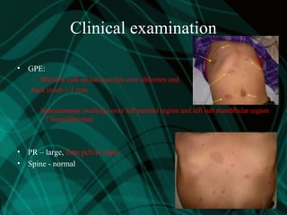
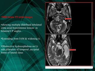
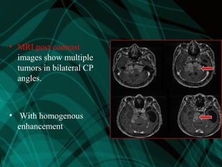
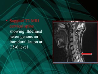
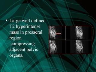
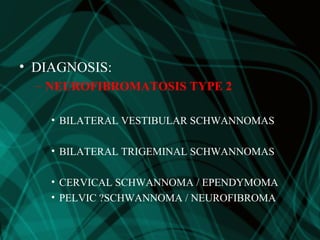
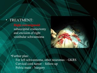
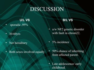
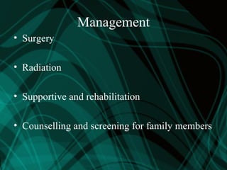
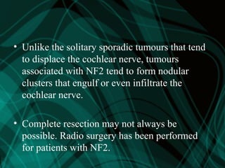
Patients with vestibular
schwannomas associated with
neurofibromatosis 2 represent a
special challenge because of the
risk of complete deafness.
Case details
A 23yrs male,presented to
ENT dept ,NMCH, with
• B/L impaired hearing since 4yrs
• Frequent falls since 4 years
• Ringing sensation in both ears since 4yrs
• Difficulty in walking 4 months
Clinical history
• 6 months back had one episode of tonic-clonic
seizures and controlled on anti-epileptic
medications
• Following seizures he had loss of
consciousness and post-ichtal headache
• Progressive weakness of all four limbs
Clinical history cont…
• No history of
– Vomiting
– Blurring of vision
– Diminution of vision
• No significant personal history
• He had a swelling over scalp and for the same
he was operated 2 years back
• All siblings and parents are healthy
Clinical examination
• GPE:
– Multiple café au lait macules over abdomen and
back (each 1-3 cm)
– Subcutaneous swellings over left parietal region and left sub mandibular region
- ? Neurofibromas
• PR – large, firm pelvic mass
• Spine - normal
– ENT examination – revealed
– RNNIES- B/L AC>BC
– WEBERS-NO LATERALISATION
– ABC- REDUCED
NEUROLOGICAL EXAMINATION
• V – corneal reflex absent b/l,
• Motor:
– Bulk – N
– Tone - spasticity in lower limbs
– Power:
• 3-4 in right sided limbs
• 4-5 in left sided limbs
• Sensory – normal
• Cerebellar signs:
– Axial and appendicular ataxia
• Gait – wide based, swaying to either side
• AUDIOMETRY
• PTA:
– Right ear loss 75 dB (SNHL)
– Left ear loss 60 dB
• SRT – 90 dB
• SDS – very poor (out of proportion to
PTA loss)
• BERA-absent waves
• ACOUSTIC REFLEX- absent
• OAE-absent
• VEMP-absent
•MRI brain T2 axial images
•showing multiple illdefined lobulated
extra axial hyperintense lesions in
bilateral CP angles .
•Extending from IAM & widening it.
•Obstructive hydrocephalous on Lt
side,dilatation of temporal ,occipital
horns of lateral sinus
• MRI post contrast
images show multiple
tumors in bilateral CP
angles.
• With homogenous
enhancement
• Saggital T2 MRI
cervical spine
showing illdefined
heterogenous an
intradural lesion at
C5-6 level
• Large well defined
T2 hyperintense
mass in presacral
region
,compressing
adjacent pelvic
organs.
• DIAGNOSIS:
– NEUROFIBROMATOSIS TYPE 2
• BILATERAL VESTIBULAR SCHWANNOMAS
• BILATERAL TRIGEMINAL SCHWANNOMAS
• CERVICAL SCHWANNOMA / EPENDYMOMA
• PELVIC ?SCHWANNOMA / NEUROFIBROMA
• TREATMENT:
– Right retrosigmoid
suboccipital craniectomy
and excision of right
vestibular schwannoma
•Further plan:
For left schwannoma, other neuromas – GKRS
Cervical cord tumor – follow-up
Pelvic mass - ?surgery
DISCUSSION
U/L VS
• sporadic ,95%
• 30-60yrs
• Not hereditary
• Both sexes involved equally
B/L VS
• a/w NF2 genetic disorder
with fault in chrom22.
• 5% incidence
• 50% chance of inheriting
from affected parent.
• Late adolescence/ early
childhood.
Management
• Surgery
• Radiation
• Supportive and rehabilitation
• Counselling and screening for family members
• Unlike the solitary sporadic tumours that tend
to displace the cochlear nerve, tumours
associated with NF2 tend to form nodular
clusters that engulf or even infiltrate the
cochlear nerve.
• Complete resection may not always be
possible. Radio surgery has been performed
for patients with NF2.
CONCLUSION
• Though B/L vestibular schwannoma is a rare
presentation ,if pt present with b/l progressive
SNHL, balancing disorder ,skin lesions suspect in
the view of NF2
• Rule out family history
• Early diagnosis &management is necessary to
prevent complete deafness & serious
consequences
• Post operatively the patient had no
deterioration in hearing revealed by PTA
• Pt was followed uP for 1 yr & he is doing well
Case report vs with nf2

Case report vs with nf2

  • 3.
    Patients with vestibular schwannomasassociated with neurofibromatosis 2 represent a special challenge because of the risk of complete deafness.
  • 4.
    Case details A 23yrsmale,presented to ENT dept ,NMCH, with • B/L impaired hearing since 4yrs • Frequent falls since 4 years • Ringing sensation in both ears since 4yrs • Difficulty in walking 4 months
  • 5.
    Clinical history • 6months back had one episode of tonic-clonic seizures and controlled on anti-epileptic medications • Following seizures he had loss of consciousness and post-ichtal headache • Progressive weakness of all four limbs
  • 6.
    Clinical history cont… •No history of – Vomiting – Blurring of vision – Diminution of vision • No significant personal history
  • 7.
    • He hada swelling over scalp and for the same he was operated 2 years back • All siblings and parents are healthy
  • 8.
    Clinical examination • GPE: –Multiple café au lait macules over abdomen and back (each 1-3 cm) – Subcutaneous swellings over left parietal region and left sub mandibular region - ? Neurofibromas • PR – large, firm pelvic mass • Spine - normal
  • 9.
    – ENT examination– revealed – RNNIES- B/L AC>BC – WEBERS-NO LATERALISATION – ABC- REDUCED NEUROLOGICAL EXAMINATION • V – corneal reflex absent b/l,
  • 10.
    • Motor: – Bulk– N – Tone - spasticity in lower limbs – Power: • 3-4 in right sided limbs • 4-5 in left sided limbs • Sensory – normal • Cerebellar signs: – Axial and appendicular ataxia • Gait – wide based, swaying to either side
  • 11.
    • AUDIOMETRY • PTA: –Right ear loss 75 dB (SNHL) – Left ear loss 60 dB • SRT – 90 dB • SDS – very poor (out of proportion to PTA loss) • BERA-absent waves • ACOUSTIC REFLEX- absent • OAE-absent • VEMP-absent
  • 12.
    •MRI brain T2axial images •showing multiple illdefined lobulated extra axial hyperintense lesions in bilateral CP angles . •Extending from IAM & widening it. •Obstructive hydrocephalous on Lt side,dilatation of temporal ,occipital horns of lateral sinus
  • 13.
    • MRI postcontrast images show multiple tumors in bilateral CP angles. • With homogenous enhancement
  • 14.
    • Saggital T2MRI cervical spine showing illdefined heterogenous an intradural lesion at C5-6 level
  • 15.
    • Large welldefined T2 hyperintense mass in presacral region ,compressing adjacent pelvic organs.
  • 16.
    • DIAGNOSIS: – NEUROFIBROMATOSISTYPE 2 • BILATERAL VESTIBULAR SCHWANNOMAS • BILATERAL TRIGEMINAL SCHWANNOMAS • CERVICAL SCHWANNOMA / EPENDYMOMA • PELVIC ?SCHWANNOMA / NEUROFIBROMA
  • 17.
    • TREATMENT: – Rightretrosigmoid suboccipital craniectomy and excision of right vestibular schwannoma •Further plan: For left schwannoma, other neuromas – GKRS Cervical cord tumor – follow-up Pelvic mass - ?surgery
  • 18.
    DISCUSSION U/L VS • sporadic,95% • 30-60yrs • Not hereditary • Both sexes involved equally B/L VS • a/w NF2 genetic disorder with fault in chrom22. • 5% incidence • 50% chance of inheriting from affected parent. • Late adolescence/ early childhood.
  • 19.
    Management • Surgery • Radiation •Supportive and rehabilitation • Counselling and screening for family members
  • 20.
    • Unlike thesolitary sporadic tumours that tend to displace the cochlear nerve, tumours associated with NF2 tend to form nodular clusters that engulf or even infiltrate the cochlear nerve. • Complete resection may not always be possible. Radio surgery has been performed for patients with NF2.
  • 21.
    CONCLUSION • Though B/Lvestibular schwannoma is a rare presentation ,if pt present with b/l progressive SNHL, balancing disorder ,skin lesions suspect in the view of NF2 • Rule out family history • Early diagnosis &management is necessary to prevent complete deafness & serious consequences
  • 22.
    • Post operativelythe patient had no deterioration in hearing revealed by PTA • Pt was followed uP for 1 yr & he is doing well

Editor's Notes

  • #12 PTA – PURE TONE AUDIOMETRY, SRT – speech reception threshold, SDS- speech discrimination score
  • #13 MRI brain T2 axial images – showing multiple illdefined lobulated extra axial hyperintense lesions in bilateral CP angles (R>L), causing braistem compression and distortion. Extending from IAM & widening it. Obstructive hydrocephalous on Lt side,dilatation of temoral ,occipital horns of lateral sinus.
  • #14 MRI postcontrast images show multiple tumors in bilateral CP angles. With homogenous enhancement The upper tumor is extending along the Meckel’s cave into the middle fossa. The lower tumors are widening and extending into the IAM
  • #15 Saggital T2 MRI cervical spine showing illdefined heterogenous an intradural lesion at C5-6 level
  • #16 Large well defined T2 hyperintense mass in presacral region ,compressing adjacent pelvic organs.
  • #22 We followed pt for 6 months ,now pt is doing well…