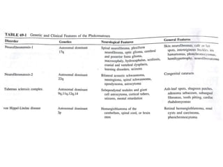
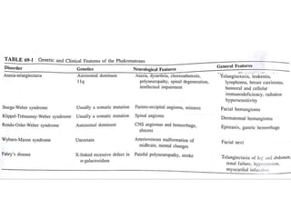
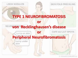
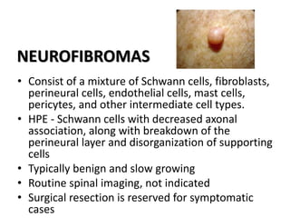
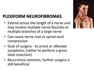
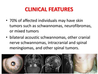
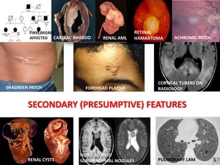
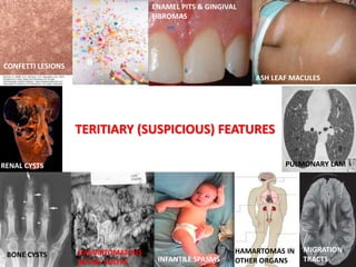
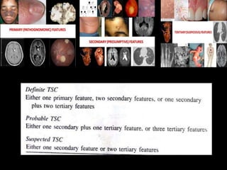
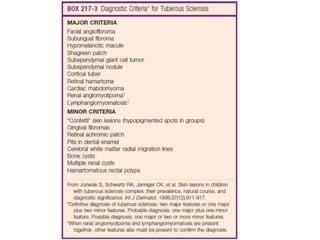
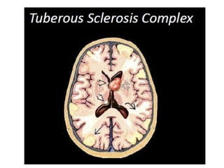
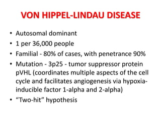
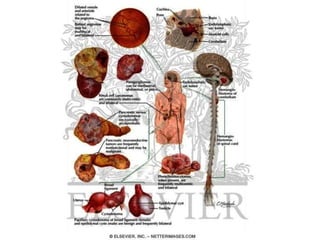
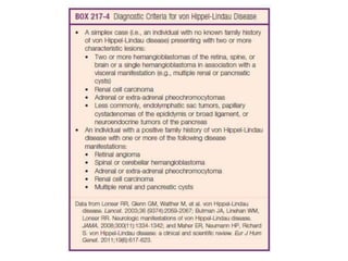
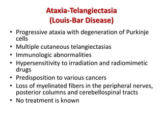
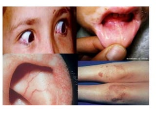
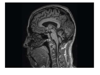
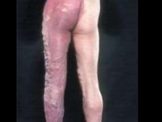
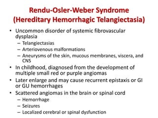
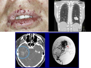
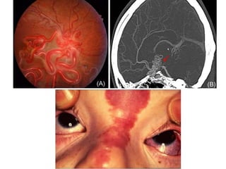
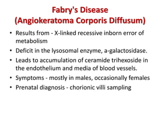
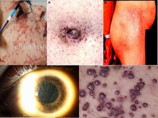
INTRODUCTION
• Phakomatoses – Van der Hoeve (1920)
• Phakos – Birthmark
• Heterogenous group of hereditary disorders
with cutaneous, ocular and neurological
manifestations
PHAKOMATOSES
• Neurofibromatosis
• Tuberous sclerosis complex (Bourneville's
disease)
• Von Hippel-Lindau disease (retinocerebellar
angiomatosis)
• Neurocutaneous angiomatoses
– Ataxia-telangiectasia
– Sturge-weber
– Klippel trénaunay-weber
– Rendu-Osler-Weber syndrome
– Wyburn-Mason syndrome
– Fabry's disease
TYPE 1 NEUROFIBROMATOSIS
or
von Recklinghausen’s disease
or
Peripheral Neurofibromatosis
TYPE 1 NEUROFIBROMATOSIS
• Most common of the neurocutaneous tumor syndromes
• Neurofibromin protein, chromosome 17q11.2
• RAS pathway - Loss of function
• 97% of patients with NF1 meet diagnostic criteria by age 8
1
2 or more
6 or more
2 or more
or
• Multidisciplinary care team
• The lesions necessitating neurosurgical care
– Plexiform or other neurofibromas
– Optic pathway gliomas
– Brainstem gliomas
– Cerebellar gliomas
NEUROFIBROMAS
• Consist of a mixture of Schwann cells, fibroblasts,
perineural cells, endothelial cells, mast cells,
pericytes, and other intermediate cell types.
• HPE - Schwann cells with decreased axonal
association, along with breakdown of the
perineural layer and disorganization of supporting
cells
• Typically benign and slow growing
• Routine spinal imaging, not indicated
• Surgical resection is reserved for symptomatic
cases
PLEXIFORM NEUROFIBROMAS
• Extend across the length of a nerve and
may involve multiple nerve fascicles or
multiple branches of a large nerve
• Can cause nerve root or spinal cord
compression
• Goal of surgery - to arrest or alleviate
symptoms, (rather to perform a gross
total resection)
• Recurrence common, further surgery is
still beneficial
MALIGNANT PERIPHERAL NERVE
SHEATH TUMORS (MPNSTs)
• Neurofibromas degenerate to MPNTs
• The lifetime risk with NF1 - 6% to 13%
• Primarily from nodular neurofibromas or
plexiform neurofibromas.
• Pain or rapid enlargement of a known mass is
a classic symptom
MPNSTs
• Mostly affects adults
(15% in <17 years)
• Mean length of
survival - 2 to 3 years
• Treatment - radical
surgical excision with
radiotherapy as
adjuvant or
neoadjuvant , and
chemotherapy for
high-grade, systemic
disease.
OPTIC PATHWAY GLIOMAS (OPGs)
• 15% of patients with NF1
• Typically affect the anterior optic apparatus
(optic nerve or chiasm)
• Typically low grade (most commonly pilocytic
astrocytomas), but some are higher grade
lesions
• Symptoms - headache, visual changes,
proptosis, endocrine abnormalities, and
symptoms of hydrocephalus
• Non-enhancing OPGs typically do not progress
• Surgical intervention for tissue diagnosis for
patients with an atypical tumor appearance on
MRI
• Progression of either symptoms or imaging
findings - Tumor debulking
• Adjuvant therapy - chemotherapy, with
vincristine and carboplatin as first-line agents.
• Radiation therapy - also a consideration
BRAINSTEM GLIOMAS
• Indolent course in NF-1
• Intervention is reserved for cases of clinical or
radiographic progression.
CEREBELLAR GLIOMAS
• Small percentage of NF1 patients
• Most commonly low grade (pilocytic
astrocytoma)
• Tend to be at subependymal white matter of
the fourth ventricle
• More malignant
• Should be managed aggressively with surgical
resection - curative
TYPE 2 NEUROFIBROMATOSIS
or
Central Neurofibromatosis
TYPE 2 NEUROFIBROMATOSIS
• Autosomal dominant
• Penetrance nearly 100%.
• >50% - de novo mutations
• Present in late adolescence or early adulthood
• Mutations of the NF2 ,chromosome 22(protein-
merlin), regulates contact-mediated growth
inhibition
• Bilateral acoustic schwannomas, intracranial and
spinal meningiomas, and other spinal cord
tumors.
DIAGNOSIS
NIH CRITERIA
• Bilateral acoustic schwannomas
• First-degree family relative with NF2 and unilateral
acoustic schwannoma or any one of the following:
meningioma, schwannoma, glioma, neurofibroma,
juvenile posterior subcapsular lens opacity(MSGNJ)
MANCHESTER CRITERIA
• Bilateral acoustic schwannomas
• First-degree family relative with NF2 and unilateral
acoustic schwannoma or any two of the following:
MSGNJ
• Unilateral acoustic schwannoma and any two of the
following: MSGNJ
• Multiple meningiomas and unilateral acoustic
schwannoma or any two of the following: SGNJ
Baser criteria -
Sensitivity up to 79%
and specificity of 100%
SCREENING
Screening for NF2 should occur in at-risk
individuals even before satisfaction of diagnostic
criteria
Accepted criteria for NF2 screening
• First-degree relative with NF2
• Acoustic schwannomas before the age of 30
• Spinal tumor or meningioma before the age of 20
• Cutaneous schwannomas
• Multiple spinal tumors
SCREENING
• Detailed personal and family histories, cutaneous and
ocular examinations, and MRI of the neuraxis.
• Interval screening - For those with a confirmed NF2
– Annual hearing evaluation with BERA
– Annual ophthalmologic examinations
– Annual dermatologic examinations
– MRI of the neuraxis beginning at age 10, repeated every 2 years up to
age 20, then repeated every 3 to 5 years afterward
CLINICAL FEATURES
• 70% of affected individuals may have skin
tumors such as schwannomas, neurofibromas,
or mixed tumors
• bilateral acoustic schwannomas, other cranial
nerve schwannomas, intracranial and spinal
meningiomas, and other spinal tumors.
ACOUSTIC SCHWANNOMAS
• Benign tumors, when bilateral, are
pathognomonic for NF2 (upto 95%)
• Hearing preservation rates are highest when
tumors are small, but it is difficult to predict
which tumors will grow.
• Tumors are often multifocal and may grow to
involve facial nerve fibers.
• Microsurgical resection - significant tumor
recurrence
• Stereotactic radiosurgery - successful in unilateral
• Development of secondary malignancies in NF2
Intracranial meningiomas (50% of NF2 patients)
• Earlier onset and higher grade than sporadic
Spinal tumors (90% of NF2 patients)
• MC - meningiomas or schwannomas,
Intramedullary tumors such as ependymomas are
also common
• Surgical resection - radiographic progression or
neurological decline.
TUBEROUS SCLEROSIS COMPLEX
• Autosomal dominant
• Affects the CNS, heart,
kidneys, liver, skin, and
lungs.
• De novo mutations -
80%
• Major cause of
epilepsy, autism, and
neurocognitive deficits
• Mutations - 9q34
(TSC1) hamartin or
16p13 (TSC2) tuberin
DIAGNOSIS
• Vogt’s Triad - epilepsy, low intelligence and
adenoma sebaceum, “Epiloia” (less than half
of cases)
RENAL ANGIOMYOLIPOMAS
CARDIAC RHABDOMYOMAS
PULMONARY
LYMPHANGIOMYOMATOSIS
RENAL CELL CARCINOMA OR CYSTS
FACIAL ANGIOFIBROMAS
ADENOMA SEBACEUM LEAF SPOTS
SUBUNGUAL FIBROMAS SHAGREEN PATCHES GINGIVAL FIBROMAS
CORTICAL TUBERS SUBEPENDYMAL NODULES
SUBEPENDYMAL GIANT CELL ASTROCYTOMAS
(SEGAS)
DIAGNOSTIC CRITERIA
FOR
TUBEROUS SCLEROSIS COMPLEX
PRIMARY (PATHOGNOMONIC) FEATURES
FACIAL
ANGIOFIBROMAS
MULTIPLE UNGUAL FIBROMAS
CORTICAL
TUBERS ON HPE
CALCIFIED
SUBEPENDYMAL NODULES
M/L RETINAL
ASTROCYTOMAS
SEGA
SECONDARY (PRESUMPTIVE) FEATURES
FIRST DEGREE
AFFECTED CARDIAC RHABDO
RETINAL
HAMARTOMA ACHROMIC PATCH
RENAL AML
SHAGREEN PATCH FOREHEAD PLAQUE
CORTICAL TUBERS ON
RADIOLOGY
RENAL CYSTS
NON-CALCIFIED
SUBEPENDYMAL NODULES PULMONARY LAM
TERITIARY (SUSPICIOUS) FEATURES
CONFETTI LESIONS
ASH LEAF MACULES
ENAMEL PITS & GINGIVAL
FIBROMAS
RENAL CYSTS PULMONARY LAM
HAMARTOMATOUS
RECTAL POLYPS
BONE CYSTS
INFANTILE SPASMS
HAMARTOMAS IN
OTHER ORGANS
MIGRATION
TRACTS
CNS MANIFESTATIONS
• Smooth, firm, pale gliotic plaques typically distributed
along GW matter jn
• Hamartomas of abnormal neurons and glia
• Can act as seizure foci
CORTICAL TUBERS
• other intracranial abnormalities, such as
cortical dysplasia and white matter linear
migration lines, are visible on imaging.
SUBEPENDYMAL NODULES
• Small, usually asymptomatic calcifications projecting
from the ependymal surface of the lateral ventricles.
• Pathologically similar to SEGAs but do not show
enhancement with contrast material
SEGAs
• Manifest during the first two decades
• Benign tumors of neuroglial origin (5% to
20% of TSC)
• Slow-growing tumors, typically found at
the caudothalamic groove adjacent to
the foramen of Monro
CURRENT SCREENING GUIDELINES
Contrast-enhanced MRI every 1 to 3 years until age 25
• If no additional SEGA is identified during this period,
then some practitioners cease obtaining imaging.
• Some advice to receive serial imaging after this age,
but the interval may be significantly prolonged.
• Any patient who develops new symptoms or
demonstrates a history of serial growth of a SEGA
should promptly undergo imaging, regardless of age.
Management of SEGAs
• Watchful waiting, surgical resection, and
pharmacotherapy with mTOR inhibitors.
• The choice is case dependent
• Incidental SEGAs without hydrocephalus and who
do not demonstrate tumor growth on repeat
imaging - monitoring
• Surgery – If, intratumoral hemorrhage or
symptomatic hydrocephalus
• Gross total resection or radical subtotal resection
may be curative
VON HIPPEL-LINDAU DISEASE
• Autosomal dominant
• 1 per 36,000 people
• Familial - 80% of cases, with penetrance 90%
• Mutation - 3p25 - tumor suppressor protein
pVHL (coordinates multiple aspects of the cell
cycle and facilitates angiogenesis via hypoxia-
inducible factor 1-alpha and 2-alpha)
• “Two-hit” hypothesis
HEMANGIOBLASTOMAS
• Most frequently observed in
VHL
• Sporadic hemangioblastomas
vs VHL disease–associated
• Mean age at presentation is 29
years
• Unpredictable growth pattern - “stuttering”
• Surgery for symptomatic or rapidly growing
lesions
• Preoperative embolization may help to facilitate
surgery by decreasing tumor vascularity
• Hemangioblastomas of the brainstem and spinal
cord can be resected safely
• Preoperative neurological function - best
predictor of postoperative neurological outcome
• Retinal capillary hemangioblastomas and
endolymphatic sac tumors, do not typically
necessitate neurosurgical care
NEUROCUTANEOUS ANGIOMATOSIS
Ataxia-Telangiectasia
(Louis-Bar Disease)
• Progressive ataxia with degeneration of Purkinje
cells
• Multiple cutaneous telangiectasias
• Immunologic abnormalities
• Hypersensitivity to irradiation and radiomimetic
drugs
• Predisposition to various cancers
• Loss of myelinated fibers in the peripheral nerves,
posterior columns and cerebellospinal tracts
• No treatment is known
• Neonatal period - may be asymptomatic
• Toddler stage - walking is clumsy and unsteady
• By 3 years of age and later - cutaneous
telangiectasias appear
• By school age - ataxia, choreoathetosis and
dysarthria may be noted
• By age 10 or so - intellectual impairment and a
mild polyneuropathy may appear
• Death usually occurs in the second decade
from infection or neoplasia
Sturge-Weber Syndrome
(Encephalotrigeminal Angiomatosis)
• Sturge-Weber syndrome - skin and the nervous system
involved
• Most cases, however, seem to be caused by a somatic
mutation
– Unilateral facial angioma (port-wine stain)
– Ipsilateral parieto-occipital leptomeningeal venous angiomatosis
– Underlying cortical atrophy
– Calcifications in cortical layers
– Cystic and enlarged choroid plexus.
– Angiomas occasionally in the eye
– Increased scleral venous pressure reduces outflow of aqueous
humor and may produce glaucoma or buphthalmos
• Neurological abnormalities
– Seizures
– Spastic hemiparesis
– Hemianopia contralateral to the angiomatosis
– Subarachnoid hemorrhage (uncommon)
TREATMENT
• Skin lesion - Argon laser
• Neurosurgical intervention in patients with
medically intractable seizures
– Hemispherectomy
– Occipital lobectomy
– Local excision of the pial angioma, calcified cortex, or
corpus callosotomy
Klippel-Trénaunay-Weber Syndrome
(Spinal Cutaneous Angiomatosis)
• Spinal variant of Sturge-Weber syndrome
– Extensive skin hemangiomas in dermatomal
pattern
– Hemangiomas of the spinal cord in the same
dermatomal distribution
– Osseous or muscular hypertrophy of the involved
area
• Both SW and KTW could be caused by a
similar sporadic mutation affecting different
dermatomes during development
Rendu-Osler-Weber Syndrome
(Hereditary Hemorrhagic Telangiectasia)
• Uncommon disorder of systemic fibrovascular
dysplasia
– Telangiectasias
– Arteriovenous malformations
– Aneurysms of the skin, mucous membranes, viscera, and
CNS
• In childhood, diagnosed from the development of
multiple small red or purple angiomas
• Later enlarge and may cause recurrent epistaxis or GI
or GU hemorrhages
• Scattered angiomas in the brain or spinal cord
– Hemorrhage
– Seizures
– Localized cerebral or spinal dysfunction
• Subarachnoid hemorrhage (SAH) in
association with multiple arterio-venous
malformations, cutaneous telangiectasias,
repeated epistaxis or a family history of SAH
• Septic emboli to brain because of these
vascular defects in the lung
Wyburn-Mason Syndrome
(Bulbar-Facial Angiomatosis)
• Rare disorder
• Hereditary aspects - uncertain
Fabry's Disease
(Angiokeratoma Corporis Diffusum)
• Results from - X-linked recessive inborn error of
metabolism
• Deficit in the lysosomal enzyme, a-galactosidase.
• Leads to accumulation of ceramide trihexoside in
the endothelium and media of blood vessels.
• Symptoms - mostly in males, occasionally females
• Prenatal diagnosis - chorionic villi sampling
• Vascular involvement is usually more diffuse
and can lead to impaired renal function,
hypertension, myocardial infarction, and
cerebrovascular insufficiency.
• Painful polyneuropathy due to ceramide
trihexoside deposition
THANK YOU

Phakomatoses

  • 2.
    INTRODUCTION • Phakomatoses –Van der Hoeve (1920) • Phakos – Birthmark • Heterogenous group of hereditary disorders with cutaneous, ocular and neurological manifestations
  • 3.
    PHAKOMATOSES • Neurofibromatosis • Tuberoussclerosis complex (Bourneville's disease) • Von Hippel-Lindau disease (retinocerebellar angiomatosis) • Neurocutaneous angiomatoses – Ataxia-telangiectasia – Sturge-weber – Klippel trénaunay-weber – Rendu-Osler-Weber syndrome – Wyburn-Mason syndrome – Fabry's disease
  • 6.
    TYPE 1 NEUROFIBROMATOSIS or vonRecklinghausen’s disease or Peripheral Neurofibromatosis
  • 7.
    TYPE 1 NEUROFIBROMATOSIS •Most common of the neurocutaneous tumor syndromes • Neurofibromin protein, chromosome 17q11.2 • RAS pathway - Loss of function • 97% of patients with NF1 meet diagnostic criteria by age 8
  • 8.
    1 2 or more 6or more 2 or more or
  • 10.
    • Multidisciplinary careteam • The lesions necessitating neurosurgical care – Plexiform or other neurofibromas – Optic pathway gliomas – Brainstem gliomas – Cerebellar gliomas
  • 11.
    NEUROFIBROMAS • Consist ofa mixture of Schwann cells, fibroblasts, perineural cells, endothelial cells, mast cells, pericytes, and other intermediate cell types. • HPE - Schwann cells with decreased axonal association, along with breakdown of the perineural layer and disorganization of supporting cells • Typically benign and slow growing • Routine spinal imaging, not indicated • Surgical resection is reserved for symptomatic cases
  • 13.
    PLEXIFORM NEUROFIBROMAS • Extendacross the length of a nerve and may involve multiple nerve fascicles or multiple branches of a large nerve • Can cause nerve root or spinal cord compression • Goal of surgery - to arrest or alleviate symptoms, (rather to perform a gross total resection) • Recurrence common, further surgery is still beneficial
  • 14.
    MALIGNANT PERIPHERAL NERVE SHEATHTUMORS (MPNSTs) • Neurofibromas degenerate to MPNTs • The lifetime risk with NF1 - 6% to 13% • Primarily from nodular neurofibromas or plexiform neurofibromas. • Pain or rapid enlargement of a known mass is a classic symptom
  • 15.
    MPNSTs • Mostly affectsadults (15% in <17 years) • Mean length of survival - 2 to 3 years • Treatment - radical surgical excision with radiotherapy as adjuvant or neoadjuvant , and chemotherapy for high-grade, systemic disease.
  • 16.
    OPTIC PATHWAY GLIOMAS(OPGs) • 15% of patients with NF1 • Typically affect the anterior optic apparatus (optic nerve or chiasm) • Typically low grade (most commonly pilocytic astrocytomas), but some are higher grade lesions • Symptoms - headache, visual changes, proptosis, endocrine abnormalities, and symptoms of hydrocephalus
  • 17.
    • Non-enhancing OPGstypically do not progress • Surgical intervention for tissue diagnosis for patients with an atypical tumor appearance on MRI • Progression of either symptoms or imaging findings - Tumor debulking • Adjuvant therapy - chemotherapy, with vincristine and carboplatin as first-line agents. • Radiation therapy - also a consideration
  • 18.
    BRAINSTEM GLIOMAS • Indolentcourse in NF-1 • Intervention is reserved for cases of clinical or radiographic progression.
  • 19.
    CEREBELLAR GLIOMAS • Smallpercentage of NF1 patients • Most commonly low grade (pilocytic astrocytoma) • Tend to be at subependymal white matter of the fourth ventricle • More malignant • Should be managed aggressively with surgical resection - curative
  • 20.
  • 21.
    TYPE 2 NEUROFIBROMATOSIS •Autosomal dominant • Penetrance nearly 100%. • >50% - de novo mutations • Present in late adolescence or early adulthood • Mutations of the NF2 ,chromosome 22(protein- merlin), regulates contact-mediated growth inhibition • Bilateral acoustic schwannomas, intracranial and spinal meningiomas, and other spinal cord tumors.
  • 22.
  • 23.
    NIH CRITERIA • Bilateralacoustic schwannomas • First-degree family relative with NF2 and unilateral acoustic schwannoma or any one of the following: meningioma, schwannoma, glioma, neurofibroma, juvenile posterior subcapsular lens opacity(MSGNJ) MANCHESTER CRITERIA • Bilateral acoustic schwannomas • First-degree family relative with NF2 and unilateral acoustic schwannoma or any two of the following: MSGNJ • Unilateral acoustic schwannoma and any two of the following: MSGNJ • Multiple meningiomas and unilateral acoustic schwannoma or any two of the following: SGNJ
  • 24.
    Baser criteria - Sensitivityup to 79% and specificity of 100%
  • 25.
    SCREENING Screening for NF2should occur in at-risk individuals even before satisfaction of diagnostic criteria Accepted criteria for NF2 screening • First-degree relative with NF2 • Acoustic schwannomas before the age of 30 • Spinal tumor or meningioma before the age of 20 • Cutaneous schwannomas • Multiple spinal tumors
  • 26.
    SCREENING • Detailed personaland family histories, cutaneous and ocular examinations, and MRI of the neuraxis. • Interval screening - For those with a confirmed NF2 – Annual hearing evaluation with BERA – Annual ophthalmologic examinations – Annual dermatologic examinations – MRI of the neuraxis beginning at age 10, repeated every 2 years up to age 20, then repeated every 3 to 5 years afterward
  • 27.
    CLINICAL FEATURES • 70%of affected individuals may have skin tumors such as schwannomas, neurofibromas, or mixed tumors • bilateral acoustic schwannomas, other cranial nerve schwannomas, intracranial and spinal meningiomas, and other spinal tumors.
  • 28.
    ACOUSTIC SCHWANNOMAS • Benigntumors, when bilateral, are pathognomonic for NF2 (upto 95%) • Hearing preservation rates are highest when tumors are small, but it is difficult to predict which tumors will grow. • Tumors are often multifocal and may grow to involve facial nerve fibers.
  • 29.
    • Microsurgical resection- significant tumor recurrence • Stereotactic radiosurgery - successful in unilateral • Development of secondary malignancies in NF2 Intracranial meningiomas (50% of NF2 patients) • Earlier onset and higher grade than sporadic Spinal tumors (90% of NF2 patients) • MC - meningiomas or schwannomas, Intramedullary tumors such as ependymomas are also common • Surgical resection - radiographic progression or neurological decline.
  • 30.
  • 31.
    TUBEROUS SCLEROSIS COMPLEX •Autosomal dominant • Affects the CNS, heart, kidneys, liver, skin, and lungs. • De novo mutations - 80% • Major cause of epilepsy, autism, and neurocognitive deficits • Mutations - 9q34 (TSC1) hamartin or 16p13 (TSC2) tuberin
  • 32.
    DIAGNOSIS • Vogt’s Triad- epilepsy, low intelligence and adenoma sebaceum, “Epiloia” (less than half of cases)
  • 33.
  • 34.
    FACIAL ANGIOFIBROMAS ADENOMA SEBACEUMLEAF SPOTS SUBUNGUAL FIBROMAS SHAGREEN PATCHES GINGIVAL FIBROMAS
  • 35.
    CORTICAL TUBERS SUBEPENDYMALNODULES SUBEPENDYMAL GIANT CELL ASTROCYTOMAS (SEGAS)
  • 36.
  • 37.
    PRIMARY (PATHOGNOMONIC) FEATURES FACIAL ANGIOFIBROMAS MULTIPLEUNGUAL FIBROMAS CORTICAL TUBERS ON HPE CALCIFIED SUBEPENDYMAL NODULES M/L RETINAL ASTROCYTOMAS SEGA
  • 38.
    SECONDARY (PRESUMPTIVE) FEATURES FIRSTDEGREE AFFECTED CARDIAC RHABDO RETINAL HAMARTOMA ACHROMIC PATCH RENAL AML SHAGREEN PATCH FOREHEAD PLAQUE CORTICAL TUBERS ON RADIOLOGY RENAL CYSTS NON-CALCIFIED SUBEPENDYMAL NODULES PULMONARY LAM
  • 39.
    TERITIARY (SUSPICIOUS) FEATURES CONFETTILESIONS ASH LEAF MACULES ENAMEL PITS & GINGIVAL FIBROMAS RENAL CYSTS PULMONARY LAM HAMARTOMATOUS RECTAL POLYPS BONE CYSTS INFANTILE SPASMS HAMARTOMAS IN OTHER ORGANS MIGRATION TRACTS
  • 42.
  • 44.
    • Smooth, firm,pale gliotic plaques typically distributed along GW matter jn • Hamartomas of abnormal neurons and glia • Can act as seizure foci CORTICAL TUBERS
  • 45.
    • other intracranialabnormalities, such as cortical dysplasia and white matter linear migration lines, are visible on imaging.
  • 46.
    SUBEPENDYMAL NODULES • Small,usually asymptomatic calcifications projecting from the ependymal surface of the lateral ventricles. • Pathologically similar to SEGAs but do not show enhancement with contrast material
  • 47.
    SEGAs • Manifest duringthe first two decades • Benign tumors of neuroglial origin (5% to 20% of TSC) • Slow-growing tumors, typically found at the caudothalamic groove adjacent to the foramen of Monro
  • 48.
    CURRENT SCREENING GUIDELINES Contrast-enhancedMRI every 1 to 3 years until age 25 • If no additional SEGA is identified during this period, then some practitioners cease obtaining imaging. • Some advice to receive serial imaging after this age, but the interval may be significantly prolonged. • Any patient who develops new symptoms or demonstrates a history of serial growth of a SEGA should promptly undergo imaging, regardless of age.
  • 49.
    Management of SEGAs •Watchful waiting, surgical resection, and pharmacotherapy with mTOR inhibitors. • The choice is case dependent • Incidental SEGAs without hydrocephalus and who do not demonstrate tumor growth on repeat imaging - monitoring • Surgery – If, intratumoral hemorrhage or symptomatic hydrocephalus • Gross total resection or radical subtotal resection may be curative
  • 50.
  • 51.
    VON HIPPEL-LINDAU DISEASE •Autosomal dominant • 1 per 36,000 people • Familial - 80% of cases, with penetrance 90% • Mutation - 3p25 - tumor suppressor protein pVHL (coordinates multiple aspects of the cell cycle and facilitates angiogenesis via hypoxia- inducible factor 1-alpha and 2-alpha) • “Two-hit” hypothesis
  • 55.
    HEMANGIOBLASTOMAS • Most frequentlyobserved in VHL • Sporadic hemangioblastomas vs VHL disease–associated • Mean age at presentation is 29 years
  • 56.
    • Unpredictable growthpattern - “stuttering” • Surgery for symptomatic or rapidly growing lesions • Preoperative embolization may help to facilitate surgery by decreasing tumor vascularity • Hemangioblastomas of the brainstem and spinal cord can be resected safely • Preoperative neurological function - best predictor of postoperative neurological outcome
  • 59.
    • Retinal capillaryhemangioblastomas and endolymphatic sac tumors, do not typically necessitate neurosurgical care
  • 60.
  • 61.
    Ataxia-Telangiectasia (Louis-Bar Disease) • Progressiveataxia with degeneration of Purkinje cells • Multiple cutaneous telangiectasias • Immunologic abnormalities • Hypersensitivity to irradiation and radiomimetic drugs • Predisposition to various cancers • Loss of myelinated fibers in the peripheral nerves, posterior columns and cerebellospinal tracts • No treatment is known
  • 64.
    • Neonatal period- may be asymptomatic • Toddler stage - walking is clumsy and unsteady • By 3 years of age and later - cutaneous telangiectasias appear • By school age - ataxia, choreoathetosis and dysarthria may be noted • By age 10 or so - intellectual impairment and a mild polyneuropathy may appear • Death usually occurs in the second decade from infection or neoplasia
  • 65.
    Sturge-Weber Syndrome (Encephalotrigeminal Angiomatosis) •Sturge-Weber syndrome - skin and the nervous system involved • Most cases, however, seem to be caused by a somatic mutation – Unilateral facial angioma (port-wine stain) – Ipsilateral parieto-occipital leptomeningeal venous angiomatosis – Underlying cortical atrophy – Calcifications in cortical layers – Cystic and enlarged choroid plexus. – Angiomas occasionally in the eye – Increased scleral venous pressure reduces outflow of aqueous humor and may produce glaucoma or buphthalmos
  • 67.
    • Neurological abnormalities –Seizures – Spastic hemiparesis – Hemianopia contralateral to the angiomatosis – Subarachnoid hemorrhage (uncommon) TREATMENT • Skin lesion - Argon laser • Neurosurgical intervention in patients with medically intractable seizures – Hemispherectomy – Occipital lobectomy – Local excision of the pial angioma, calcified cortex, or corpus callosotomy
  • 68.
    Klippel-Trénaunay-Weber Syndrome (Spinal CutaneousAngiomatosis) • Spinal variant of Sturge-Weber syndrome – Extensive skin hemangiomas in dermatomal pattern – Hemangiomas of the spinal cord in the same dermatomal distribution – Osseous or muscular hypertrophy of the involved area • Both SW and KTW could be caused by a similar sporadic mutation affecting different dermatomes during development
  • 70.
    Rendu-Osler-Weber Syndrome (Hereditary HemorrhagicTelangiectasia) • Uncommon disorder of systemic fibrovascular dysplasia – Telangiectasias – Arteriovenous malformations – Aneurysms of the skin, mucous membranes, viscera, and CNS • In childhood, diagnosed from the development of multiple small red or purple angiomas • Later enlarge and may cause recurrent epistaxis or GI or GU hemorrhages • Scattered angiomas in the brain or spinal cord – Hemorrhage – Seizures – Localized cerebral or spinal dysfunction
  • 71.
    • Subarachnoid hemorrhage(SAH) in association with multiple arterio-venous malformations, cutaneous telangiectasias, repeated epistaxis or a family history of SAH • Septic emboli to brain because of these vascular defects in the lung
  • 73.
    Wyburn-Mason Syndrome (Bulbar-Facial Angiomatosis) •Rare disorder • Hereditary aspects - uncertain
  • 75.
    Fabry's Disease (Angiokeratoma CorporisDiffusum) • Results from - X-linked recessive inborn error of metabolism • Deficit in the lysosomal enzyme, a-galactosidase. • Leads to accumulation of ceramide trihexoside in the endothelium and media of blood vessels. • Symptoms - mostly in males, occasionally females • Prenatal diagnosis - chorionic villi sampling
  • 77.
    • Vascular involvementis usually more diffuse and can lead to impaired renal function, hypertension, myocardial infarction, and cerebrovascular insufficiency. • Painful polyneuropathy due to ceramide trihexoside deposition
  • 78.

Editor's Notes

  • #20 Tend to be at subependymal white matter of the fourth ventricle, (opposed to sporadic tumors, which tend to occur in the vermis or hemispheres) They are actually more malignant, but not all case series confirm that finding
  • #32 unaffected patients, these proteins dimerize to form a protein that suppresses the mammalian target of rapamycin (mTOR) signaling pathway, which regulates cell proliferation via guanosine triphosphatase (GTPase) activity in the Ras signaling pathway. When either protein is dysfunctional, the heterodimer cannot be formed, which results in abnormal mTOR activation and unchecked cell proliferation, which in turn leads to cellular overgrowth and hamartoma (tuber) or tumor formation
  • #48 SEGAs that are extraventricular, bilateral, and invasive are extremely rare.
  • #54 Numerous visceral oncologic lesions may be observed in VHL disease, including renal cell carcinomas, pheochromocytomas, neuroendocrine tumors, and epididymal cystadenocarcinomas
  • #56 Sporadic hemangioblastomas tend to be solitary and to occur in older patients, VHL disease–associated hemangioblastomas tend to be multiple and occur in young adults Well-circumscribed, benign, vascular tumors usually found in the posterior fossa or spinal cord and frequently consist of a mural nodule with an adjacent cyst
  • #62 lymphomas, leukemias, and breast cancer The immunodeficiency -recurrent sinopulmonary infections
  • #65 By 3 years of age and later - cutaneous telangiectasias appear and are most prominent over the ears, nose, cheeks, conjuctivae, exposed parts of the neck, and flexion creases of the forearm
  • #66 Most cases, however, seem to be caused by a somatic mutation rather than to a germ cell mutation in that they are sporadic rather than inherited.
  • #67 Unilateral facial angioma (port-wine stain) Ipsilateral parieto-occipital leptomeningeal venous angiomatosis The ipsilateral brain underlying the meningeal abnormality shows cortical atrophy, with calcifications in the second and third cortical layers, and often a cystic and enlarged choroid plexus. Angiomas occasionally may be seen in the eye as well as in other parts of the body along the distribution of sensory nerves. In addition, increased scleral venous pressure reduces outflow of aqueous humor and may produce glaucoma or buphthalmos.
  • #69 Some cases suggest autosomal dominant inheritance with incomplete penetrance, in most patients hereditary patterns are not obvious
  • #70 Extensive skin hemangiomas in dermatomal pattern Hemangiomas of the spinal cord in the same dermatomal distribution Osseous or muscular hypertrophy of the involved area
  • #72 This diagnosis should be excluded in all patients with subarachnoid hemorrhage (SAH) in association with multiple arterio-venous malformations, cutaneous telangiectasias, repeated epistaxis or a family history of SAH. Septic emboli that reach the brain because of these vascular defects in the lung are another manifestation of this disease that may present to the neurosurgeon as a brain abscess.
  • #73 Telangiectasias Arteriovenous malformations Aneurysms of the skin, mucous membranes, viscera, and CNS
  • #75 Arteriovenous malformation of one or both sides of the midbrain associated with unilateral or bilateral retinal arteriovenous malformations, facial nevi, and mental changes
  • #76 Although not usually considered among the phakomatoses, Fabry's disease is a genetic abnormality with nervous system, skin,and systemic manifestations that should be considered with the other angiomatoses. For the most part, heterozygous females are asymptomatic carriers who can be diagnosed by assaying a-galactosidase activity.
  • #77 Characterized by multiple, small, flat or slightly raised telangiectasias on the abdomen and lower extremities.
  • #78 Cerebrovascular involvement may lead to cerebral thromboembolic lesions during the teenage years or early adulthood. Neurological involvement also includes a painful polyneuropathy caused by ceramide trihexoside deposition both perineurally and intraneurally. Pain relief is often obtained with phenytoin or carbamazepine.