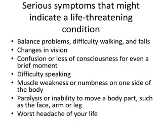
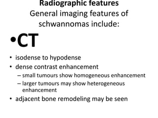
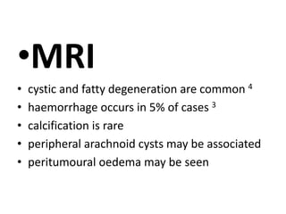
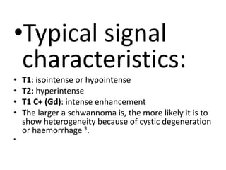
schwannoma
Name : Mustafa Ahmed
Group:509
What schwannoma is ?
• A schwannoma (sh-won-oma) is a tumour of
the tissue that covers nerves, called the nerve
sheath. These tumours develop from a type of
cell called a Schwann cell, which gives them
their name. Schwannomas are often not
cancerous (benign). The most common type of
benign schwannoma is the acoustic neuroma.
This can cause deafness because it grows on a
nerve called the 8th cranial nerve, which
controls hearing
cranial nerves: any cranial nerve may
be involved, except CNI and
CNII, which lack sheaths composed of
schwann cells. The most commonly
involved cranial nerves are sensory
nerves with pure motor nerves rarely
involved; these include :
CN VIII (acoustic neuroma), most commonly the
superior portion of vestibular nerve (most
common~>90%)
– CN V (2nd most common)
– CN VII (3rd most common)
– Jugular foramen (CN IX, X, XI)
– CN XII (hypoglossal schwannomas are very rare)
• spinal nerve roots
• intercostal nerves: intercostal nerve schwannoma
• flexor surfaces of upper and lower limbs
(specifically ulnar and peroneal nerves)
•
posterior mediastinum
• retroperitoneum
• Intracerebral (very rare)
Epidemiology
• Peak presentation is in the 5th-6th decades.
When they occur in patients
withneurofibromatosis type 2 (NF2),
schwannomas usually present by the
3rd decade4. There is no sex predilection.
Associations
• Most schwannomas are solitary (90%) 2 and
sporadic, however there is an association
with NF2 (abnormality of chromosome 22).
Multiple schwannomas are characteristic of NF2.
Approximately 18% of solitary schwannomas
occur in patients with NF2 4.
• There is also schwannomatosis, which
consists of multiple schwannomas without
concomitant involvement of cranial nerve VIII.
Clinical presentation
Presentation depends on location of
the tumour
Pathology
• Schwannomas are benign encapsulated
neoplasms of schwann cells (WHO grade I).
They arise eccentrically from their parent
nerve. They are composed of two cell types:
Antoni A and Antoni B. The Antoni A cells are
densely packed and arranged in fascicles;
acellular areas lie between opposing rows of
parallel nuclei (Verocay bodies). The Antoni B
cells are less compact and are prone to cystic
degeneration.
• Pathologic variants include:
• conventional (most common type)
• cellular schwannomas (predominantly Antoni
A tissue without Verocay bodies)
• melanotic schwannomas (dense melanin
pigment)
• plexiform schwannomas (do not undergo
malignant change, unlike plexiform
neurofibromas)
What are the symptoms of
schwannoma?
• Because acoustic schwannomas cause damage
to the fibers of the nerve of the inner ear,
irritation and other ear-related symptoms are
common. Schwannomas may also affect other
nerves, and the symptoms experienced are
associated with the specific nerve involved.
The symptoms vary in intensity.
Common symptoms of
schwannoma
Balance problems, difficulty walking, and
falls
• Difficulty hearing on one side
• Dizziness or vertigo
• Headache
• Numbness in the face
• Ringing in the ear (tinnitus)
Less common symptoms
of schwannoma
• Numbness
• Pain, including shooting pain (if the nerve root
is affected)
• Paralysis
• Weakness (loss of strength), including facial or
muscular weakness
Serious symptoms that might
indicate a life-threatening
condition
• Balance problems, difficulty walking, and falls
• Changes in vision
• Confusion or loss of consciousness for even a
brief moment
• Difficulty speaking
• Muscle weakness or numbness on one side of
the body
• Paralysis or inability to move a body part, such
as the face, arm or leg
• Worst headache of your life
Radiographic features
General imaging features of
schwannomas include:
•CT
• isodense to hypodense
• dense contrast enhancement
– small tumours show homogeneous enhancement
– larger tumours may show heterogeneous
enhancement
• adjacent bone remodeling may be seen
•MRI
• cystic and fatty degeneration are common 4
• haemorrhage occurs in 5% of cases 3
• calcification is rare
• peripheral arachnoid cysts may be associated
• peritumoural oedema may be seen
•Typical signal
characteristics:
• T1: isointense or hypointense
• T2: hyperintense
• T1 C+ (Gd): intense enhancement
• The larger a schwannoma is, the more likely it is to
show heterogeneity because of cystic degeneration
or haemorrhage 3.
•
Treatment and prognosis
• Schwannomas are slow-growing lesions.
Surgery is the treatment of choice. As
schwannomas do not infiltrate the parent
nerve, they can usually be separated from it.
Recurrence is unusual, even after complete
resection. They almost never undergo
malignant change.

Schwannoma

  • 1.
  • 2.
    What schwannoma is? • A schwannoma (sh-won-oma) is a tumour of the tissue that covers nerves, called the nerve sheath. These tumours develop from a type of cell called a Schwann cell, which gives them their name. Schwannomas are often not cancerous (benign). The most common type of benign schwannoma is the acoustic neuroma. This can cause deafness because it grows on a nerve called the 8th cranial nerve, which controls hearing
  • 3.
    cranial nerves: anycranial nerve may be involved, except CNI and CNII, which lack sheaths composed of schwann cells. The most commonly involved cranial nerves are sensory nerves with pure motor nerves rarely involved; these include :
  • 4.
    CN VIII (acousticneuroma), most commonly the superior portion of vestibular nerve (most common~>90%) – CN V (2nd most common) – CN VII (3rd most common) – Jugular foramen (CN IX, X, XI) – CN XII (hypoglossal schwannomas are very rare) • spinal nerve roots • intercostal nerves: intercostal nerve schwannoma • flexor surfaces of upper and lower limbs (specifically ulnar and peroneal nerves) • posterior mediastinum • retroperitoneum • Intracerebral (very rare)
  • 5.
    Epidemiology • Peak presentationis in the 5th-6th decades. When they occur in patients withneurofibromatosis type 2 (NF2), schwannomas usually present by the 3rd decade4. There is no sex predilection.
  • 6.
    Associations • Most schwannomasare solitary (90%) 2 and sporadic, however there is an association with NF2 (abnormality of chromosome 22). Multiple schwannomas are characteristic of NF2. Approximately 18% of solitary schwannomas occur in patients with NF2 4. • There is also schwannomatosis, which consists of multiple schwannomas without concomitant involvement of cranial nerve VIII.
  • 7.
  • 8.
    Pathology • Schwannomas arebenign encapsulated neoplasms of schwann cells (WHO grade I). They arise eccentrically from their parent nerve. They are composed of two cell types: Antoni A and Antoni B. The Antoni A cells are densely packed and arranged in fascicles; acellular areas lie between opposing rows of parallel nuclei (Verocay bodies). The Antoni B cells are less compact and are prone to cystic degeneration.
  • 9.
    • Pathologic variantsinclude: • conventional (most common type) • cellular schwannomas (predominantly Antoni A tissue without Verocay bodies) • melanotic schwannomas (dense melanin pigment) • plexiform schwannomas (do not undergo malignant change, unlike plexiform neurofibromas)
  • 10.
    What are thesymptoms of schwannoma? • Because acoustic schwannomas cause damage to the fibers of the nerve of the inner ear, irritation and other ear-related symptoms are common. Schwannomas may also affect other nerves, and the symptoms experienced are associated with the specific nerve involved. The symptoms vary in intensity.
  • 11.
    Common symptoms of schwannoma Balanceproblems, difficulty walking, and falls • Difficulty hearing on one side • Dizziness or vertigo • Headache • Numbness in the face • Ringing in the ear (tinnitus)
  • 12.
    Less common symptoms ofschwannoma • Numbness • Pain, including shooting pain (if the nerve root is affected) • Paralysis • Weakness (loss of strength), including facial or muscular weakness
  • 13.
    Serious symptoms thatmight indicate a life-threatening condition • Balance problems, difficulty walking, and falls • Changes in vision • Confusion or loss of consciousness for even a brief moment • Difficulty speaking • Muscle weakness or numbness on one side of the body • Paralysis or inability to move a body part, such as the face, arm or leg • Worst headache of your life
  • 14.
    Radiographic features General imagingfeatures of schwannomas include: •CT • isodense to hypodense • dense contrast enhancement – small tumours show homogeneous enhancement – larger tumours may show heterogeneous enhancement • adjacent bone remodeling may be seen
  • 15.
    •MRI • cystic andfatty degeneration are common 4 • haemorrhage occurs in 5% of cases 3 • calcification is rare • peripheral arachnoid cysts may be associated • peritumoural oedema may be seen
  • 16.
    •Typical signal characteristics: • T1:isointense or hypointense • T2: hyperintense • T1 C+ (Gd): intense enhancement • The larger a schwannoma is, the more likely it is to show heterogeneity because of cystic degeneration or haemorrhage 3. •
  • 17.
    Treatment and prognosis •Schwannomas are slow-growing lesions. Surgery is the treatment of choice. As schwannomas do not infiltrate the parent nerve, they can usually be separated from it. Recurrence is unusual, even after complete resection. They almost never undergo malignant change.