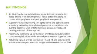
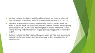
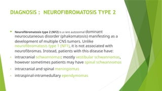
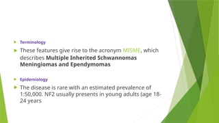
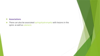
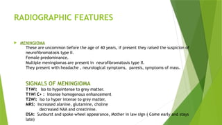
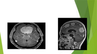
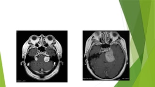
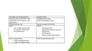
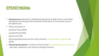
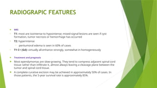
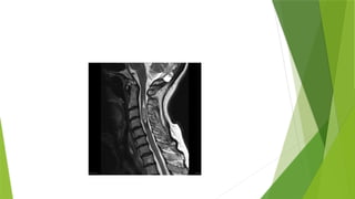
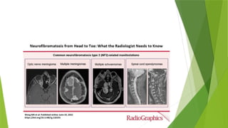
CASE PRESENTATION
 A 22 year old girl came with complain of eclampsia associated with right sided
vision loss after her pregnancy and left eye vision loss after her second
pregnancy . c/o headache.
 She was advised MRI with IV contrast.
 Her MRI no . 10752565
MRI FINDINGS
 An ill defined extra axial altered signal intensity mass lesion
noted arising from left trigeminal nerve extending along its
course with ganglionic and post ganglionic component.
 Superiorly it is compressing left optic nerve and optic chiasma
anteriorly extending into bilateral posterior ethmoid air cells
pushing posterior wall of left maxillary sinus anteriorly and
causing proptosis of left eye ball
 Posteriorly extending up to the level of interpeduncular cistern
compressing left sided midbrain and pons towards opposite side.
 Returning signals are iso intense on T1 and T2 and showing avid
enhancement on post contrast images and no restriction on DWI.
 Multiple variable sized extra axial broad base lesion are noted in bilateral
Para falx region, frontal and parietal lobes with average size of 1.3 x 1 cm.
 Two other altered signal intensity lesion arising from 7th
and 8th
nerve are
noted in left CP angle and protruding into left internal acoustic meatus giving
appearance of ICE CREAM CONE. Returning signals are iso intense on T1 and
T2 and showing avid enhancement on post contrast images and no restriction
on DWI.
 Multiple variable sized cervicomedullary and upper cervical cord shows intra
medullary enhancing lesions having average size of 8 x4 mm suggestive of
ependymoma.
DIAGNOSIS : NEUROFIBROMATOSIS TYPE 2
 Neurofibromatosis type 2 (NF2) is a rare autosomal dominant
neurocutaneous disorder (phakomatosis) manifesting as a
development of multiple CNS tumors. Unlike
neurofibromatosis type 1 (NF1), it is not associated with
neurofibromas. Instead, patients with this disease have:
• intracranial schwannomas: mostly vestibular schwannomas,
however sometimes patients may have spinal schwannomas
• intracranial and spinal meningiomas
• intraspinal-intramedullary ependymomas
 Terminology
 These features give rise to the acronym MISME, which
describes Multiple Inherited Schwannomas
Meningiomas and Ependymomas
 Epidemiology
 The disease is rare with an estimated prevalence of
1:50,000. NF2 usually presents in young adults (age 18-
24 years
 Associations
 There can also be associated syringohydromyelia with lesions in the
spine as well as cataracts
RADIOGRAPHIC FEATURES
 MENINGIOMA
These are uncommon before the age of 40 years, if present they raised the suspicion of
neurofibromatosis type II.
Female predominance.
Multiple meningiomas are present in neurofibromatosis type II.
They present with headache , neurological symptoms, paresis, symptoms of mass.
SIGNALS OF MENINGIOMA
T1WI: Iso to hypointense to grey matter.
T1WI C+ : Intense homogenous enhancement
T2WI: Iso to hyper intense to grey matter,
MRS: Increased alanine, glutamine, choline
decreased NAA and creatinine.
DSA: Sunburst and spoke wheel appearance, Mother in law sign ( Come early and stays
late)
SCHWANOMAS
 Schwannomas, less commonly called neurinomas or neurilemmomas, are
benign tumors of Schwann cell origin and are the most common tumor of
peripheral nerves, including cranial nerves.
When they occur in patients with neurofibromatosis type 2 (NF2), schwannomas
usually present by the 3rd
decade
Multiple schwannomas are characteristic of neurofibromatosis type 2.
Approximately 18% of solitary schwannomas occur in patients with
neurofibromatosis type 2
CLINICAL PRESENTATION
Presentation depends on the location of the tumor but generally, symptoms are
due to local mass effect or dysfunction of the nerve they arise from.
RADIOGRAPHIC SIGNALS
 MRI
 Schwannomas have fairly predictable signal characteristics 7
:
• T1: isointense or hypointense
• T1 C+ (Gd): intense enhancement
• T2: heterogeneously hyperintense cystic degenerative areas may be present, especially in
larger tumors
 Treatment and prognosis
 Schwannomas are slow-growing lesions. Surgery is the treatment of choice. As
schwannomas do not infiltrate the parent nerve, they can usually be separated
from it. Recurrence is unusual after complete resection. They rarely undergo
malignant change.
EPENDYMOMA
 Ependymomas represent a relatively broad group of glial tumors most often
arising from the lining of the ventricles of the brain or the central canal of
the spinal cord.
 They are present in
• posterior fossa (60%)
• supratentorial (30%)
• spinal cord (10%
• Spinal ependymomas are the most common intramedullary neoplasm in
adults
 Clinical presentation is similar to that of other intramedullary spinal tumors
, with pain, weakness, and sensory changes common.
RADIOGRAPIC FEATURES
 MRI
• T1: most are isointense to hypointense; mixed-signal lesions are seen if cyst
formation, tumor necrosis or hemorrhage has occurred
• T2: hyperintense
• peritumoral edema is seen in 60% of cases
• T1 C+ (Gd): virtually all enhance strongly, somewhat in homogeneously
 Treatment and prognosis
 Most ependymomas are slow-growing. They tend to compress adjacent spinal cord
tissue rather than infiltrate it, almost always leaving a cleavage plane between the
tumor and spinal cord tissue.
 A complete curative excision may be achieved in approximately 50% of cases. In
those patients, the 5-year survival rate is approximately 85%.
THANK YOU

Neurofibromatosis type 2 presentation.pptx

  • 1.
  • 2.
     A 22year old girl came with complain of eclampsia associated with right sided vision loss after her pregnancy and left eye vision loss after her second pregnancy . c/o headache.  She was advised MRI with IV contrast.  Her MRI no . 10752565
  • 3.
    MRI FINDINGS  Anill defined extra axial altered signal intensity mass lesion noted arising from left trigeminal nerve extending along its course with ganglionic and post ganglionic component.  Superiorly it is compressing left optic nerve and optic chiasma anteriorly extending into bilateral posterior ethmoid air cells pushing posterior wall of left maxillary sinus anteriorly and causing proptosis of left eye ball  Posteriorly extending up to the level of interpeduncular cistern compressing left sided midbrain and pons towards opposite side.  Returning signals are iso intense on T1 and T2 and showing avid enhancement on post contrast images and no restriction on DWI.
  • 4.
     Multiple variablesized extra axial broad base lesion are noted in bilateral Para falx region, frontal and parietal lobes with average size of 1.3 x 1 cm.  Two other altered signal intensity lesion arising from 7th and 8th nerve are noted in left CP angle and protruding into left internal acoustic meatus giving appearance of ICE CREAM CONE. Returning signals are iso intense on T1 and T2 and showing avid enhancement on post contrast images and no restriction on DWI.  Multiple variable sized cervicomedullary and upper cervical cord shows intra medullary enhancing lesions having average size of 8 x4 mm suggestive of ependymoma.
  • 5.
    DIAGNOSIS : NEUROFIBROMATOSISTYPE 2  Neurofibromatosis type 2 (NF2) is a rare autosomal dominant neurocutaneous disorder (phakomatosis) manifesting as a development of multiple CNS tumors. Unlike neurofibromatosis type 1 (NF1), it is not associated with neurofibromas. Instead, patients with this disease have: • intracranial schwannomas: mostly vestibular schwannomas, however sometimes patients may have spinal schwannomas • intracranial and spinal meningiomas • intraspinal-intramedullary ependymomas
  • 6.
     Terminology  Thesefeatures give rise to the acronym MISME, which describes Multiple Inherited Schwannomas Meningiomas and Ependymomas  Epidemiology  The disease is rare with an estimated prevalence of 1:50,000. NF2 usually presents in young adults (age 18- 24 years
  • 7.
     Associations  Therecan also be associated syringohydromyelia with lesions in the spine as well as cataracts
  • 8.
    RADIOGRAPHIC FEATURES  MENINGIOMA Theseare uncommon before the age of 40 years, if present they raised the suspicion of neurofibromatosis type II. Female predominance. Multiple meningiomas are present in neurofibromatosis type II. They present with headache , neurological symptoms, paresis, symptoms of mass. SIGNALS OF MENINGIOMA T1WI: Iso to hypointense to grey matter. T1WI C+ : Intense homogenous enhancement T2WI: Iso to hyper intense to grey matter, MRS: Increased alanine, glutamine, choline decreased NAA and creatinine. DSA: Sunburst and spoke wheel appearance, Mother in law sign ( Come early and stays late)
  • 10.
    SCHWANOMAS  Schwannomas, lesscommonly called neurinomas or neurilemmomas, are benign tumors of Schwann cell origin and are the most common tumor of peripheral nerves, including cranial nerves. When they occur in patients with neurofibromatosis type 2 (NF2), schwannomas usually present by the 3rd decade Multiple schwannomas are characteristic of neurofibromatosis type 2. Approximately 18% of solitary schwannomas occur in patients with neurofibromatosis type 2 CLINICAL PRESENTATION Presentation depends on the location of the tumor but generally, symptoms are due to local mass effect or dysfunction of the nerve they arise from.
  • 11.
    RADIOGRAPHIC SIGNALS  MRI Schwannomas have fairly predictable signal characteristics 7 : • T1: isointense or hypointense • T1 C+ (Gd): intense enhancement • T2: heterogeneously hyperintense cystic degenerative areas may be present, especially in larger tumors  Treatment and prognosis  Schwannomas are slow-growing lesions. Surgery is the treatment of choice. As schwannomas do not infiltrate the parent nerve, they can usually be separated from it. Recurrence is unusual after complete resection. They rarely undergo malignant change.
  • 14.
    EPENDYMOMA  Ependymomas representa relatively broad group of glial tumors most often arising from the lining of the ventricles of the brain or the central canal of the spinal cord.  They are present in • posterior fossa (60%) • supratentorial (30%) • spinal cord (10% • Spinal ependymomas are the most common intramedullary neoplasm in adults  Clinical presentation is similar to that of other intramedullary spinal tumors , with pain, weakness, and sensory changes common.
  • 15.
    RADIOGRAPIC FEATURES  MRI •T1: most are isointense to hypointense; mixed-signal lesions are seen if cyst formation, tumor necrosis or hemorrhage has occurred • T2: hyperintense • peritumoral edema is seen in 60% of cases • T1 C+ (Gd): virtually all enhance strongly, somewhat in homogeneously  Treatment and prognosis  Most ependymomas are slow-growing. They tend to compress adjacent spinal cord tissue rather than infiltrate it, almost always leaving a cleavage plane between the tumor and spinal cord tissue.  A complete curative excision may be achieved in approximately 50% of cases. In those patients, the 5-year survival rate is approximately 85%.
  • 18.