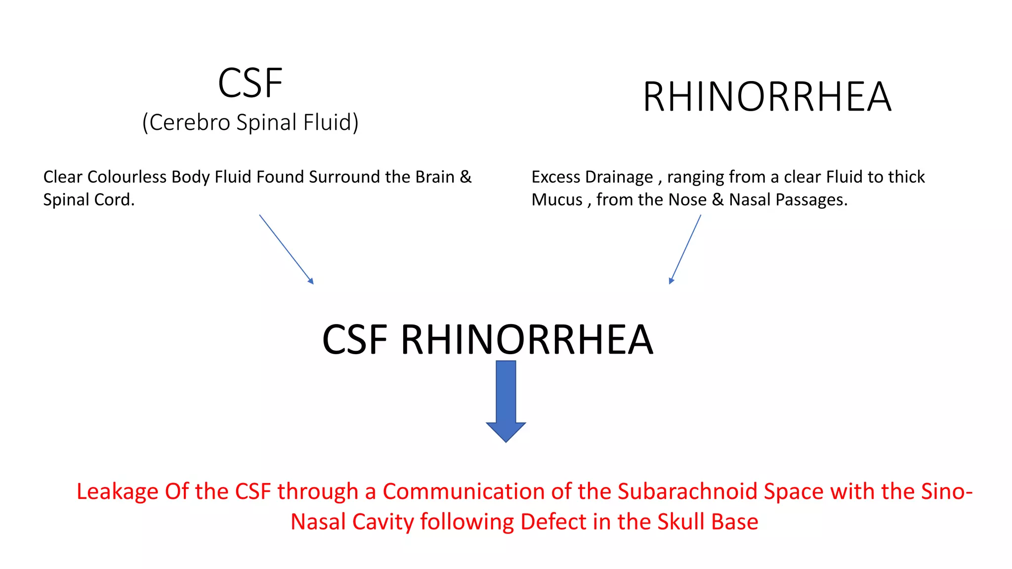
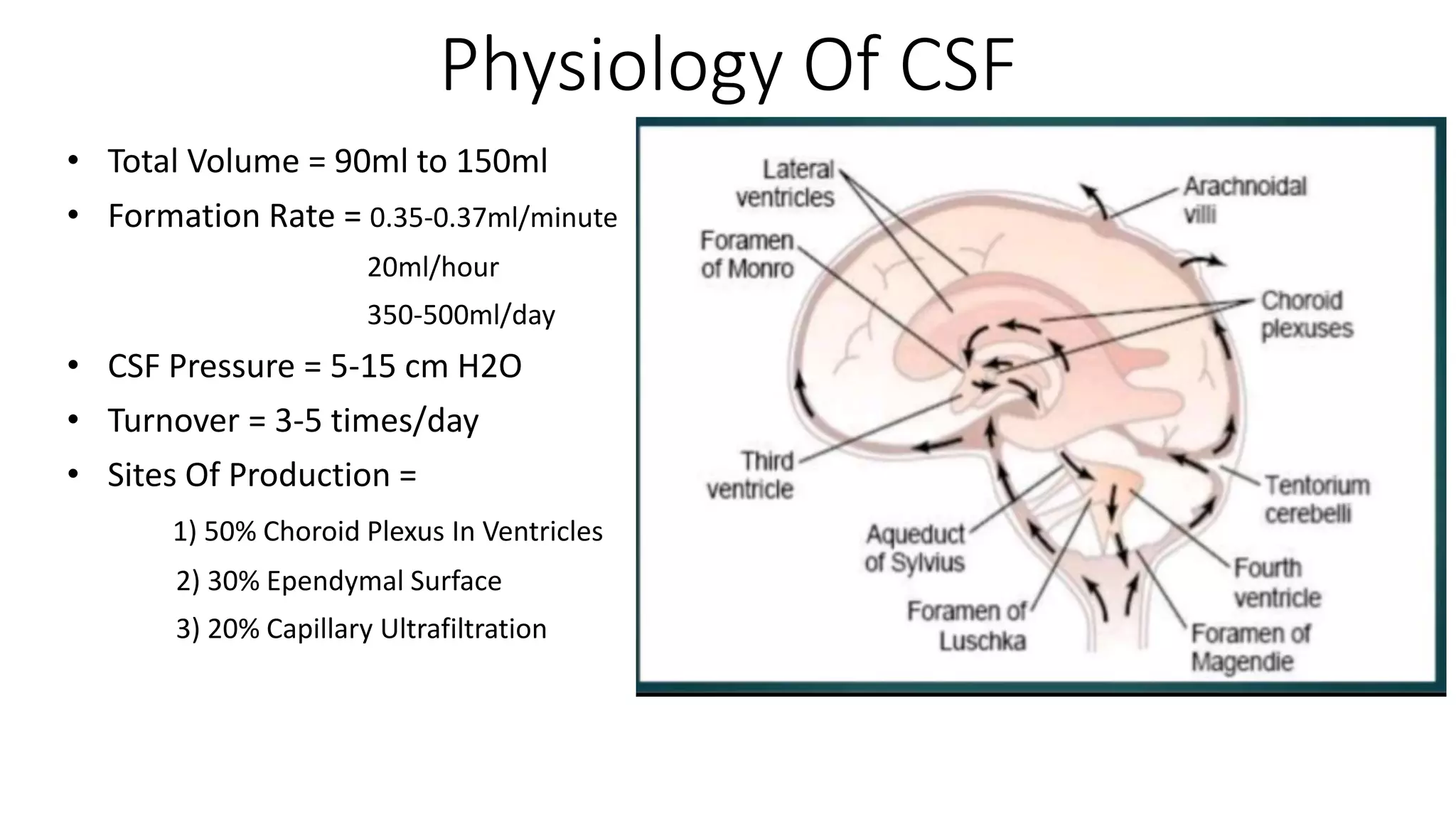
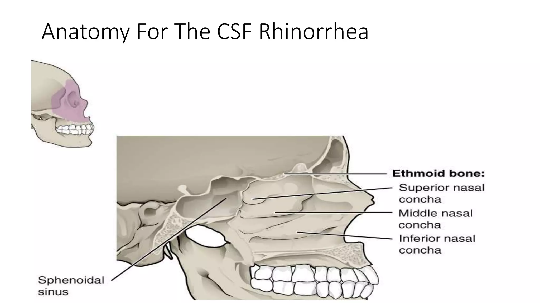
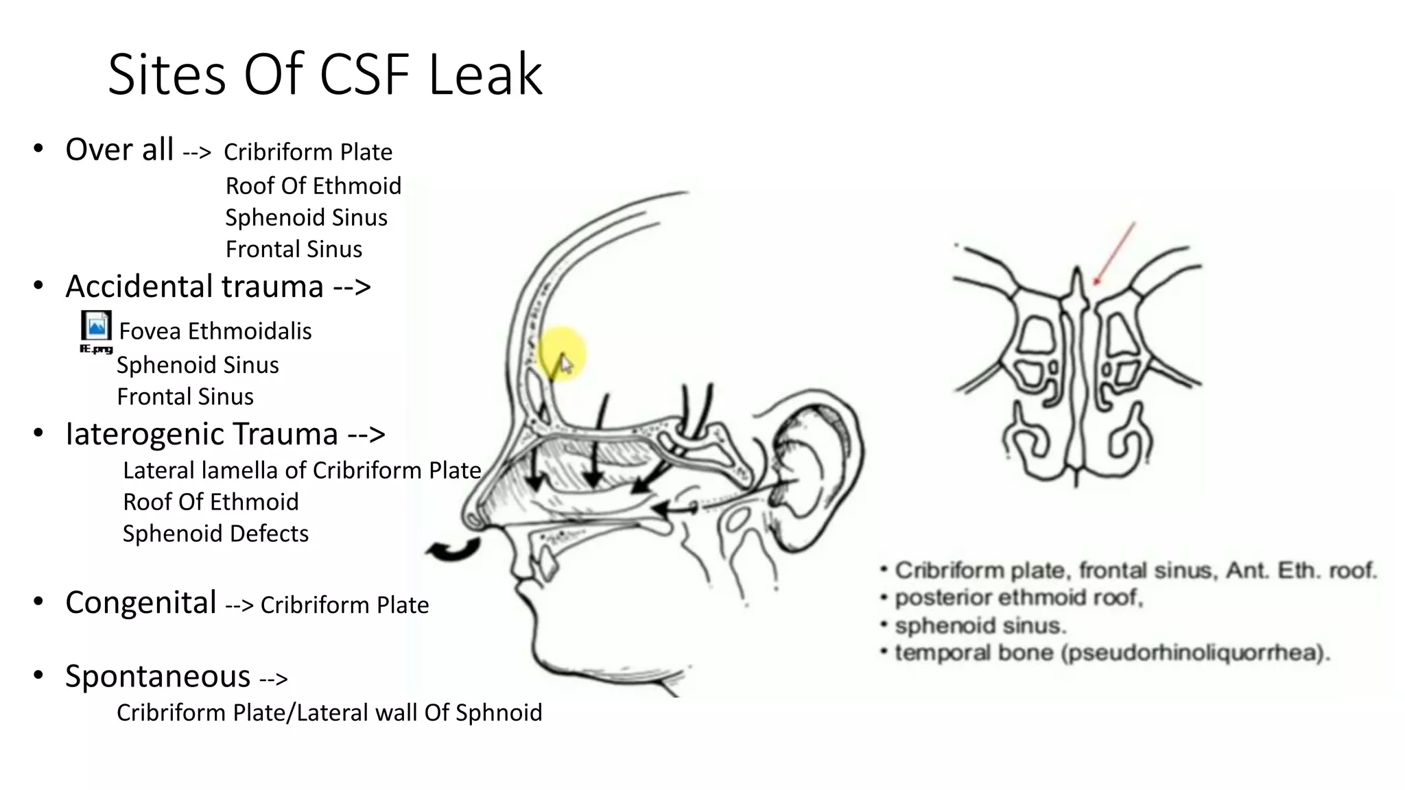
CSF RHINORRHEA
Dr. Jeet M. Amin
Resident Doctor, ENT Department, PDU Medical College & Hospital, Rajkot
Dr. Manoj G. Amin[M. S., ENT]
JEET ENT Hospital, Deesa
HOD, ENT, AASHKA Multispecility Hospital, Gandhinagar
CSF
(Cerebro Spinal Fluid)
RHINORRHEA
Clear Colourless Body Fluid Found Surround the Brain &
Spinal Cord.
Excess Drainage , ranging from a clear Fluid to thick
Mucus , from the Nose & Nasal Passages.
CSF RHINORRHEA
Leakage Of the CSF through a Communication of the Subarachnoid Space with the Sino-
Nasal Cavity following Defect in the Skull Base
Physiology Of CSF
• Total Volume = 90ml to 150ml
• Formation Rate = 0.35-0.37ml/minute
20ml/hour
350-500ml/day
• CSF Pressure = 5-15 cm H2O
• Turnover = 3-5 times/day
• Sites Of Production =
1) 50% Choroid Plexus In Ventricles
2) 30% Ependymal Surface
3) 20% Capillary Ultrafiltration
Anatomy For The CSF Rhinorrhea
Etiology of CSF Rhinorrhea
Sites Of CSF Leak
• Over all --> Cribriform Plate
Roof Of Ethmoid
Sphenoid Sinus
Frontal Sinus
• Accidental trauma -->
Fovea Ethmoidalis
Sphenoid Sinus
Frontal Sinus
• Iaterogenic Trauma -->
Lateral lamella of Cribriform Plate
Roof Of Ethmoid
Sphenoid Defects
• Congenital --> Cribriform Plate
• Spontaneous -->
Cribriform Plate/Lateral wall Of Sphnoid
Clinically
• Watery Nasal Discharge [unilateral >> bilateral]
• Headache
• Anosmia
• Vertigo +/-
• Pulsatile Tinnitus
Lately  Meningitis, Seizures, Fever
Reservoir’s Sign/Teapot Sign
Halo Sign
Blood Stained Nasal Discharge
Take It On Filter Paper
Pale Surrounding
Mostly in Traumatic CSF Leak
Handkerchief Test
Nasal Secretion
Take it on Handkerchief
Non – Sticky
CSF
Sticky
Nasal
Secretion
(Mucin)
Difference
Investigation
•Biochemical
CBC (total count)
Nasal Secretion Routine Micro
Nasal Secretion Sugar & Protein
ß2 Transferrin
ß Trace Protein
•Radiological
HRCT PNS (1-2 mm cut)
T2 Weighted MRI
CT/MR Cisternography
Radionuclide Cisternography
Intrathecal Fluorescein
ß2 Transferrin
• Highly Sensitive & Specific
• CSF , Perilymph , Vitreous
• ß1 – cerebral neuraminidase – >ß2
• False –ve = high mucus content
• False +ve = Chronic Alcohol
Chronic Hepatic
Glycoprotein Disorder
Ca Rectum
• 91% Sensitive & 99.9% Specific
• CSF , Body Fluids including
Serum
• False +ve = Renal Insufficiency
ß Trace Protein
HRCT Para Nasal Sinus(to look for leak)
• False +ve = h/o Skull Base Surgery
• Difficulty = To distinguish between mucosal thickening , meningocele
& CSF Leak from Distal side with collection in sinus
MRI Brain with Skull Base & PNS(to look for leak)
• Coronal T2 Weighted
• To Differentiate between Inflammation & Meningoencephalocele
without radiation exposure.
CT Cisternography
• Used in Doubts.
• Intrathecal Radio-Opaque dye = iohexol &iopamidol ; metrizamide(older & toxic)
• C/I in Active Meningitis & High ICP
Active Leak
Detect
No Active Leak
Put Cotton Pledges in Nose
Wait for Sometime
Check the Pledges
If Dye Present = > Diagnosis √
BUT SITE CAN NOT BE DETECTED
Intrathecal Fluorescein
• Intra Nasal Pledges followed by Intrathecal Injection of Radio-Nuclide
Agent = Tc99
• Limitation = High False +ve Rate, Variable Sensitivity , Low Specificity
Radionuclide Cisternography
• Pre-Operatively Intrathecal injection of 10ml CSF + 0.5ml of 5%
Fluorescein
• Green – Yellow Fluid Gives Site Of Leakage
• Blue light Filter Improves the Result
• Multiple Complications = Seizures , Dizziness , Nausea , Vomiting
Plan Of Radiological Investigation
Conservative Management
• Bed Rest
• Head Elevation (15 Degree)
• Stool Softeners
• Diuretics [Azetazolamide]
• Avoid Coughing, Sneezing , Blowing
• Prophylactic Antibiotics
• Lumber Drain for High Pressure Leak(Hourly 10 ml drainage)
• Mostly for 7-10 Days.
Post-Traumatic CSF Leak
Endoscopic Repair >>>>>> Craniotomy
Supine Position
Nasal Packing
Part Prepare
• Failure of Conservative Management
• Delayed Post-Traumatic Leak
• Very Large Defect
• Persistent Pneumocephalus
• SPONTANEOUS CSF LEAK
Indication Surgical Management
Cribriform Plate , Roof Of Ethmoid
1)Medial Lamella
Lateralize Middle Turbinate
Decongestion Medial Space
2)Lateral Lamella &
Roof Of Ethmoid
Trimming/Medialize Middle Turbinate
Complete Ethmoidectomy
Sphenoid Sinus
Transpterygoid
ParaSaggittal
Transethmoidal
Trimming/Medialize Middle
Turbinate
Complete Ethmoidectomy
Frontal Sinus
• Roof And Posterior Wall = External Approach
• Frontal Recess & Other Part Of Frontal Sinus = Endoscopic Approach
• Awkward Region Endoscopically
Points To Look For
• Remove Whole Skull Base Mucosa
• Expose the Bone to adhere The Graft
• Abrade the Bone for Raw Surface
• Multiple Defects In Sphenoid – Fat Obliteration
• Bipolar Cautery Only.
• Graft Slightly Larger than the Defect
Grafts
• Hadad Flap(Naso-Septal Artery)
• Free Grafts = Conchal Cartilage
Temporalis Facia
Facia Lata
Rectus Sheath
Abdominal Fat
Types Of Reconstruction
Under Lay Over Lay
Small Defects < 5mm
Combined
Moderate (5-10 mm)
Large (>10mm)
Bath Plug Technique
3-0 Absorbable Suture
along Length Of the
Fat Plug And Knot it at
the Inferior End
Insert The Whole Fat
Plug
Gentle Pull Inferiorly
Fibrine Glu = Fibrinogen + Thrombin + Calcium
Factor
Gelatin Glu
Microfibrillar Collage
Surgicel with Abgel
Facia Cartilage
Sandwich
Technique
Post-Operative Care
• Nasal Packing
• Complete Bed Rest for At least 5 Days
• Higher Antibiotics
• Diuretics [Azetazolamide] to maintain the ICP
• FOLLOW UP CT SCAN @ 3 MONTHS to look for the Sinus Blockage
Conclusion
• Diagnosis = Beta 2 transferrin
• Radiology = Cisternography
• Endoscopic Repair Preferable.
• Complete Bed Rest.
Thank You

CSF Rhinorrhea

  • 1.
    CSF RHINORRHEA Dr. JeetM. Amin Resident Doctor, ENT Department, PDU Medical College & Hospital, Rajkot Dr. Manoj G. Amin[M. S., ENT] JEET ENT Hospital, Deesa HOD, ENT, AASHKA Multispecility Hospital, Gandhinagar
  • 2.
    CSF (Cerebro Spinal Fluid) RHINORRHEA ClearColourless Body Fluid Found Surround the Brain & Spinal Cord. Excess Drainage , ranging from a clear Fluid to thick Mucus , from the Nose & Nasal Passages. CSF RHINORRHEA Leakage Of the CSF through a Communication of the Subarachnoid Space with the Sino- Nasal Cavity following Defect in the Skull Base
  • 3.
    Physiology Of CSF •Total Volume = 90ml to 150ml • Formation Rate = 0.35-0.37ml/minute 20ml/hour 350-500ml/day • CSF Pressure = 5-15 cm H2O • Turnover = 3-5 times/day • Sites Of Production = 1) 50% Choroid Plexus In Ventricles 2) 30% Ependymal Surface 3) 20% Capillary Ultrafiltration
  • 4.
    Anatomy For TheCSF Rhinorrhea
  • 5.
    Etiology of CSFRhinorrhea
  • 6.
    Sites Of CSFLeak • Over all --> Cribriform Plate Roof Of Ethmoid Sphenoid Sinus Frontal Sinus • Accidental trauma --> Fovea Ethmoidalis Sphenoid Sinus Frontal Sinus • Iaterogenic Trauma --> Lateral lamella of Cribriform Plate Roof Of Ethmoid Sphenoid Defects • Congenital --> Cribriform Plate • Spontaneous --> Cribriform Plate/Lateral wall Of Sphnoid
  • 7.
    Clinically • Watery NasalDischarge [unilateral >> bilateral] • Headache • Anosmia • Vertigo +/- • Pulsatile Tinnitus Lately  Meningitis, Seizures, Fever
  • 8.
  • 9.
    Halo Sign Blood StainedNasal Discharge Take It On Filter Paper Pale Surrounding Mostly in Traumatic CSF Leak
  • 10.
    Handkerchief Test Nasal Secretion Takeit on Handkerchief Non – Sticky CSF Sticky Nasal Secretion (Mucin)
  • 11.
  • 12.
    Investigation •Biochemical CBC (total count) NasalSecretion Routine Micro Nasal Secretion Sugar & Protein ß2 Transferrin ß Trace Protein •Radiological HRCT PNS (1-2 mm cut) T2 Weighted MRI CT/MR Cisternography Radionuclide Cisternography Intrathecal Fluorescein
  • 13.
    ß2 Transferrin • HighlySensitive & Specific • CSF , Perilymph , Vitreous • ß1 – cerebral neuraminidase – >ß2 • False –ve = high mucus content • False +ve = Chronic Alcohol Chronic Hepatic Glycoprotein Disorder Ca Rectum • 91% Sensitive & 99.9% Specific • CSF , Body Fluids including Serum • False +ve = Renal Insufficiency ß Trace Protein
  • 14.
    HRCT Para NasalSinus(to look for leak) • False +ve = h/o Skull Base Surgery • Difficulty = To distinguish between mucosal thickening , meningocele & CSF Leak from Distal side with collection in sinus
  • 15.
    MRI Brain withSkull Base & PNS(to look for leak) • Coronal T2 Weighted • To Differentiate between Inflammation & Meningoencephalocele without radiation exposure.
  • 16.
    CT Cisternography • Usedin Doubts. • Intrathecal Radio-Opaque dye = iohexol &iopamidol ; metrizamide(older & toxic) • C/I in Active Meningitis & High ICP Active Leak Detect No Active Leak Put Cotton Pledges in Nose Wait for Sometime Check the Pledges If Dye Present = > Diagnosis √ BUT SITE CAN NOT BE DETECTED
  • 17.
    Intrathecal Fluorescein • IntraNasal Pledges followed by Intrathecal Injection of Radio-Nuclide Agent = Tc99 • Limitation = High False +ve Rate, Variable Sensitivity , Low Specificity Radionuclide Cisternography • Pre-Operatively Intrathecal injection of 10ml CSF + 0.5ml of 5% Fluorescein • Green – Yellow Fluid Gives Site Of Leakage • Blue light Filter Improves the Result • Multiple Complications = Seizures , Dizziness , Nausea , Vomiting
  • 18.
    Plan Of RadiologicalInvestigation
  • 19.
    Conservative Management • BedRest • Head Elevation (15 Degree) • Stool Softeners • Diuretics [Azetazolamide] • Avoid Coughing, Sneezing , Blowing • Prophylactic Antibiotics • Lumber Drain for High Pressure Leak(Hourly 10 ml drainage) • Mostly for 7-10 Days. Post-Traumatic CSF Leak
  • 20.
    Endoscopic Repair >>>>>>Craniotomy Supine Position Nasal Packing Part Prepare • Failure of Conservative Management • Delayed Post-Traumatic Leak • Very Large Defect • Persistent Pneumocephalus • SPONTANEOUS CSF LEAK Indication Surgical Management
  • 21.
    Cribriform Plate ,Roof Of Ethmoid 1)Medial Lamella Lateralize Middle Turbinate Decongestion Medial Space 2)Lateral Lamella & Roof Of Ethmoid Trimming/Medialize Middle Turbinate Complete Ethmoidectomy
  • 23.
  • 24.
    Frontal Sinus • RoofAnd Posterior Wall = External Approach • Frontal Recess & Other Part Of Frontal Sinus = Endoscopic Approach • Awkward Region Endoscopically
  • 25.
    Points To LookFor • Remove Whole Skull Base Mucosa • Expose the Bone to adhere The Graft • Abrade the Bone for Raw Surface • Multiple Defects In Sphenoid – Fat Obliteration • Bipolar Cautery Only. • Graft Slightly Larger than the Defect
  • 26.
    Grafts • Hadad Flap(Naso-SeptalArtery) • Free Grafts = Conchal Cartilage Temporalis Facia Facia Lata Rectus Sheath Abdominal Fat
  • 27.
    Types Of Reconstruction UnderLay Over Lay Small Defects < 5mm Combined Moderate (5-10 mm) Large (>10mm)
  • 28.
    Bath Plug Technique 3-0Absorbable Suture along Length Of the Fat Plug And Knot it at the Inferior End Insert The Whole Fat Plug Gentle Pull Inferiorly
  • 29.
    Fibrine Glu =Fibrinogen + Thrombin + Calcium Factor Gelatin Glu Microfibrillar Collage Surgicel with Abgel
  • 30.
  • 31.
    Post-Operative Care • NasalPacking • Complete Bed Rest for At least 5 Days • Higher Antibiotics • Diuretics [Azetazolamide] to maintain the ICP • FOLLOW UP CT SCAN @ 3 MONTHS to look for the Sinus Blockage
  • 32.
    Conclusion • Diagnosis =Beta 2 transferrin • Radiology = Cisternography • Endoscopic Repair Preferable. • Complete Bed Rest.
  • 33.