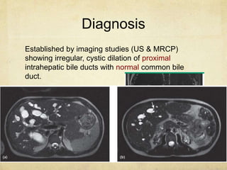
Caroli 
disease 
Ahmed Abdulghany
In 1958, Jacques Caroli first described a rare 
congenital condition in which there was a non-obstructive 
saccular dilatation of large intrahepatic 
bile ducts. 
Caroli Syndrome: more common variable in which 
bile duct dilatation is associated with congenital 
hepatic fibrosis
Genetics 
Incompletely understood however gene for 
autosomal recessive polycystic kidney disease has 
been linked to caroli disease coding for Fibrocystin.
Clinical manifestations 
There are several modes of presentations 
depending on the age of onset and predominance of 
hepatic or renal involvement. 
Biliary dilation predispose to biliary stagnation and 
stone formation. 
Bacterial cholangitis occur frequently and may be 
complicated by septicemia.
Diagnosis 
Established by imaging studies (US & MRCP) 
showing irregular, cystic dilation of proximal 
intrahepatic bile ducts with normal common bile 
duct.
Liver functions show mild cholestatic pattern 
Liver biopsy is rarely required for diagnosis
Differential diagnosis 
1ry biliary cirrhosis 
1ry sclerosing 
cholangitis 
Drug induced 
cholestasis 
Intrahepatic 
cholestasis of 
pregnancy 
Sarcoidosis 
Amyloidosis 
Infiltrating neoplasms
Treatment 
Supportive 
Complications as cholangitis and portal 
hypertension 
Liver transplantation for recurrent cholangitis
Prognosis 
Variable depending upon severity of the disease 
and presence of coexisting renal dysfunction. 
Although risk of cholangiocarcinoma is up to 7%, 
surveillance is unclear. Suspect if there is 
unexplained clinical deterioration or development 
of new biliary strictures.
Thank you

Caroli syndrome

  • 1.
  • 2.
    In 1958, JacquesCaroli first described a rare congenital condition in which there was a non-obstructive saccular dilatation of large intrahepatic bile ducts. Caroli Syndrome: more common variable in which bile duct dilatation is associated with congenital hepatic fibrosis
  • 3.
    Genetics Incompletely understoodhowever gene for autosomal recessive polycystic kidney disease has been linked to caroli disease coding for Fibrocystin.
  • 4.
    Clinical manifestations Thereare several modes of presentations depending on the age of onset and predominance of hepatic or renal involvement. Biliary dilation predispose to biliary stagnation and stone formation. Bacterial cholangitis occur frequently and may be complicated by septicemia.
  • 5.
    Diagnosis Established byimaging studies (US & MRCP) showing irregular, cystic dilation of proximal intrahepatic bile ducts with normal common bile duct.
  • 6.
    Liver functions showmild cholestatic pattern Liver biopsy is rarely required for diagnosis
  • 7.
    Differential diagnosis 1rybiliary cirrhosis 1ry sclerosing cholangitis Drug induced cholestasis Intrahepatic cholestasis of pregnancy Sarcoidosis Amyloidosis Infiltrating neoplasms
  • 8.
    Treatment Supportive Complicationsas cholangitis and portal hypertension Liver transplantation for recurrent cholangitis
  • 9.
    Prognosis Variable dependingupon severity of the disease and presence of coexisting renal dysfunction. Although risk of cholangiocarcinoma is up to 7%, surveillance is unclear. Suspect if there is unexplained clinical deterioration or development of new biliary strictures.
  • 10.

Editor's Notes

  • #6 CENTRAL DOT SIGN: small foci of strong contrast enhancement
  • #8  ----- Meeting Notes (10/26/14 22:00) ----- Supportive: cholangitis fat sol vitamens portal htn