SlideShare a Scribd company logo
Dr. Kamal Motawei                          HIMT          Introduction to Anatomy (ANAT 215)

Unit 7: The cardiovascular system
      :

The cardiovascular system is formed of the heart and the blood vessels

                                      A. The HEART


The heart is a hollow muscular pump formed of four chambers (2atria and 2
ventricles).

Position:
It lies obliquely in the lower part o the
                                    of
mediastinum of the thorax (one third of the
heart lies to the right of the midline, while 2/3
lies to the left).
Shape:
The heart is pyramidal in shape. So, it has an
apex,      base      and   surfaces   which       are:
sternocostal, diaphragmatic, right and left
surfaces.
The apex:
It is formed of the left ventricle only.
It is directed downwards, forwards and to the
left.
It lies opposite a point in the left 5th intercostal
space 9cm away from the midline.
The Base:
It is formed by the 2 atria (mainly the left
atrium).
It is directed upwards, backwards and to the
right.
It lies opposite the middle 4 thoracic vertebrae
(separated from them by the oesophagus, the
       ted
descending aorta, the trachea and bronchi)
                ,                 bronchi).
The major blood vessels are connected to the
heart via the base.


                                                                                    49
Dr. Kamal Motawei                         HIMT                        Introduction to Anatomy (ANAT 215)

The sternocostal surface:
It is related to sternum and costal wall. It is formed of the left and right ventricles.
The diaphragmatic surface:
It lies on the diaphragm. It is formed of the 2 ventricles
The right surface:
It is formed by the right atrium It is related to the right lung.
                          atrium.
The left surface:
It is formed of the left ventricle. It is related to the left lung.


Chambers of the heart and their connections:
The heart is formed of 4 chambers, Rt. & Lt. atria and Rt. & Lt. ventricles. The 2 atria
are demarcated from the 2 ventricles by a groove called the coronary groove, while
the 2 ventricles are separated from each other by the anterior and posterior
interventricular grooves.
There is no direct connection between the right side and the left side of the heart.
                                                                  side


The right atrium:
It forms the right surface of the heart.
It is separated from the left atrium by the
interatrial septum.
During intrauterine life, this septum shows
an oval foramen to allow passage of blood
from the right atrium directl into the left
                      directly
atrium (as there is no respiration). This
foramen is closed after birth.
Connections:
The right atrium receives non oxygenated blood from the whole body via:
                          non-oxygenated
   1. Superior vena cava (SVC) which collects venous blood from the upper half of
      the body.
   2. Inferior vena cava (IVC) which collects venous blood from the lower half of the
      body.
   3. Coronary sinus that collects venous blood from the heart itself.
The right atrium pumps its blood content to the right ventricle through the right
   atrioventricular orifice (which is guarded by the tricuspid valve).
            ricular


                                                                                                 50
Dr. Kamal Motawei                         HIMT                     Introduction to Anatomy (ANAT 215)


The right ventricle:
It forms most of the costo-diaphragmatic surface of the heart.
It is separated from the left ventricle by the interventricular septum that bulges into
the cavity of the right ventricle.
Connections:
It receives venous blood from the right atrium via the tricuspid valve.
It pumps its blood content, via the pulmonary trunk, to the lungs.
(The pulmonary valve prevents regurgitation of blood from the pulmonary trunk to the
right ventricle).


The left atrium:
It forms most of the base of the heart.
It receives two pulmonary veins from each lung.
It pumps its blood content to the left ventricle via the left atrioventricular orifice
(guarded by the mitral valve).


The left ventricle:
Its wall is thicker than that of the right ventricle.
It receives oxygenated blood from the left atrium via the mitral valve.
It pumps its blood content, via the aorta, to the whole body.
(The aortic valve prevents regurgitation of blood from the aorta to the left ventricle).


                                                                                              51
Dr. Kamal Motawei                     HIMT             Introduction to Anatomy (ANAT 215)


Blood Supply of the Heart:


The heart is supplied with arterial blood by
the right and the left coronary arteries,
which are branches from the ascending
aorta.
A large vein called coronary sinus drains
most of the heart, then it opens into the right
atrium.


Surface Anatomy of the heart:


The heart can be projected to the surface by determining 4 points and connecting
them together by four lines:
First point (A)(The apex of the
    heart): it is a point drawn in the
    left 5th intercostal space, 9cm
    from the midline.
Second point (B): on the 2nd left
    costal cartilage, 1.5 cm to the left
    of the sternal edge.
Third point (C): on the 3rd right
    costal cartilage, 1.5 cm to the
    right of the sternal edge.
Fourth point (D): on the 6th right
    costal cartilage, 1.5 cm to the
    right of the sternal edge.


                                                                                  52
Dr. Kamal Motawei                     HIMT                  Introduction to Anatomy (ANAT 215)


Coverings of the heart:
The heart lies within a fibrous tissue envelope called the fibrous pericardium
                                                                   pericardium.
The fibrous pericardium is continuos with the adventitia of the great vessels
connected to the heart.
A serous sac intervenes between the heart and its
fibrous pericardium to prevent friction. This serous
sac is called serous pericardium It is formed of
                     pericardium.
two layers and a cavity in between, visceral layer
covering the heart surface and pari
                               parietal layer lining
the fibrous pericardium. The cavity of the serous
pericardium contains nothing but few drops of fluid
for lubrication.


Structure of the Heart:
The heart is formed of two myocardial muscles, one for the atria and another one for
the ventricles. Both muscles are attached to the corresponding side of the fibrous
        ricles.
rings surrounding the atrioventricular orifices. The atrial and ventricular muscles are
completely separated from each other except for at the atrioventricular bundle, which
represents the only communication between both muscles.
       nts
The heart cavity is lined with endothelial layer.
The conducting system: this is formed of some specialized cardiac muscle fibers,
which are able to initialize and conduct cardiac impulses. It comprises the following
structures:
Sino-atrial node (pacemaker of the heart) in the right atrium close to the opening of
the SVC
Atrioventricular node: in the lower part of the interatrial septum.
The atrioventricular bundle: It represents the only possible communicat
                     bundle:                                 communication
between the atrial and ventricular muscles. It is divided into to two (Rt. &Lt.)
branches for the Rt. and Lt. Ventricles.


                                                                                       53
Dr. Kamal Motawei                          HIMT              Introduction to Anatomy (ANAT 215)


                                         B. The Blood Vessels


   The blood vessels are of three types: arteries, veins, and capillaries.
1. The arteries: They convey blood from the heart and distribute it to the body tissues.
   The smallest arteries (less than 0.1 mm in diameter, are called arterioles.
   The wall of an artery is formed of three layers;
     a) Tunica intema: it is formed of simple squamous endothelium.
     b) Tunica Media: It is formed of smooth muscle fibers and elastic fibers. Medium
           sized and small sized arteries contain more smooth muscles fibers than elastic
           fibers. On the other hand, Large sized arteries contain more elastic fibers than
           muscle fibers.
     c) Tunica adventitia: it is formed of fibrous connective tissue.
2. The veins: They convey blood back to the heart. The smallest veins are called
    venules. The wall of the vien is formed of the same layers as the artery but it is
    comparatively thin and very poor in muscle and elastic fibers.
   Veins may be deep or superficial. Medium-sized deep arteries are usually
   accompanied            by      two
   veins, one on each side,
   called venae comitantes.
   The superficial veins lie
   under      the      skin,      they
   clinically important as they
   are used for intravenous
   injections.
3. The capillaries: they are
   microscopic                 vessels
   arranged in a network-form
   connecting the arterioles to
   the venules. The wall of the
   blood capillary is formed of
   a single layer of squamous
   cells on a basement membrane.


                                                                                           54
Dr. Kamal Motawei                     HIMT                    Introduction to Anatomy (ANAT 215)


                                Major arteries of the body
 The Aorta: it is the largest artery in the body. It conveys blood from the left ventricle of
       the heart to the whole body. It has three distinct parts: Ascending aor
                                                                           aorta, it gives
       left and right coronary arteries to the heart.
       Arch of the aorta, it giv three main arteries: brachiocephalic left common
                        , gives                       brachiocephalic,
       carotid & left subclavian arteries to the head, neck and upper limbs.
       Descending aorta, the part of the descending aorta
                       ,
       that lies above the diaphragm is called thoracic
       aorta. The part below the diaphra
            .                    diaphragm is the
       abdominal aorta. The abominal aorta ends by
                      .
       dividing into right and left common iliac arteries.
The Common iliac arteries: Each one divides into Internal
       iliac artery (for the pelvis) and external iliac arteries
       (for the lower limbs).
Arteries of the upper limb: The subclavian artery when
       passing the axilla is called axillary artery. It
       continues its course in the upper arm as the brachial
       artery, which is a superficial artery. The brachial
       artery divides into radial and ulnar arteries in the
       forearm (they are superficial in the lower part of the forearm.
           arm
Arteries of the lower limb: The external iliac artery
       continues on the front aspect of the thigh as the
       femoral artery, then the artery passes behind the
       Knee joint as the popliteal artey The poplitea
                                   artey.
       artery divides in the back of the leg into anterior
       and posterior tibial arteries.
Arterial pulse
Arterial pulse can be palpated at the superficial arteries
       especially those related to bones, e.g. the radial
       artery at the lower end of the radius, the common
       carotid artery in the neck and the femoral artery at
       the upper part of the front of the thigh.


                                                                                              55
Dr. Kamal Motawei                        HIMT                   Introduction to Anatomy (ANAT 215)


                                      The major deep veins
The Superior vena Cava (SVC): it drains the upper part of the body. It is formed by the
        union of the the right and left brachiocephalic veins
The right and left brachio cephalic veins: Each of which is formed by the union of the
        corresponding internal jugular and subclavian vein.
The inferior vena cava (IVC): It drains the lower part of the body. It is formed in the
        abdomen by the union of the right and left common iliac veins.


     Superficial veins of clinical importance:
External Jugular Vein: it lies in the neck extending obliquely backwards from the angle
        of the mandible to the middle of the clavicle. It is used as a monitor for the heart
        function. Also, it can be used for intravenous injections.
Cephalic vein of the upper limb: It lies constantly under the skin immediately behind
        the styloid process of the radius.
Median cubital vein: it lies obliquely in the cubital fossa (in front of the elbow joint).
The great saphenous vein of the lower limb: It has a constant position in front of the
        medial malleolus. It is liable to be varicose (elongated and tortuous).


                                                                                               56
Dr. Kamal Motawei        HIMT        Introduction to Anatomy (ANAT 215)

                    Major Arteries


                                                                57
Dr. Kamal Motawei    HIMT         Introduction to Anatomy (ANAT 215)

                    Major Veins


                                                             58

More Related Content

What's hot

Cardiac cycle physiology_4_dpt
Cardiac cycle physiology_4_dptCardiac cycle physiology_4_dpt
Cardiac cycle physiology_4_dpt
Student
 
The coronary circulation
The coronary circulationThe coronary circulation
The coronary circulation
Idris Siddiqui
 
The valves of the heart
The valves of the heartThe valves of the heart
The valves of the heart
Idris Siddiqui
 
Coronary artery anatomy
Coronary artery anatomyCoronary artery anatomy
Coronary artery anatomy
Dr. Yash Kumar Achantani
 
Cardiovascular system ppt
Cardiovascular system pptCardiovascular system ppt
Holter monitor and information does it provide
Holter monitor and information does it provideHolter monitor and information does it provide
Holter monitor and information does it provide
Progressive Cardiac Services
 
Heart anatomy
Heart anatomyHeart anatomy
Heart anatomy
Dr. Waqas Nawaz
 
Conducting system of the heart
Conducting system of the heartConducting system of the heart
Conducting system of the heart
Dr. Manish Kharel
 
Coronary circulation
Coronary circulationCoronary circulation
Coronary circulation
DrChintansinh Parmar
 
Conduction system of the heart
Conduction system of the heartConduction system of the heart
Conduction system of the heart
Raghu Kishore Galla
 
Heart Anatomy and Physiology:Review
Heart Anatomy and Physiology:ReviewHeart Anatomy and Physiology:Review
Heart Anatomy and Physiology:Review
Tosca Torres
 
Anatomy and function of the coronary arteries
Anatomy and function of the coronary arteriesAnatomy and function of the coronary arteries
Anatomy and function of the coronary arteries
Society for Heart Attack Prevention and Eradication
 
Pulmonary circulation
Pulmonary circulationPulmonary circulation
Pulmonary circulation
Dr Nilesh Kate
 
Blood Pressure Measurement (2011)
Blood Pressure Measurement (2011)Blood Pressure Measurement (2011)
Blood Pressure Measurement (2011)
Liesl Brown
 
MEASUREMENT OF CARDIAC OUTPUT
MEASUREMENT OF CARDIAC OUTPUTMEASUREMENT OF CARDIAC OUTPUT
MEASUREMENT OF CARDIAC OUTPUT
AmudhaLakshmi1
 
ECG leads
ECG leadsECG leads
ECG leads
meducationdotnet
 
Cardiac contractility
Cardiac contractilityCardiac contractility
Cardiac contractility
Kiran Goushika
 
Heart
HeartHeart
3. internal structure of the heart
3. internal structure of the heart3. internal structure of the heart
3. internal structure of the heart
Dr. Mohammad Mahmoud
 
cardiac output
 cardiac output cardiac output
cardiac output
Athulya Lakshmanan
 

What's hot (20)

Cardiac cycle physiology_4_dpt
Cardiac cycle physiology_4_dptCardiac cycle physiology_4_dpt
Cardiac cycle physiology_4_dpt
 
The coronary circulation
The coronary circulationThe coronary circulation
The coronary circulation
 
The valves of the heart
The valves of the heartThe valves of the heart
The valves of the heart
 
Coronary artery anatomy
Coronary artery anatomyCoronary artery anatomy
Coronary artery anatomy
 
Cardiovascular system ppt
Cardiovascular system pptCardiovascular system ppt
Cardiovascular system ppt
 
Holter monitor and information does it provide
Holter monitor and information does it provideHolter monitor and information does it provide
Holter monitor and information does it provide
 
Heart anatomy
Heart anatomyHeart anatomy
Heart anatomy
 
Conducting system of the heart
Conducting system of the heartConducting system of the heart
Conducting system of the heart
 
Coronary circulation
Coronary circulationCoronary circulation
Coronary circulation
 
Conduction system of the heart
Conduction system of the heartConduction system of the heart
Conduction system of the heart
 
Heart Anatomy and Physiology:Review
Heart Anatomy and Physiology:ReviewHeart Anatomy and Physiology:Review
Heart Anatomy and Physiology:Review
 
Anatomy and function of the coronary arteries
Anatomy and function of the coronary arteriesAnatomy and function of the coronary arteries
Anatomy and function of the coronary arteries
 
Pulmonary circulation
Pulmonary circulationPulmonary circulation
Pulmonary circulation
 
Blood Pressure Measurement (2011)
Blood Pressure Measurement (2011)Blood Pressure Measurement (2011)
Blood Pressure Measurement (2011)
 
MEASUREMENT OF CARDIAC OUTPUT
MEASUREMENT OF CARDIAC OUTPUTMEASUREMENT OF CARDIAC OUTPUT
MEASUREMENT OF CARDIAC OUTPUT
 
ECG leads
ECG leadsECG leads
ECG leads
 
Cardiac contractility
Cardiac contractilityCardiac contractility
Cardiac contractility
 
Heart
HeartHeart
Heart
 
3. internal structure of the heart
3. internal structure of the heart3. internal structure of the heart
3. internal structure of the heart
 
cardiac output
 cardiac output cardiac output
cardiac output
 

Similar to Cardiovascular

Human heart
Human heartHuman heart
Human heart
Sheen Khan
 
Heart (Its Structure & Function).pptx
Heart (Its Structure & Function).pptxHeart (Its Structure & Function).pptx
Heart (Its Structure & Function).pptx
Vipin Chandran
 
Cardiovascular system of goat
Cardiovascular system of goatCardiovascular system of goat
Cardiovascular system of goat
Dr. Waqas Nawaz
 
Cardiovascular system.pdf
Cardiovascular system.pdfCardiovascular system.pdf
Cardiovascular system.pdf
AderawAlemie
 
3-pericardium & Heart.pptx
3-pericardium & Heart.pptx3-pericardium & Heart.pptx
3-pericardium & Heart.pptx
muhammadsumair19
 
Aorta&its branches . pptx.pptx
Aorta&its branches . pptx.pptxAorta&its branches . pptx.pptx
Aorta&its branches . pptx.pptx
KannurLion
 
Surgery 5th year, 9th, 10th &11th lectures (Dr. Ahmed Al-Azzawi)
Surgery 5th year, 9th, 10th &11th lectures (Dr. Ahmed Al-Azzawi)Surgery 5th year, 9th, 10th &11th lectures (Dr. Ahmed Al-Azzawi)
Surgery 5th year, 9th, 10th &11th lectures (Dr. Ahmed Al-Azzawi)
College of Medicine, Sulaymaniyah
 
Cardiovascular System & Antihypertensives drugs.pptx
Cardiovascular System & Antihypertensives drugs.pptxCardiovascular System & Antihypertensives drugs.pptx
Cardiovascular System & Antihypertensives drugs.pptx
Shahid Khan
 
heart anatomy
heart anatomyheart anatomy
heart anatomy
Jay Patel
 
"THE HEART"
"THE HEART""THE HEART"
"THE HEART"
UmairKhan463
 
Cardiovascular Anatomy.pptx
Cardiovascular Anatomy.pptxCardiovascular Anatomy.pptx
Cardiovascular Anatomy.pptx
xamdathisbad
 
Cardiovascular Anatomy.pptx
Cardiovascular Anatomy.pptxCardiovascular Anatomy.pptx
Cardiovascular Anatomy.pptx
xamdathisbad
 
Heart.pptx
Heart.pptxHeart.pptx
Heart.pptx
SpandanaRavikanti
 
Circulatory anes 16.pptx
Circulatory anes 16.pptxCirculatory anes 16.pptx
Circulatory anes 16.pptx
Wasihun Aragie
 
Cardiovascular system
Cardiovascular systemCardiovascular system
Cardiovascular system
deepaingawale21
 
Anatomy of Human Heart along with internal and external features.
Anatomy of Human Heart along with internal and external features.Anatomy of Human Heart along with internal and external features.
Anatomy of Human Heart along with internal and external features.
ssuser7bf0a2
 
CARDIOVASCULAR SYSTEM
CARDIOVASCULAR SYSTEMCARDIOVASCULAR SYSTEM
CARDIOVASCULAR SYSTEM
sandeep chandakavate
 
INTRODUCTION TO CVS PHYSIOLOGY-BSCN.pptx
INTRODUCTION TO CVS PHYSIOLOGY-BSCN.pptxINTRODUCTION TO CVS PHYSIOLOGY-BSCN.pptx
INTRODUCTION TO CVS PHYSIOLOGY-BSCN.pptx
JamesMwaura15
 
Cardiovascular system
Cardiovascular systemCardiovascular system
Cardiovascular system
s shariff
 
Anatomy of heart .pptx
Anatomy of heart .pptxAnatomy of heart .pptx
Anatomy of heart .pptx
sonipranshu810
 

Similar to Cardiovascular (20)

Human heart
Human heartHuman heart
Human heart
 
Heart (Its Structure & Function).pptx
Heart (Its Structure & Function).pptxHeart (Its Structure & Function).pptx
Heart (Its Structure & Function).pptx
 
Cardiovascular system of goat
Cardiovascular system of goatCardiovascular system of goat
Cardiovascular system of goat
 
Cardiovascular system.pdf
Cardiovascular system.pdfCardiovascular system.pdf
Cardiovascular system.pdf
 
3-pericardium & Heart.pptx
3-pericardium & Heart.pptx3-pericardium & Heart.pptx
3-pericardium & Heart.pptx
 
Aorta&its branches . pptx.pptx
Aorta&its branches . pptx.pptxAorta&its branches . pptx.pptx
Aorta&its branches . pptx.pptx
 
Surgery 5th year, 9th, 10th &11th lectures (Dr. Ahmed Al-Azzawi)
Surgery 5th year, 9th, 10th &11th lectures (Dr. Ahmed Al-Azzawi)Surgery 5th year, 9th, 10th &11th lectures (Dr. Ahmed Al-Azzawi)
Surgery 5th year, 9th, 10th &11th lectures (Dr. Ahmed Al-Azzawi)
 
Cardiovascular System & Antihypertensives drugs.pptx
Cardiovascular System & Antihypertensives drugs.pptxCardiovascular System & Antihypertensives drugs.pptx
Cardiovascular System & Antihypertensives drugs.pptx
 
heart anatomy
heart anatomyheart anatomy
heart anatomy
 
"THE HEART"
"THE HEART""THE HEART"
"THE HEART"
 
Cardiovascular Anatomy.pptx
Cardiovascular Anatomy.pptxCardiovascular Anatomy.pptx
Cardiovascular Anatomy.pptx
 
Cardiovascular Anatomy.pptx
Cardiovascular Anatomy.pptxCardiovascular Anatomy.pptx
Cardiovascular Anatomy.pptx
 
Heart.pptx
Heart.pptxHeart.pptx
Heart.pptx
 
Circulatory anes 16.pptx
Circulatory anes 16.pptxCirculatory anes 16.pptx
Circulatory anes 16.pptx
 
Cardiovascular system
Cardiovascular systemCardiovascular system
Cardiovascular system
 
Anatomy of Human Heart along with internal and external features.
Anatomy of Human Heart along with internal and external features.Anatomy of Human Heart along with internal and external features.
Anatomy of Human Heart along with internal and external features.
 
CARDIOVASCULAR SYSTEM
CARDIOVASCULAR SYSTEMCARDIOVASCULAR SYSTEM
CARDIOVASCULAR SYSTEM
 
INTRODUCTION TO CVS PHYSIOLOGY-BSCN.pptx
INTRODUCTION TO CVS PHYSIOLOGY-BSCN.pptxINTRODUCTION TO CVS PHYSIOLOGY-BSCN.pptx
INTRODUCTION TO CVS PHYSIOLOGY-BSCN.pptx
 
Cardiovascular system
Cardiovascular systemCardiovascular system
Cardiovascular system
 
Anatomy of heart .pptx
Anatomy of heart .pptxAnatomy of heart .pptx
Anatomy of heart .pptx
 

More from Dr Motawei

Lec.10 mechanics of resp
Lec.10 mechanics of respLec.10 mechanics of resp
Lec.10 mechanics of resp
Dr Motawei
 
Lec.9 diaphragm pt&rt
Lec.9 diaphragm pt&rtLec.9 diaphragm pt&rt
Lec.9 diaphragm pt&rt
Dr Motawei
 
Lec.8 lungs pt&rc
Lec.8 lungs pt&rcLec.8 lungs pt&rc
Lec.8 lungs pt&rc
Dr Motawei
 
Lec.7,intercostal spaces
Lec.7,intercostal spacesLec.7,intercostal spaces
Lec.7,intercostal spaces
Dr Motawei
 
Lec.6,larynx pt&rc
Lec.6,larynx pt&rcLec.6,larynx pt&rc
Lec.6,larynx pt&rc
Dr Motawei
 
Lec.5,pharynx pt&rc
Lec.5,pharynx pt&rcLec.5,pharynx pt&rc
Lec.5,pharynx pt&rc
Dr Motawei
 
Lec.4,nose pt&rc
Lec.4,nose pt&rcLec.4,nose pt&rc
Lec.4,nose pt&rc
Dr Motawei
 
Lec.1 3,descriptive anatomical-terms-pt-rc
Lec.1 3,descriptive anatomical-terms-pt-rcLec.1 3,descriptive anatomical-terms-pt-rc
Lec.1 3,descriptive anatomical-terms-pt-rc
Dr Motawei
 
Blood
BloodBlood
Blood
Dr Motawei
 
Blood
BloodBlood
Blood
Dr Motawei
 
The gastrointestinal system
The gastrointestinal systemThe gastrointestinal system
The gastrointestinal system
Dr Motawei
 
Respiratory system
Respiratory systemRespiratory system
Respiratory system
Dr Motawei
 

More from Dr Motawei (12)

Lec.10 mechanics of resp
Lec.10 mechanics of respLec.10 mechanics of resp
Lec.10 mechanics of resp
 
Lec.9 diaphragm pt&rt
Lec.9 diaphragm pt&rtLec.9 diaphragm pt&rt
Lec.9 diaphragm pt&rt
 
Lec.8 lungs pt&rc
Lec.8 lungs pt&rcLec.8 lungs pt&rc
Lec.8 lungs pt&rc
 
Lec.7,intercostal spaces
Lec.7,intercostal spacesLec.7,intercostal spaces
Lec.7,intercostal spaces
 
Lec.6,larynx pt&rc
Lec.6,larynx pt&rcLec.6,larynx pt&rc
Lec.6,larynx pt&rc
 
Lec.5,pharynx pt&rc
Lec.5,pharynx pt&rcLec.5,pharynx pt&rc
Lec.5,pharynx pt&rc
 
Lec.4,nose pt&rc
Lec.4,nose pt&rcLec.4,nose pt&rc
Lec.4,nose pt&rc
 
Lec.1 3,descriptive anatomical-terms-pt-rc
Lec.1 3,descriptive anatomical-terms-pt-rcLec.1 3,descriptive anatomical-terms-pt-rc
Lec.1 3,descriptive anatomical-terms-pt-rc
 
Blood
BloodBlood
Blood
 
Blood
BloodBlood
Blood
 
The gastrointestinal system
The gastrointestinal systemThe gastrointestinal system
The gastrointestinal system
 
Respiratory system
Respiratory systemRespiratory system
Respiratory system
 

Recently uploaded

South African Journal of Science: Writing with integrity workshop (2024)
South African Journal of Science: Writing with integrity workshop (2024)South African Journal of Science: Writing with integrity workshop (2024)
South African Journal of Science: Writing with integrity workshop (2024)
Academy of Science of South Africa
 
The Diamonds of 2023-2024 in the IGRA collection
The Diamonds of 2023-2024 in the IGRA collectionThe Diamonds of 2023-2024 in the IGRA collection
The Diamonds of 2023-2024 in the IGRA collection
Israel Genealogy Research Association
 
Your Skill Boost Masterclass: Strategies for Effective Upskilling
Your Skill Boost Masterclass: Strategies for Effective UpskillingYour Skill Boost Masterclass: Strategies for Effective Upskilling
Your Skill Boost Masterclass: Strategies for Effective Upskilling
Excellence Foundation for South Sudan
 
Main Java[All of the Base Concepts}.docx
Main Java[All of the Base Concepts}.docxMain Java[All of the Base Concepts}.docx
Main Java[All of the Base Concepts}.docx
adhitya5119
 
CACJapan - GROUP Presentation 1- Wk 4.pdf
CACJapan - GROUP Presentation 1- Wk 4.pdfCACJapan - GROUP Presentation 1- Wk 4.pdf
CACJapan - GROUP Presentation 1- Wk 4.pdf
camakaiclarkmusic
 
Smart-Money for SMC traders good time and ICT
Smart-Money for SMC traders good time and ICTSmart-Money for SMC traders good time and ICT
Smart-Money for SMC traders good time and ICT
simonomuemu
 
How to Manage Your Lost Opportunities in Odoo 17 CRM
How to Manage Your Lost Opportunities in Odoo 17 CRMHow to Manage Your Lost Opportunities in Odoo 17 CRM
How to Manage Your Lost Opportunities in Odoo 17 CRM
Celine George
 
ISO/IEC 27001, ISO/IEC 42001, and GDPR: Best Practices for Implementation and...
ISO/IEC 27001, ISO/IEC 42001, and GDPR: Best Practices for Implementation and...ISO/IEC 27001, ISO/IEC 42001, and GDPR: Best Practices for Implementation and...
ISO/IEC 27001, ISO/IEC 42001, and GDPR: Best Practices for Implementation and...
PECB
 
Pollock and Snow "DEIA in the Scholarly Landscape, Session One: Setting Expec...
Pollock and Snow "DEIA in the Scholarly Landscape, Session One: Setting Expec...Pollock and Snow "DEIA in the Scholarly Landscape, Session One: Setting Expec...
Pollock and Snow "DEIA in the Scholarly Landscape, Session One: Setting Expec...
National Information Standards Organization (NISO)
 
PCOS corelations and management through Ayurveda.
PCOS corelations and management through Ayurveda.PCOS corelations and management through Ayurveda.
PCOS corelations and management through Ayurveda.
Dr. Shivangi Singh Parihar
 
Top five deadliest dog breeds in America
Top five deadliest dog breeds in AmericaTop five deadliest dog breeds in America
Top five deadliest dog breeds in America
Bisnar Chase Personal Injury Attorneys
 
Exploiting Artificial Intelligence for Empowering Researchers and Faculty, In...
Exploiting Artificial Intelligence for Empowering Researchers and Faculty, In...Exploiting Artificial Intelligence for Empowering Researchers and Faculty, In...
Exploiting Artificial Intelligence for Empowering Researchers and Faculty, In...
Dr. Vinod Kumar Kanvaria
 
How to Add Chatter in the odoo 17 ERP Module
How to Add Chatter in the odoo 17 ERP ModuleHow to Add Chatter in the odoo 17 ERP Module
How to Add Chatter in the odoo 17 ERP Module
Celine George
 
Assessment and Planning in Educational technology.pptx
Assessment and Planning in Educational technology.pptxAssessment and Planning in Educational technology.pptx
Assessment and Planning in Educational technology.pptx
Kavitha Krishnan
 
The basics of sentences session 5pptx.pptx
The basics of sentences session 5pptx.pptxThe basics of sentences session 5pptx.pptx
The basics of sentences session 5pptx.pptx
heathfieldcps1
 
বাংলাদেশ অর্থনৈতিক সমীক্ষা (Economic Review) ২০২৪ UJS App.pdf
বাংলাদেশ অর্থনৈতিক সমীক্ষা (Economic Review) ২০২৪ UJS App.pdfবাংলাদেশ অর্থনৈতিক সমীক্ষা (Economic Review) ২০২৪ UJS App.pdf
বাংলাদেশ অর্থনৈতিক সমীক্ষা (Economic Review) ২০২৪ UJS App.pdf
eBook.com.bd (প্রয়োজনীয় বাংলা বই)
 
Digital Artefact 1 - Tiny Home Environmental Design
Digital Artefact 1 - Tiny Home Environmental DesignDigital Artefact 1 - Tiny Home Environmental Design
Digital Artefact 1 - Tiny Home Environmental Design
amberjdewit93
 
RPMS TEMPLATE FOR SCHOOL YEAR 2023-2024 FOR TEACHER 1 TO TEACHER 3
RPMS TEMPLATE FOR SCHOOL YEAR 2023-2024 FOR TEACHER 1 TO TEACHER 3RPMS TEMPLATE FOR SCHOOL YEAR 2023-2024 FOR TEACHER 1 TO TEACHER 3
RPMS TEMPLATE FOR SCHOOL YEAR 2023-2024 FOR TEACHER 1 TO TEACHER 3
IreneSebastianRueco1
 
PIMS Job Advertisement 2024.pdf Islamabad
PIMS Job Advertisement 2024.pdf IslamabadPIMS Job Advertisement 2024.pdf Islamabad
PIMS Job Advertisement 2024.pdf Islamabad
AyyanKhan40
 
How to Fix the Import Error in the Odoo 17
How to Fix the Import Error in the Odoo 17How to Fix the Import Error in the Odoo 17
How to Fix the Import Error in the Odoo 17
Celine George
 

Recently uploaded (20)

South African Journal of Science: Writing with integrity workshop (2024)
South African Journal of Science: Writing with integrity workshop (2024)South African Journal of Science: Writing with integrity workshop (2024)
South African Journal of Science: Writing with integrity workshop (2024)
 
The Diamonds of 2023-2024 in the IGRA collection
The Diamonds of 2023-2024 in the IGRA collectionThe Diamonds of 2023-2024 in the IGRA collection
The Diamonds of 2023-2024 in the IGRA collection
 
Your Skill Boost Masterclass: Strategies for Effective Upskilling
Your Skill Boost Masterclass: Strategies for Effective UpskillingYour Skill Boost Masterclass: Strategies for Effective Upskilling
Your Skill Boost Masterclass: Strategies for Effective Upskilling
 
Main Java[All of the Base Concepts}.docx
Main Java[All of the Base Concepts}.docxMain Java[All of the Base Concepts}.docx
Main Java[All of the Base Concepts}.docx
 
CACJapan - GROUP Presentation 1- Wk 4.pdf
CACJapan - GROUP Presentation 1- Wk 4.pdfCACJapan - GROUP Presentation 1- Wk 4.pdf
CACJapan - GROUP Presentation 1- Wk 4.pdf
 
Smart-Money for SMC traders good time and ICT
Smart-Money for SMC traders good time and ICTSmart-Money for SMC traders good time and ICT
Smart-Money for SMC traders good time and ICT
 
How to Manage Your Lost Opportunities in Odoo 17 CRM
How to Manage Your Lost Opportunities in Odoo 17 CRMHow to Manage Your Lost Opportunities in Odoo 17 CRM
How to Manage Your Lost Opportunities in Odoo 17 CRM
 
ISO/IEC 27001, ISO/IEC 42001, and GDPR: Best Practices for Implementation and...
ISO/IEC 27001, ISO/IEC 42001, and GDPR: Best Practices for Implementation and...ISO/IEC 27001, ISO/IEC 42001, and GDPR: Best Practices for Implementation and...
ISO/IEC 27001, ISO/IEC 42001, and GDPR: Best Practices for Implementation and...
 
Pollock and Snow "DEIA in the Scholarly Landscape, Session One: Setting Expec...
Pollock and Snow "DEIA in the Scholarly Landscape, Session One: Setting Expec...Pollock and Snow "DEIA in the Scholarly Landscape, Session One: Setting Expec...
Pollock and Snow "DEIA in the Scholarly Landscape, Session One: Setting Expec...
 
PCOS corelations and management through Ayurveda.
PCOS corelations and management through Ayurveda.PCOS corelations and management through Ayurveda.
PCOS corelations and management through Ayurveda.
 
Top five deadliest dog breeds in America
Top five deadliest dog breeds in AmericaTop five deadliest dog breeds in America
Top five deadliest dog breeds in America
 
Exploiting Artificial Intelligence for Empowering Researchers and Faculty, In...
Exploiting Artificial Intelligence for Empowering Researchers and Faculty, In...Exploiting Artificial Intelligence for Empowering Researchers and Faculty, In...
Exploiting Artificial Intelligence for Empowering Researchers and Faculty, In...
 
How to Add Chatter in the odoo 17 ERP Module
How to Add Chatter in the odoo 17 ERP ModuleHow to Add Chatter in the odoo 17 ERP Module
How to Add Chatter in the odoo 17 ERP Module
 
Assessment and Planning in Educational technology.pptx
Assessment and Planning in Educational technology.pptxAssessment and Planning in Educational technology.pptx
Assessment and Planning in Educational technology.pptx
 
The basics of sentences session 5pptx.pptx
The basics of sentences session 5pptx.pptxThe basics of sentences session 5pptx.pptx
The basics of sentences session 5pptx.pptx
 
বাংলাদেশ অর্থনৈতিক সমীক্ষা (Economic Review) ২০২৪ UJS App.pdf
বাংলাদেশ অর্থনৈতিক সমীক্ষা (Economic Review) ২০২৪ UJS App.pdfবাংলাদেশ অর্থনৈতিক সমীক্ষা (Economic Review) ২০২৪ UJS App.pdf
বাংলাদেশ অর্থনৈতিক সমীক্ষা (Economic Review) ২০২৪ UJS App.pdf
 
Digital Artefact 1 - Tiny Home Environmental Design
Digital Artefact 1 - Tiny Home Environmental DesignDigital Artefact 1 - Tiny Home Environmental Design
Digital Artefact 1 - Tiny Home Environmental Design
 
RPMS TEMPLATE FOR SCHOOL YEAR 2023-2024 FOR TEACHER 1 TO TEACHER 3
RPMS TEMPLATE FOR SCHOOL YEAR 2023-2024 FOR TEACHER 1 TO TEACHER 3RPMS TEMPLATE FOR SCHOOL YEAR 2023-2024 FOR TEACHER 1 TO TEACHER 3
RPMS TEMPLATE FOR SCHOOL YEAR 2023-2024 FOR TEACHER 1 TO TEACHER 3
 
PIMS Job Advertisement 2024.pdf Islamabad
PIMS Job Advertisement 2024.pdf IslamabadPIMS Job Advertisement 2024.pdf Islamabad
PIMS Job Advertisement 2024.pdf Islamabad
 
How to Fix the Import Error in the Odoo 17
How to Fix the Import Error in the Odoo 17How to Fix the Import Error in the Odoo 17
How to Fix the Import Error in the Odoo 17
 

Cardiovascular

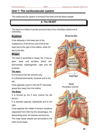
  • 1. Dr. Kamal Motawei HIMT Introduction to Anatomy (ANAT 215) Unit 7: The cardiovascular system : The cardiovascular system is formed of the heart and the blood vessels A. The HEART The heart is a hollow muscular pump formed of four chambers (2atria and 2 ventricles). Position: It lies obliquely in the lower part o the of mediastinum of the thorax (one third of the heart lies to the right of the midline, while 2/3 lies to the left). Shape: The heart is pyramidal in shape. So, it has an apex, base and surfaces which are: sternocostal, diaphragmatic, right and left surfaces. The apex: It is formed of the left ventricle only. It is directed downwards, forwards and to the left. It lies opposite a point in the left 5th intercostal space 9cm away from the midline. The Base: It is formed by the 2 atria (mainly the left atrium). It is directed upwards, backwards and to the right. It lies opposite the middle 4 thoracic vertebrae (separated from them by the oesophagus, the ted descending aorta, the trachea and bronchi) , bronchi). The major blood vessels are connected to the heart via the base. 49
  • 2. Dr. Kamal Motawei HIMT Introduction to Anatomy (ANAT 215) The sternocostal surface: It is related to sternum and costal wall. It is formed of the left and right ventricles. The diaphragmatic surface: It lies on the diaphragm. It is formed of the 2 ventricles The right surface: It is formed by the right atrium It is related to the right lung. atrium. The left surface: It is formed of the left ventricle. It is related to the left lung. Chambers of the heart and their connections: The heart is formed of 4 chambers, Rt. & Lt. atria and Rt. & Lt. ventricles. The 2 atria are demarcated from the 2 ventricles by a groove called the coronary groove, while the 2 ventricles are separated from each other by the anterior and posterior interventricular grooves. There is no direct connection between the right side and the left side of the heart. side The right atrium: It forms the right surface of the heart. It is separated from the left atrium by the interatrial septum. During intrauterine life, this septum shows an oval foramen to allow passage of blood from the right atrium directl into the left directly atrium (as there is no respiration). This foramen is closed after birth. Connections: The right atrium receives non oxygenated blood from the whole body via: non-oxygenated 1. Superior vena cava (SVC) which collects venous blood from the upper half of the body. 2. Inferior vena cava (IVC) which collects venous blood from the lower half of the body. 3. Coronary sinus that collects venous blood from the heart itself. The right atrium pumps its blood content to the right ventricle through the right atrioventricular orifice (which is guarded by the tricuspid valve). ricular 50
  • 3. Dr. Kamal Motawei HIMT Introduction to Anatomy (ANAT 215) The right ventricle: It forms most of the costo-diaphragmatic surface of the heart. It is separated from the left ventricle by the interventricular septum that bulges into the cavity of the right ventricle. Connections: It receives venous blood from the right atrium via the tricuspid valve. It pumps its blood content, via the pulmonary trunk, to the lungs. (The pulmonary valve prevents regurgitation of blood from the pulmonary trunk to the right ventricle). The left atrium: It forms most of the base of the heart. It receives two pulmonary veins from each lung. It pumps its blood content to the left ventricle via the left atrioventricular orifice (guarded by the mitral valve). The left ventricle: Its wall is thicker than that of the right ventricle. It receives oxygenated blood from the left atrium via the mitral valve. It pumps its blood content, via the aorta, to the whole body. (The aortic valve prevents regurgitation of blood from the aorta to the left ventricle). 51
  • 4. Dr. Kamal Motawei HIMT Introduction to Anatomy (ANAT 215) Blood Supply of the Heart: The heart is supplied with arterial blood by the right and the left coronary arteries, which are branches from the ascending aorta. A large vein called coronary sinus drains most of the heart, then it opens into the right atrium. Surface Anatomy of the heart: The heart can be projected to the surface by determining 4 points and connecting them together by four lines: First point (A)(The apex of the heart): it is a point drawn in the left 5th intercostal space, 9cm from the midline. Second point (B): on the 2nd left costal cartilage, 1.5 cm to the left of the sternal edge. Third point (C): on the 3rd right costal cartilage, 1.5 cm to the right of the sternal edge. Fourth point (D): on the 6th right costal cartilage, 1.5 cm to the right of the sternal edge. 52
  • 5. Dr. Kamal Motawei HIMT Introduction to Anatomy (ANAT 215) Coverings of the heart: The heart lies within a fibrous tissue envelope called the fibrous pericardium pericardium. The fibrous pericardium is continuos with the adventitia of the great vessels connected to the heart. A serous sac intervenes between the heart and its fibrous pericardium to prevent friction. This serous sac is called serous pericardium It is formed of pericardium. two layers and a cavity in between, visceral layer covering the heart surface and pari parietal layer lining the fibrous pericardium. The cavity of the serous pericardium contains nothing but few drops of fluid for lubrication. Structure of the Heart: The heart is formed of two myocardial muscles, one for the atria and another one for the ventricles. Both muscles are attached to the corresponding side of the fibrous ricles. rings surrounding the atrioventricular orifices. The atrial and ventricular muscles are completely separated from each other except for at the atrioventricular bundle, which represents the only communication between both muscles. nts The heart cavity is lined with endothelial layer. The conducting system: this is formed of some specialized cardiac muscle fibers, which are able to initialize and conduct cardiac impulses. It comprises the following structures: Sino-atrial node (pacemaker of the heart) in the right atrium close to the opening of the SVC Atrioventricular node: in the lower part of the interatrial septum. The atrioventricular bundle: It represents the only possible communicat bundle: communication between the atrial and ventricular muscles. It is divided into to two (Rt. &Lt.) branches for the Rt. and Lt. Ventricles. 53
  • 6. Dr. Kamal Motawei HIMT Introduction to Anatomy (ANAT 215) B. The Blood Vessels The blood vessels are of three types: arteries, veins, and capillaries. 1. The arteries: They convey blood from the heart and distribute it to the body tissues. The smallest arteries (less than 0.1 mm in diameter, are called arterioles. The wall of an artery is formed of three layers; a) Tunica intema: it is formed of simple squamous endothelium. b) Tunica Media: It is formed of smooth muscle fibers and elastic fibers. Medium sized and small sized arteries contain more smooth muscles fibers than elastic fibers. On the other hand, Large sized arteries contain more elastic fibers than muscle fibers. c) Tunica adventitia: it is formed of fibrous connective tissue. 2. The veins: They convey blood back to the heart. The smallest veins are called venules. The wall of the vien is formed of the same layers as the artery but it is comparatively thin and very poor in muscle and elastic fibers. Veins may be deep or superficial. Medium-sized deep arteries are usually accompanied by two veins, one on each side, called venae comitantes. The superficial veins lie under the skin, they clinically important as they are used for intravenous injections. 3. The capillaries: they are microscopic vessels arranged in a network-form connecting the arterioles to the venules. The wall of the blood capillary is formed of a single layer of squamous cells on a basement membrane. 54
  • 7. Dr. Kamal Motawei HIMT Introduction to Anatomy (ANAT 215) Major arteries of the body The Aorta: it is the largest artery in the body. It conveys blood from the left ventricle of the heart to the whole body. It has three distinct parts: Ascending aor aorta, it gives left and right coronary arteries to the heart. Arch of the aorta, it giv three main arteries: brachiocephalic left common , gives brachiocephalic, carotid & left subclavian arteries to the head, neck and upper limbs. Descending aorta, the part of the descending aorta , that lies above the diaphragm is called thoracic aorta. The part below the diaphra . diaphragm is the abdominal aorta. The abominal aorta ends by . dividing into right and left common iliac arteries. The Common iliac arteries: Each one divides into Internal iliac artery (for the pelvis) and external iliac arteries (for the lower limbs). Arteries of the upper limb: The subclavian artery when passing the axilla is called axillary artery. It continues its course in the upper arm as the brachial artery, which is a superficial artery. The brachial artery divides into radial and ulnar arteries in the forearm (they are superficial in the lower part of the forearm. arm Arteries of the lower limb: The external iliac artery continues on the front aspect of the thigh as the femoral artery, then the artery passes behind the Knee joint as the popliteal artey The poplitea artey. artery divides in the back of the leg into anterior and posterior tibial arteries. Arterial pulse Arterial pulse can be palpated at the superficial arteries especially those related to bones, e.g. the radial artery at the lower end of the radius, the common carotid artery in the neck and the femoral artery at the upper part of the front of the thigh. 55
  • 8. Dr. Kamal Motawei HIMT Introduction to Anatomy (ANAT 215) The major deep veins The Superior vena Cava (SVC): it drains the upper part of the body. It is formed by the union of the the right and left brachiocephalic veins The right and left brachio cephalic veins: Each of which is formed by the union of the corresponding internal jugular and subclavian vein. The inferior vena cava (IVC): It drains the lower part of the body. It is formed in the abdomen by the union of the right and left common iliac veins. Superficial veins of clinical importance: External Jugular Vein: it lies in the neck extending obliquely backwards from the angle of the mandible to the middle of the clavicle. It is used as a monitor for the heart function. Also, it can be used for intravenous injections. Cephalic vein of the upper limb: It lies constantly under the skin immediately behind the styloid process of the radius. Median cubital vein: it lies obliquely in the cubital fossa (in front of the elbow joint). The great saphenous vein of the lower limb: It has a constant position in front of the medial malleolus. It is liable to be varicose (elongated and tortuous). 56
  • 9. Dr. Kamal Motawei HIMT Introduction to Anatomy (ANAT 215) Major Arteries 57
  • 10. Dr. Kamal Motawei HIMT Introduction to Anatomy (ANAT 215) Major Veins 58