The Anatomy and Function of the Heart
•
9 likes•5,529 views

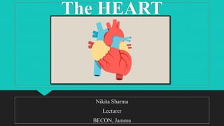
The heart is a hollow muscular organ located in the thoracic cavity between the lungs. It is composed of three layers: the outer pericardium, middle myocardium layer of muscle, and inner endocardium lining. The heart is further divided into four chambers - right and left atria on top which receive blood and right and left ventricles on bottom which pump blood out. It has a specialized conduction system including the sinoatrial node which initiates impulses and pacemaking, atrioventricular node which conducts impulses to ventricles, and Purkinje fibers which transmit the impulse through the ventricles to contract in a coordinated way. The heart is supplied by coronary arteries and drained

Recommended
More Related Content
What's hot
What's hot (20)
Similar to The Anatomy and Function of the Heart
Similar to The Anatomy and Function of the Heart (20)
More from Nikita Sharma
More from Nikita Sharma (20)
Recently uploaded
Recently uploaded (20)
The Anatomy and Function of the Heart
- 2. INTRODUCTION The heart is a roughly cone-shaped hollow muscular organ. It is about 10 cm long and is about the size of the owner’s fist. It weighs about 225 g in women and is heavier in men (about 310g)
- 3. POSITION The heart lies in the thoracic cavity in the mediastinum (space b/w the lungs). It lies obliquely, a little more to the left than the right & present a base above and an apex below. The apex is about 9cm to the left of the midline at the level of 5th intercostal space, i.e. a little below the nipple and slightly nearer the midline. The base extends to the level of the 2nd rib.
- 4. POSITION OF THE HEART IN THE THORAX
- 5. ORGANS ASSOCIATED WITH THE HEART INFERIORLY- the apex rests on the central tendon of the diaphragm SUPERIORLY- the great blood vessels i.e. the aorta, superior vena cava, Pulmonary artery and pulmonary veins POSTERIORLY- the esophagus, trachea, left and right bronchus, descending aorta, inferior vena cava and thoracic vertebrae. LATERALLY- the lungs- the left lung overlaps the left side of the heart. ANTERIORLY- the sternum, ribs & intercostal muscles.
- 6. ORGANS ASSOCIATED WITH THE HEART
- 7. STRUCTURE (THE HEART WALL) The heart wall is composed of 3 layers of tissue i.e. 1. Pericardium 2. Myocardium 3. Endocardium
- 9. PERICARDIUM Outermost layer of the heart It is made up of 2 sacs i.e. fibrous pericardium (outer) & serous pericardium (inner) The outer sac consists of fibrous tissue and the inner of a continuous double layer of serous membrane. The fibrous pericardium is continuous with the tunica adventitia of the great blood vessels above and is adherent to the diaphragm below. Its inelastic & fibrous nature prevents over distension of the heart.
- 10. PERICARDIUM
- 11. The outer layer of the serous pericardium , the parietal pericardium, lines the fibrous pericardium. The inner layer, the visceral pericardium, which is continuous with the parietal pericardium, is adherent to the heart muscle. The serous membrane consist of flattened epithelial cells. It secretes serous fluid, called pericardial fluid, into the space between the visceral and parietal layers, which allows the smooth movement between them when the heart beats. PERICARDIUM
- 12. The space between the parietal and visceral pericardium is only a potential space. In health the two layers lie closely together, with only the thin film of pericardium fluid between them. PERICARDIUM
- 13. It is composed of specialized cardiac muscle found only in the heart. It is striated, like skeletal muscle, but is not under voluntary control. Each fibre has a nucleus and one or more branches. The ends of the cells & their branches are in very close contact with the ends & branches of adjacent cells. Microscopically these joints, or intercalated discs, are thicker, darker lines than striations. This arrangement gives cardiac muscle the appearance of being a sheet of muscle rather than a very large no. of individual cells. MYOCARDIUM
- 15. Each one of these muscles don’t need separate nerve supply. The sheet arrangement of the myocardium enables the atria and ventricles to contract in a contracted & efficient manner. Running through the myocardium is also the network of specialized conducting fibres responsible for transmitting the heart’s electrical signals. Myocardium is thickest at the apex and thin out towards the base. MYOCARDIUM
- 16. This reflects the amount of work each chamber contributes to the pumping of blood. It is thickest in the left ventricle, which has the highest workload. Specialized muscle cells in the walls of the atria secrete atrial natriuretic peptide (ANP). MYOCARDIUM
- 17. The myocardium is supported by a network of fine fibres that run through all the heart muscle. This is called the fibrous skeleton of the heart. The atria and ventricles are separated by a ring of fibrous tissue, which does not conduct electrical impulses. When a wave of electrical activity passes over the atrial muscle, it can only spread to the ventricles through the conducting system that bridges the fibrous ring from the atria to ventricles. FIBROUS TISSUE IN THE HEART
- 18. ENDOCARDIUM This lines the chambers ad valves of the heart. it is a thin, smooth membrane to ensure smooth flow of blood through the heart. It consist of flattened epithelial cells & it is continuous with the endothelium lining the blood vessels.
- 19. INTERIOR OF THE HEART The heart is divided into right and left side by the sternum, a partition consisting of myocardium covered by endocardium. Each side is divided by an atrioventricular (AV) valve in to upper atrium and the ventricle below. The atrioventricular valves are formed by double folds of endocardium strengthened by a little fibrous tissue. The right AV (tricuspid) valve has 3 flaps or cusps & the left AV (mitral) valve has 2 cusps.
- 20. INTERIOR OF THE HEART
- 21. MITRAL VALVE
- 22. Flow of the blood in the heart is one way; blood enters the heart via the atria and passes into the ventricles below. The valves between the atria and ventricles open & close passively according to changes in pressure in the chambers. They open when the pressure in the atria is greater than that in the ventricles. During ventricular sysytole, the pressure in the ventricles rise above than in the atria and the valves snap shut, preventing backward flow of blood. INTERIOR OF THE HEART
- 23. The valves preventing from opening upwards into the atria by tendinous cords, called chordae tendineae, which extend from the inferior surface of the cusps to little projections of myocardium into the ventricles, covered with endothelium, called papillary muscles. INTERIOR OF THE HEART
- 24. PULONARY AND SYSTEMIC CIRCULATION
- 25. FLOW OF BLOOD THROUGH THE HEART
- 26. BLOOD SUPPLY TO THE HEART ARTERIAL SUPPLY: the heart is supplied with Arterial blood by right & left coronary arteries, Which branch from the aorta immediately distal To the aortic valve.
- 27. VENOUS DRAINAGE: most of the venous blood is collected into a number of cardiac veins that join to form the coronary sinus, which opens into right atrium. The remainder passes directly into the heart chambers through little venous channels. BLOOD SUPPLY TO THE HEART
- 28. CONDUCTING SYSTEM OF THE HEART The heart possess the property of autorhythmicity, which means it generates its own electrical impulses and beats independently of nervous or hormonal control, i.e. it is not reliant on external mechanisms to initiate each heart beat. It is supplies with both sympathetic and parasympathetic nerve fibres, which increase & decrease respectively the intrinsic heart rate. Heart responds to a number of circulating hormones, including adrenaline and thyroxine.
- 29. Small group of specialized neuromuscular cells in the myocardium initiate and conduct impulses, causing coordinated and synchronized contraction of the heart muscle. CONDUCTING SYSTEM OF THE HEART
- 30. SINOATRIAL NODE (SA NODE) This small mass of specialized cells lies in the wall of the right atrium near the opening of the superior vena cava. The sinoatrial cells generate these regular impulses because they are electrically unstable. This instability leads them to discharge (depolarize) regularly, usually between 60 & 80 times a minute. This depolarization is followed by recovery (repolarization), but almost immediately their instability leads them to discharge again, setting the heart rate.
- 31. Because the SA node discharges faster than any other part of the heart, it normally sets the heart rate and is called the pacemaker of the heart. Firing of the SA node triggers atrial contraction. SINOATRIAL NODE (SA NODE)
- 32. CONDUCTING SYSTEM OF THE HEART
- 33. This small mass of neuromuscular tissue is situated in the wall of the atrial septum near the AV valves. Normally, the AV node merely transmits the electrical signals from the atria into the ventricles. There is a delay here; the electrical signal takes 0.1 of a second to pass through into the ventricles. This allows the atria to finish contracting before the ventricles start. ATRIOVENTRICULAR NODE (AV NODE)
- 34. The AV node also has a secondary pacemaker function and takes over this role if there is a problem with the SA node itself or with the transmission of impulses from the atria. Its intrinsic firing rate is slower than that set by the SA node (40-60 beats/min.) ATRIOVENTRICULAR NODE (AV NODE)
- 35. ATRIOVENTRICULAR BUNDLE (AV BUNDLE OR BUNDLE OF HIS) This mass of the specialized fibres originates from the AV node. The AV bundle crosses the fibrous ring that separates atria and ventricles then, at the upper end of the ventricular septum, it divides in to right and left bundle branches. With in the ventricular myocardium the branches breakup into the fine fibres called purkinje fibres. The AV bundle, bundle branches and purkinje fibres transmit electrical impulses from the AV node to the apex of the myocardium where the wave of ventricular contraction begins, then sweeps upwards and outwards, pumping blood into the pulmonary artery and the aorta.
- 36. NERVE SUPPLY TO THE HEART The vagus nerve (parasympathetic) supplies mainly the SA & AV nodes and atrial muscles. Vagal stimulation reduces the rate at which impulses are produced, decreasing the rate and force of the heartbeat. Sympathetic nerve supply to the SA and AV nodes and the myocardium of the atria and ventricles & stimulation increases the rate and force of the heartbeat.