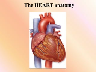
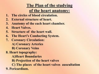
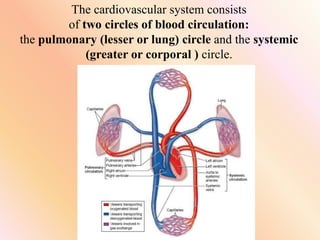
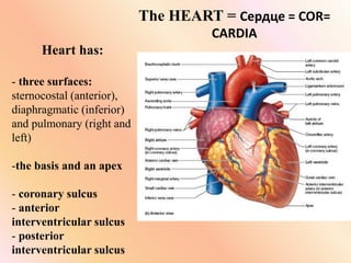
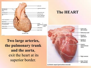
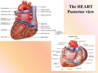
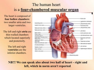
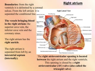
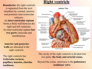
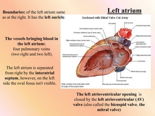
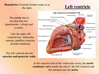
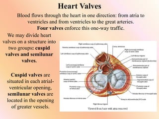
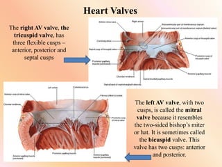
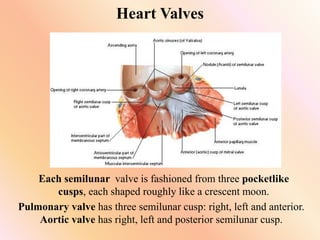
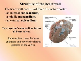
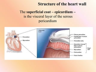
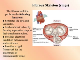
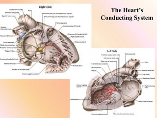
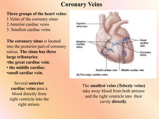
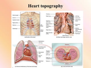
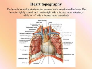
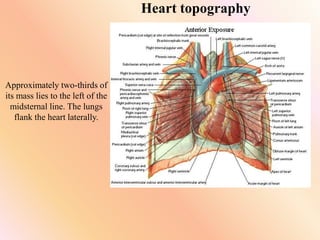
The document provides a detailed overview of heart anatomy, beginning with an outline of the key topics covered. It then describes the two circles of blood circulation - pulmonary and systemic. The four chambers of the heart are explained, including the right and left atria and ventricles. Heart valves, the heart wall structure, and conducting system are defined. Coronary circulation and the vessels are outlined. Key aspects of heart topography like boundaries and valve locations are defined. Finally, the layers of the pericardium are described.
A closed system of the heart and blood vessels
The heart pumps blood
Blood vessels allow blood to circulate to all parts of the body
The function of the cardiovascular system is to deliver oxygen and nutrients and to remove carbon dioxide and other waste products
The heart contributes to homeostasis by pumping blood through blood vessels to the tissues of the body to deliver oxygen and nutrients and remove wastes.
Blood to reach body cells and exchange materials with them, it must be pumped continuously by the heart through the body’s blood vessels.
The heart beats about 100,000 times every day, which adds up to about 35 million beats in a year, and approximately 2.5 billion times in an average lifetime.
The left side of the heart pumps blood through an estimated 100,000 km (60,000 mi) of blood vessels, which is equivalent to traveling around the earth’s equator about three times.
The right side of the heart pumps blood through the lungs, enabling blood to pick up oxygen and unload carbon dioxide.
The heart has four chambers. The two superior receiving chambers are the atria (= entry halls or chambers), and the two inferior pumping chambers are the ventricles (= little bellies).
On the anterior surface of each atrium is a wrinkled pouchlike structure called an auricle
The Roman Empire A Historical Colossus.pdfkaushalkr1407
The Roman Empire, a vast and enduring power, stands as one of history's most remarkable civilizations, leaving an indelible imprint on the world. It emerged from the Roman Republic, transitioning into an imperial powerhouse under the leadership of Augustus Caesar in 27 BCE. This transformation marked the beginning of an era defined by unprecedented territorial expansion, architectural marvels, and profound cultural influence.
The empire's roots lie in the city of Rome, founded, according to legend, by Romulus in 753 BCE. Over centuries, Rome evolved from a small settlement to a formidable republic, characterized by a complex political system with elected officials and checks on power. However, internal strife, class conflicts, and military ambitions paved the way for the end of the Republic. Julius Caesar’s dictatorship and subsequent assassination in 44 BCE created a power vacuum, leading to a civil war. Octavian, later Augustus, emerged victorious, heralding the Roman Empire’s birth.
Under Augustus, the empire experienced the Pax Romana, a 200-year period of relative peace and stability. Augustus reformed the military, established efficient administrative systems, and initiated grand construction projects. The empire's borders expanded, encompassing territories from Britain to Egypt and from Spain to the Euphrates. Roman legions, renowned for their discipline and engineering prowess, secured and maintained these vast territories, building roads, fortifications, and cities that facilitated control and integration.
The Roman Empire’s society was hierarchical, with a rigid class system. At the top were the patricians, wealthy elites who held significant political power. Below them were the plebeians, free citizens with limited political influence, and the vast numbers of slaves who formed the backbone of the economy. The family unit was central, governed by the paterfamilias, the male head who held absolute authority.
Culturally, the Romans were eclectic, absorbing and adapting elements from the civilizations they encountered, particularly the Greeks. Roman art, literature, and philosophy reflected this synthesis, creating a rich cultural tapestry. Latin, the Roman language, became the lingua franca of the Western world, influencing numerous modern languages.
Roman architecture and engineering achievements were monumental. They perfected the arch, vault, and dome, constructing enduring structures like the Colosseum, Pantheon, and aqueducts. These engineering marvels not only showcased Roman ingenuity but also served practical purposes, from public entertainment to water supply.
A Strategic Approach: GenAI in EducationPeter Windle
Artificial Intelligence (AI) technologies such as Generative AI, Image Generators and Large Language Models have had a dramatic impact on teaching, learning and assessment over the past 18 months. The most immediate threat AI posed was to Academic Integrity with Higher Education Institutes (HEIs) focusing their efforts on combating the use of GenAI in assessment. Guidelines were developed for staff and students, policies put in place too. Innovative educators have forged paths in the use of Generative AI for teaching, learning and assessments leading to pockets of transformation springing up across HEIs, often with little or no top-down guidance, support or direction.
This Gasta posits a strategic approach to integrating AI into HEIs to prepare staff, students and the curriculum for an evolving world and workplace. We will highlight the advantages of working with these technologies beyond the realm of teaching, learning and assessment by considering prompt engineering skills, industry impact, curriculum changes, and the need for staff upskilling. In contrast, not engaging strategically with Generative AI poses risks, including falling behind peers, missed opportunities and failing to ensure our graduates remain employable. The rapid evolution of AI technologies necessitates a proactive and strategic approach if we are to remain relevant.
Francesca Gottschalk - How can education support child empowerment.pptxEduSkills OECD
Francesca Gottschalk from the OECD’s Centre for Educational Research and Innovation presents at the Ask an Expert Webinar: How can education support child empowerment?
Macroeconomics- Movie Location
This will be used as part of your Personal Professional Portfolio once graded.
Objective:
Prepare a presentation or a paper using research, basic comparative analysis, data organization and application of economic information. You will make an informed assessment of an economic climate outside of the United States to accomplish an entertainment industry objective.
Model Attribute Check Company Auto PropertyCeline George
In Odoo, the multi-company feature allows you to manage multiple companies within a single Odoo database instance. Each company can have its own configurations while still sharing common resources such as products, customers, and suppliers.
Unit 8 - Information and Communication Technology (Paper I).pdfThiyagu K
This slides describes the basic concepts of ICT, basics of Email, Emerging Technology and Digital Initiatives in Education. This presentations aligns with the UGC Paper I syllabus.
Palestine last event orientationfvgnh .pptxRaedMohamed3
An EFL lesson about the current events in Palestine. It is intended to be for intermediate students who wish to increase their listening skills through a short lesson in power point.
Acetabularia Information For Class 9 .docxvaibhavrinwa19
Acetabularia acetabulum is a single-celled green alga that in its vegetative state is morphologically differentiated into a basal rhizoid and an axially elongated stalk, which bears whorls of branching hairs. The single diploid nucleus resides in the rhizoid.
2. The Plan of the studying
of the heart anatomy:
1. The circles of blood circulation.
2. External structure of heart.
3. Anatomy of the each heart chamber.
4. Heart Valves.
5. Structure of the heart wall.
6. The Heart’s Conducting System.
7. Coronary Circulation:
A) Coronary Arteries
B) Coronary Veins
8. Heart topography:
A)Heart Boundaries
B) Projection of the heart valves
C) The places of the heart valves auscultation
9. Pericardium.
3. The cardiovascular system consists
of two circles of blood circulation:
the pulmonary (lesser or lung) circle and the systemic
(greater or corporal ) circle.
4. The pulmonary (lesser or lung) circle of blood
circulation is started with the pulmonary trunk in the right
ventricle and finished in the left atrium with four
pulmonary veins (two pairs – two left and two right).
Its function is the gas exchange and blood saturation by
oxygen.
The systemic (greater or corporal) circle of blood
circulation is started with aorta in the left ventricle and
finished in the right atrium with the superior and inferior
cava veins. The aorta is divided into numerous arteries,
then the capillary course follows, then blood passes in
veins.
Function of this circle is supplementary of all organs and
tissues by oxygen.
NB!!!!
5. The HEART = Сердце = COR=
CARDIA
Heart has:
- three surfaces:
sternocostal (anterior),
diaphragmatic (inferior)
and pulmonary (right and
left)
-the basis and an apex
- coronary sulcus
- anterior
interventricular sulcus
- posterior
interventricular sulcus
6. The HEART
Two large arteries,
the pulmonary trunk
and the aorta,
exit the heart at its
superior border.
8. The human heart
is a four-chambered muscular organ
The heart is composed of
four hollow chambers:
two smaller atria and two
larger ventricles.
The left and right atria are
thin-walled chambers
which located superiorly
and posteriorly.
The left and right
ventricles are the
inferior chambers.
NB!!! We can speak also about two half of heart - right and
left, which in norm aren't reported.
9. Right atrium
Boundaries: from the right
ventricle it is delimited by a coronal
sulcus. From the left atrium it is
separated the conditional line.
The vessels bringing blood
in the right atrium: the
superior cava vein, the
inferior cava vein and the
coronary sinus.
The right atrium has the
right auricle.
The right atrium is
separated from left by an
interatrial septum
(internally).
The right atrioventricular opening is located
between the right atrium and the right ventricle.
This opening is closed by a right
atrioventricular (AV) valve (also called the
tricuspid valve)
10. Right ventricle
Boundaries: the right ventricle
is separated from the next
chambers by coronal, anterior
and posterior interventricular
sulcuses.
An interventricular septum
forms a thick wall between the
right and left ventricles.
An interventricular septum has
two parts: muscular and
membranous.
Anterior and posterior
walls are allocated in the
right ventricle.
The right ventricle has
trabeculae carneae,
papillary muscles, chordae
tendineae.
The cavity of the right ventricle is divided into
two parts: the body and arterial conus.
Beyond the conus arteriosus is the pulmonary
semilunar valve
11. Left atrium
Boundaries: of the left atrium same
as at the right. It has the left auricle.
The vessels bringing blood in
the left atrium:
four pulmonary veins
(two right and two left).
The left atrium is separated
from right by the interatrial
septum, however, on the left
side the oval fossa isn't visible.
The left atrioventricular opening is
closed by the left atrioventricular (AV)
valve (also called the bicuspid valve, the
mitral valve)
12. Left ventricle
Boundaries: External borders same as at
the right.
The cavity also is
divided into two
departments - a body and
a conus.
Like the right, left
ventricle has trabeculae
carneae, papillary muscles,
chordae tendineae.
The left ventricle has the
anterior and posterior walls.
At the superior end of the ventricular cavity, the aortic
semilunar valve marks the end of the left ventricle and
the entrance into the aorta.
13. Heart Valves
Blood flows through the heart in one direction: from atria to
ventricles and from ventricles to the great arteries.
Four valves enforce this one-way traffic.
We may divide heart
valves on a structure into
two groups: cuspid
valves and semilunar
valves.
Cuspid valves are
situated in each atrial-
ventricular opening,
semilunar valves are
located in the opening
of greater vessels.
14. Heart Valves
The right AV valve, the
tricuspid valve, has
three flexible cusps –
anterior, posterior and
septal cusps
The left AV valve, with two
cusps, is called the mitral
valve because it resembles
the two-sided bishop’s miter
or hat. It is sometimes called
the bicuspid valve. This
valve has two cusps: anterior
and posterior.
15. Heart Valves
Each semilunar valve is fashioned from three pocketlike
cusps, each shaped roughly like a crescent moon.
Pulmonary valve has three semilunar cusp: right, left and anterior.
Aortic valve has right, left and posterior semilunar cusp.
16. The heart wall consists of three distinctive coats:
- an internal endocardium,
- a middle myocardium,
- an external epicardium.
Structure of the heart wall
Two layers of endocardium forms
all heart valves.
Endocardium lines the heart
chambers and covers the fibrous
skeleton of the valves.
17. The middle coat is the myocardium (“muscle heart”)
Structure of the heart wall
The myocardium of atriums and ventricles
begins and attached to fibrous rings.
The myocardium of atriums differs from a
myocardium of ventricles.
•The myocardium of atriums is poorly
expressed and has two layers.
•The myocardium of ventricles is
considerably expressed, especially in the
left ventricle. It has three layers.
18. Structure of the heart wall
The superficial coat – epicardium –
is the visceral layer of the serous
pericardium
19. Fibrous Skeleton (rings)
The fibrous skeleton
performs the following
functions:
■ Separates the atria and
ventricles.
■ Anchors heart valves by
forming supportive rings at
their attachment points.
■ Provides electrical
insulation between atria
and ventricles.
■ Provides a rigid
framework for the
attachment of
cardiacmuscle tissue.
20. The Heart’s Conducting System
The heart exhibits autorhythmicity.
The cardiac conduction system coordinates and
synchronizes heart activity.
Functions of the Heart’s Conducting System:
1 . Changes a heart work regime under the influence of
nervous impulses.
2 . Coordinates work of atriums and ventricles.
22. The Heart’s Conducting System is presented by the
following fibers and nodes:
1. The sinoatrial (SA) node, which are located in the
posterior wall of the right atrium, adjacent to the entrance
of the superior vena cava.
2. The atrioventricular (AV) node is located in the floor of
the interatrium septum between the right AV valve and the
opening for the coronary sinus.
3. The bundle of His receives the muscle impulse from the
AV node and extends into the interventricular septum
before dividing into left and right bundles.
4. The left and right bundles. These bundles conduct the
impulse to conduction fibers called Purkinje and extend
through the walls of the ventricles.
23. Coronary Circulation
The coronary circulation, the functional blood supply of
the heart, is the shortest circulation in the body.
Coronary Arteries
The left and right coronary arteries
both arise from the base of the aorta
and encircle the heart in the coronary
sulcus.
24. Coronary Arteries
The left coronary artery
runs toward the left side of the heart
and then divides into two major
branches:
■ The anterior interventricular
artery follows the anterior
interventricular sulcus.
■ The circumflex artery passes
into the left part of a coronal sulcus.
The left coronary artery supplies:
- the left atrium,
- full anterior wall of left ventricle
and anterior papillar muscle,
- part of the anterior wall of right
ventricle,
- part of the posterior wall of the
left ventricle,
- anterior part of the interventricular
septum.
25. Coronary Arteries
The right coronary artery
courses to the right side of the heart,
where it also proceed as the
posterior interventricular artery
which runs to the heart apex and
supplies the posterior ventricular
walls.
Together the branches of the right
coronary artery supply:
• - the right atrium
• - interatrial septum
• - part of the anterior wall of right
ventricle
• - full posterior wall of the right
ventricle
• - part of the posterior wall of the
left ventricle,
• - posterior part of the
interventricular septum
• - posterior papillar muscle of the
left ventricle
26. Coronary Veins
Three groups of the heart veins:
1.Veins of the coronary sinus
2.Anterior cardiac veins
3. Smallest cardiac veins
The coronary sinus is located
into the posterior part of coronary
sulcus. The sinus has three
large tributaries:
•the great cardiac vein;
• the middle cardiac;
•small cardiac vein.
Several anterior
cardiac veins pass a
blood directly from
right ventricle into the
right atrium.
The smallest veins (Tebeziy veins)
take away blood from both atriums
and the right ventricle into their
cavity directly.
28. Heart topography
The heart is located posterior to the sternum in the anterior mediastinum. The
heart is slightly rotated such that its right side is located more anteriorly,
while its left side is located more posteriorly.
30. Heart topography
NB!!! Heart Boundaries
The superior bound passes on the upper margin of the third
ribs. To the right it acts on the 1-2cm externally from the right
sternum margin. To the left this border reaches the distance
middle between the left sternum margin and the left
middleclavical line.
The right border passes down parallel to the right sternum
margin to a cartilage of the fifth rib.
To define the left and lower borders of heart, it is necessary
to find a projection of a heart apex at first. The heart apex is
projected in the left fifth intercostals space on 1-1,5 cm
internally from the middleclavical line. The left and lower
borders of heart are agreed on a heart apex.
31. Heart topography
NB!!! In studying topography of heart valves we will allocate 1. a projection of
valves and 2. the places of their auscultation.
Projection of the heart
valves
•Semilunar valves settle down at the
level of the third ribs. Thus the
pulmonary trunk valve is behind a
cartilage of the left third rib at sternum
margin. The aorta valve is located at
the same level behind a sternum.
•Cuspid valves. The bicuspid valve
settles down behind a sternum closer to
the left third intercostals space. The
tricuspid valve is located behind a
sternum closer to a cartilage of the fifth
right rib.
32. Heart topography
The places of the heart
valves auscultation
Semilunar valves are listened in the
second intercostals space at the
sternum margin, thus the aorta valve
is listened on the right side, the
pulmonary trunk valve at the left
side.
The bicuspid valve is listened on a
heart apex. The tricuspid valve is
listened at the level of the fifth right
rib cartilage or at the basis of a
xiphoid process.
33. Pericardium
The pericardium is composed
of two parts.
The outer portion is a dense connective
tissue layer called the fibrous
pericardium. This layer is attached to
both the diaphragm and the base of the
great vessels.
The inner portion is a thin,
doublelayered serous membrane called the
serous pericardium. The serous
pericardium may be subdivided into (1) a
parietal layer and (2) a visceral layer of
serous pericardium (also called the
epicardium).
The thin space between the parietal
and visceral layers of the serous
pericardium is the pericardial cavity